Filter :

Yellow is a happy, joyous colour. It reminds us of sunflowers, sunshine and smiley faces. But when it shows up on your skin, eyes, or nails, it is not a happy sign. It is a warning sign.
This could be jaundice, and its causes and consequences vary widely from person to person. What causes jaundice in a newborn is very different from what causes it in an adult, and the treatment differs too.
In this blog, we will break down how jaundice differs between children and adults, including the causes, the symptoms, and when it is time to see a doctor.
Table of Contents
Jaundice is a symptom, not a disease. It causes the skin and the whites of the eyes to become yellow because a substance called bilirubin has built up in the blood.
Bilirubin is produced naturally when old red blood cells break down. This process releases hemoglobin, the protein that carries oxygen in the blood, which the body then converts into bilirubin. The liver normally processes it and passes it into bile, which eventually leaves the body through the stool.
Jaundice affects you when too much bilirubin is produced, the liver cannot process it properly, or bile flow is blocked. This is why the same yellow appearance can have very different causes in a newborn, an older child and an adult.
Jaundice in Newborns: It is particularly common during the newborn period. According to NICE (National Institute for Health and Care Excellence), around 60% of full-term babies and 80% of premature babies develop jaundice during the first week of life. Most cases are physiological, meaning they result from normal immaturity of the newborn’s liver and resolve without causing harm.
Jaundice in Older Child: Jaundice in an older child is less expected. But if the symptoms are particularly accompanied by fever, abdominal pain, dark urine or pale stools, it should be medically evaluated on time.
Jaundice in Adults:In adults, jaundice is not considered a normal age-related change. New yellowing of the skin or eyes should be medically evaluated because it can occur with hepatitis, alcohol-related liver disease, gallstones, medication-related liver injury, cirrhosis and other conditions.
Fatty liver disease affects a person when excess fat accumulates in the liver. Mild fatty liver disease does not usually cause jaundice, so visible yellowing should not be blamed on fatty liver alone. Jaundice may develop when the disease progresses to significant liver inflammation, advanced fibrosis or cirrhosis, which can impair the liver’s ability to process bilirubin.
A gallstone can block the flow of bile from the gallbladder into the intestine. This may cause jaundice along with dark urine, pale stools, itching or pain in the upper abdomen. Other blockages can also affect bile drainage.
Cirrhosis is advanced scarring of the liver. It can develop after years of conditions such as chronic viral hepatitis, alcohol-related liver disease or other chronic liver disorders. As liver function declines, bilirubin may accumulate.
Certain medicines can injure the liver and cause jaundice. This is why a doctor should review all prescription medicines, over-the-counter drugs and supplements when evaluating jaundice.
The causes depend strongly on the child’s age.
A newborn’s liver is still developing and may not process bilirubin as efficiently as an older child’s liver. Physiological jaundice usually appears after the first 24 hours of life that needs quick medical attention. It improves as bilirubin processing becomes more efficient. Most babies with uncomplicated jaundice improve within about 10 to 14 days.
Breastfeeding jaundice can occur when a newborn is not taking in enough milk, often during the first week. Breast milk jaundice is different and may persist for longer because substances in breast milk can affect bilirubin processing. This does not mean that you should stop breastfeeding. But, feeding adequacy and the baby’s bilirubin level should be assessed by a healthcare professional.
Sometimes the baby’s blood type does not match the mother’s, a condition called Rh or ABO incompatibility. This causes the baby’s red blood cells to break down faster than normal, leading to a rise in bilirubin and, often, noticeable jaundice.
Glucose-6-phosphate dehydrogenase (G6PD) deficiency is an inherited condition that can make red blood cells more vulnerable to breakdown. It is an important risk factor for severe neonatal hyperbilirubinemia (excess of bilirubin in the blood of a newborn).
In this rare condition, the bile ducts become scarred and blocked, stopping bile from reaching the intestine. If jaundice persists, particularly alongside pale, grey, or white stools and dark urine, it should be checked by a doctor as soon as possible.
Certain infections can also cause jaundice in newborns and children. A jaundiced child who is unwell, feverish, unusually sleepy or feeding poorly needs timely medical assessment.
The most recognisable symptom is yellowing of the whites of the eyes and skin.
In newborns, the yellow colour usually begins around the face and may extend towards the chest, abdomen, arms and legs as bilirubin rises. However, visual assessment alone is not reliable enough to determine bilirubin severity.
Other symptoms are:
A high-pitched cry, abnormal stiffness or floppiness, difficulty waking, seizures or feeding difficulty in a jaundiced newborn are warning signs requiring urgent medical attention because severe bilirubin elevation can affect the brain.
Diagnosis begins by determining how high the bilirubin is and why it has increased.
Depending on age and symptoms, evaluation may include:
For newborns, treatment decisions are based on bilirubin measurements interpreted according to the baby’s age in hours and risk factors.
Jaundice treatment in adults depends on the underlying cause. Treatment may involve:
Once the underlying problem is treated, bilirubin often falls and jaundice improves.
You should seek urgent medical care if jaundice is accompanied by confusion, severe abdominal pain, frequent vomiting, fever, vomiting blood, marked drowsiness or other signs of serious illness.
For newborns, seek urgent assessment if jaundice appears during the first 24 hours, is rapidly worsening, or occurs with poor feeding, difficulty waking, abnormal muscle tone, a high-pitched cry, fever or very dark urine and pale stools.
One warning sign that is easy to miss is pale or white stool in a jaundiced baby. It can mean bile is not reaching the intestine, and it should be checked right away, since it may point to conditions like biliary atresia (bile ducts are blocked, missing or scarred).
The yellowing may look similar, but the medical story behind it can be completely different. Jaundice in a newborn is physiological, whereas jaundice in an older child or adult generally deserves closer investigation. Age, bilirubin level, associated symptoms and the underlying cause all influence the severity.
If you or your child develops jaundice, the Paediatrics or Gastroenterology and Hepatology Department at the CK Birla Hospital can help determine the cause and guide the appropriate next steps, from diagnostic testing to treatment.

अचानक सिर घूमने लगे, कमरा जैसे नाव की तरह डोलने लगे, और पैरों तले जमीन खिसकती महसूस हो, यह अनुभव जिसने भी झेला है वह जानता है कि यह कितना डरावना हो सकता है। कोई सीढ़ी उतरते वक्त लड़खड़ा जाता है, तो किसी को सुबह बिस्तर से उठते ही ऐसा लगता है मानो पूरा कमरा घूम रहा हो। ज्यादातर लोग इसे थकान या नींद की कमी समझ कर टाल देते हैं, लेकिन जब यह बार-बार होने लगे, तो यह हमारे शरीर के संतुलन तंत्र में किसी गड़बड़ी की तरफ इशारा करता है।
चक्कर आना कोई नई बात नहीं, लगभग हर व्यक्ति ने अपनी जिंदगी में कभी न कभी इसे महसूस किया है। लेकिन जब यह बार-बार लौटकर आए, रोजमर्रा के काम में रुकावट डाले, या गिरने का डर पैदा करे, तो इसे नजरअंदाज करना सही नहीं है। अक्सर हमारी मां कुछ ऐसे लक्षण बताती हैं कि चक्कर आ रहा है और सिर घूम रहा है। आज हम उन्हीं लक्षणों के आधार पर समझेंगे कि चक्कर आना और वर्टिगो के बीच का फर्क क्या है और इसके पीछे का कारण क्या है। अगर आपको या आपके किसी अपने को बार-बार चक्कर आने की शिकायत है, तो देर न करें, हमारे अनुभवी न्यूरोलॉजिस्ट या ENT विशेषज्ञ से अपॉइंटमेंट बुक करें और सही वजह का पता लगाएं।
Table of Contents
आम बोलचाल में हम “चक्कर आना” और “वर्टिगो” को एक ही समझ लेते हैं, जबकि मेडिकल नजरिए से इन दोनों में साफ फर्क है। यह फर्क समझना इसलिए भी जरूरी है, क्योंकि इसी से डॉक्टर को असली कारण तक पहुंचने में मदद मिलती है।
यह फर्क समझना डॉक्टर और मरीज दोनों के लिए फायदेमंद है, क्योंकि इससे सही जांच और सही इलाज तक जल्दी पहुंचा जा सकता है।
डॉक्टर वर्टिगो को मुख्य रूप से दो हिस्सों में बांटते हैं।
यह वर्टिगो का सबसे सामान्य प्रकार है और यह आंतरिक कान या वेस्टिबुलर नर्व में गड़बड़ी की वजह से होता है। इसमें शामिल हैं, बेनाइन पैरॉक्सिस्मल पोजिशनल वर्टिगो (BPPV), मेनियर रोग, वेस्टिबुलर न्यूराइटिस और लैबिरिंथाइटिस। ज्यादातर मामलों में यह गंभीर नहीं होता और सही इलाज से ठीक हो जाता है।
यह कम आम है, लेकिन ज्यादा गंभीर हो सकता है। इसकी वजह दिमाग से जुड़ी समस्याएं होती हैं, जैसे माइग्रेन, स्ट्रोक, ब्रेन ट्यूमर या मल्टीपल स्क्लेरोसिस। सेंट्रल वर्टिगो में आमतौर पर लक्षण ज्यादा गंभीर होते हैं, जैसे चलने में गंभीर दिक्कत या शरीर पर से नियंत्रण खोना। इसलिए इस तरह के लक्षणों को हल्के में नहीं लेना चाहिए।
पेशाब की तरह ही चक्कर आना भी शरीर के अंदर कई अलग-अलग सिस्टम की गड़बड़ी का नतीजा हो सकता है। नीचे कुछ प्रमुख कारण दिए गए हैं।
चक्कर आने के साथ अक्सर ये लक्षण भी महसूस हो सकते हैं –
इन लक्षणों के नजर आते ही बिना देर किए हमारे अनुभवी न्यूरोलॉजिस्ट से मिलें और वर्टिगो का इलाज सफलता से कराएं।
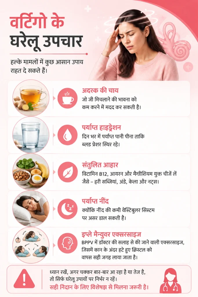
हल्के मामलों में कुछ आसान उपाय राहत दे सकते हैं, हालांकि यह किसी गंभीर वजह का इलाज नहीं है।
ध्यान रहे, अगर चक्कर बार-बार आ रहा है या तेज है, तो सिर्फ घरेलू उपायों पर निर्भर न रहें, सही निदान के लिए विशेषज्ञ से मिलना जरूरी है।
जब वर्टिगो (चक्कर आने) का संदेह होता है, तो इलाज शुरू करने से पहले BPPV की सटीक जांच (BPPV Diagnosis) की जाती है, ताकि समस्या के असली कारण की पहचान हो सके। ये जांच फिलहाल हमारे पास उपलब्ध है। जांच के बाद स्थिति के आधार पर मानक और आधुनिक उपचार दिए जाते हैं:
अगर चक्कर के साथ नीचे दिए गए लक्षणों में से कोई भी महसूस हो, तो इसे इमरजेंसी समझें और तुरंत डॉक्टर या अस्पताल पहुंचे –
यह लक्षण स्ट्रोक जैसी गंभीर स्थिति के संकेत हो सकते हैं, जहां हर मिनट कीमती है। ऐसे में देरी करना बड़ा नुकसान पहुंचा सकता है।
चक्कर आना कई बार सिर्फ थकान या पानी की कमी की वजह से होता है, तो कई बार यह आंतरिक कान, दिमाग या दिल से जुड़ी किसी गहरी समस्या का संकेत भी हो सकता है। सबसे जरूरी बात यह है कि इसे बार-बार नजरअंदाज न करें। सही समय पर सही जांच और सही विशेषज्ञ की सलाह ही इस समस्या का सबसे भरोसेमंद हल है। हमारे अस्पताल में अनुभवी न्यूरोलॉजिस्ट और ENT विशेषज्ञों की टीम मौजूद है, आप किसी से भी अपना परामर्श बुक करा कर अपनी समस्या का इलाज करा सकते हैं।
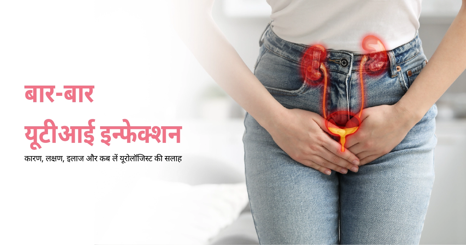
एक अंतरराष्ट्रीय रिसर्च के अनुसार करीब आधी वयस्क महिलाएं अपने जीवन में कम से कम एक बार यूटीआई का सामना करती हैं, और इनमें से लगभग एक-चौथाई महिलाओं को यह इन्फेक्शन बार-बार होता है। सही जानकारी और सही समय पर इलाज पाने वाली (या कराने वाली) महिलाएं इस चक्र से बाहर निकल सकती हैं।
इस ब्लॉग में हम बार-बार यूटीआई क्यों होता है, इसके लक्षण, कारण, इलाज के विकल्प और कब यूरोलॉजिस्ट या नेफ्रोलॉजिस्ट से मिलना जरूरी है, यह सब आसान भाषा में समझेंगे। अगर आप भी बार-बार यूटीआई से परेशान हैं, तो अकेले इससे लड़ने की जरूरत नहीं, हमारे अनुभवी यूरोलॉजी विशेषज्ञों से अभी अपॉइंटमेंट बुक करें और इस समस्या का स्थायी समाधान पाएं।
Table of Contents
यूटीआई यानी यूरिनरी ट्रैक्ट इन्फेक्शन (Urinary Tract Infection) तब होता है, जब बैक्टीरिया, ज्यादातर मामलों में E. coli नाम का बैक्टीरिया, मूत्र मार्ग में पहुंचकर संक्रमण फैला देता है। जब यह इन्फेक्शन बार-बार लौटकर आता है, तो इसे मेडिकल भाषा में “रिकरंट यूटीआई” (Recurrent UTI) कहा जाता है। ज्यादातर मामलों में हर बार अलग बैक्टीरिया की वजह से नया इन्फेक्शन होता है, यानी यह पुराना इन्फेक्शन दोबारा सिर नहीं उठाता, बल्कि हर बार एक नई लड़ाई होती है।
क्लिनिकल तौर पर इसकी परिभाषा साफ है, अगर किसी महिला को 6 महीने में 2 बार या 12 महीने में 3 बार यूटीआई हो जाए, तो इसे बार-बार होने वाला यूटीआई माना जाता है। यह क्राइटेरिया दुनिया भर के यूरोलॉजी दिशा-निर्देशों में लगभग एक जैसा है। दिलचस्प बात यह है कि 55 साल से ऊपर की महिलाओं में यह समस्या और भी ज्यादा देखी जाती है, एक अध्ययन के अनुसार इस उम्र वर्ग की करीब आधी महिलाओं को एक साल के भीतर दोबारा इन्फेक्शन हो जाता है, जबकि युवा महिलाओं में यह आंकड़ा कुछ कम रहता है।
बार-बार यूटीआई के लक्षण पहचानना इसलिए भी जरूरी है क्योंकि शुरुआती संकेतों को अक्सर मामूली समझकर टाल दिया जाता है। ध्यान देने वाले लक्षण इस प्रकार हैं –
अगर बुखार के साथ कमर दर्द जैसे लक्षण दिखें, तो इसे सामान्य यूटीआई समझकर नजरअंदाज बिल्कुल न करें, यह किडनी इन्फेक्शन की तरफ इशारा कर सकता है।
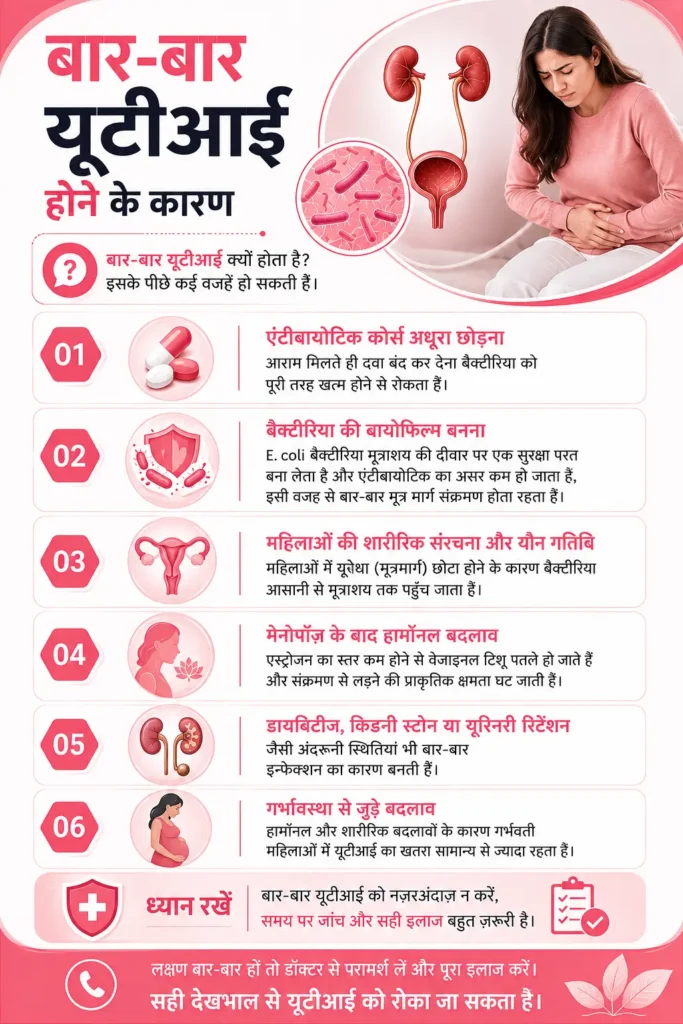
बार-बार यूटीआई क्यों होता है, यह सवाल हमारे पास आने वाली ज्यादातर महिलाओं का सबसे पहला सवाल होता है। इसके पीछे कई वजहें हो सकती हैं –
पिछले महीने हमारे यूरोलॉजी OPD में एक 42 वर्षीय महिला आईं, जो पिछले एक साल में चार बार यूटीआई से जूझ चुकी थीं। हर बार वे खुद ही तीन दिन एंटीबायोटिक लेकर छोड़ देती थीं, क्योंकि लक्षण जल्दी कम हो जाते थे। जांच में पता चला कि अधूरा कोर्स ही उनके बार-बार इन्फेक्शन का असली कारण था। यूरिन कल्चर टेस्ट और सही अवधि की एंटीबायोटिक थेरेपी शुरू करने के बाद उनकी स्थिति काफी हद तक सुधरी। ऐसे मामले बताते हैं कि दवा का कोर्स पूरा करना कितना जरूरी है, चाहे लक्षण कितनी भी जल्दी ठीक क्यों न लगें।
अगर बार-बार यूटीआई को नजरअंदाज किया जाए, तो यह सिर्फ मूत्राशय तक सीमित नहीं रहता, इसके गंभीर परिणाम हो सकते हैं जैसे कि –
इसलिए बुखार, तेज कमर दर्द या उल्टी जैसे लक्षणों के साथ आने वाले यूटीआई को कभी भी घर पर मैनेज करने की कोशिश न करें।
इलाज का तरीका इस बात पर निर्भर करता है कि इन्फेक्शन कितनी बार हो रहा है और इसकी वजह क्या है –
हर मरीज की स्थिति अलग होती है, इसलिए बिना यूरिन कल्चर टेस्ट और डॉक्टर की सलाह के खुद से एंटीबायोटिक शुरू करना या बदलना सही नहीं। दिल्ली और गुरुग्राम, दोनों जगह स्थित हमारे अस्पतालों में अनुभवी यूरोलॉजिस्ट मरीज की पूरी हिस्ट्री देखकर व्यक्तिगत इलाज योजना तैयार करते हैं, आप अपनी सुविधा के अनुसार किसी भी सेंटर में अपॉइंटमेंट ले सकते हैं।
दवाओं के साथ-साथ कुछ आदतें बार-बार यूटीआई से बचाव में बड़ी भूमिका निभाती हैं –
ये उपाय यूटीआई का घरेलू इलाज नहीं बल्कि बचाव के तरीके हैं, इंफेक्शन हो जाने पर डॉक्टरी सलाह और सही जांच जरूरी है।
अगर इनमें से कोई भी लक्षण दिखे, तो देर न करें और तुरंत यूरोलॉजिस्ट से संपर्क करें –
अक्सर लोग सोचते हैं कि यूरोलॉजिस्ट और नेफ्रोलॉजिस्ट एक ही होते हैं, जबकि दोनों की भूमिका अलग है। यूरोलॉजिस्ट मूत्र मार्ग की संरचनात्मक और संक्रमण संबंधी समस्याओं का इलाज करते हैं, जबकि नेफ्रोलॉजिस्ट किडनी की कार्यक्षमता और उससे जुड़ी बीमारियों पर फोकस करते हैं। अगर बार-बार यूटीआई किडनी को प्रभावित करने लगे, तो दोनों विशेषज्ञों की राय एक साथ ली जा सकती है।
बार-बार यूटीआई एक ऐसी समस्या है जिसे शर्म या झिझक की वजह से अक्सर छुपाया जाता है, जबकि यह पूरी तरह से एक सामान्य मेडिकल स्थिति है, जिसका सही इलाज मौजूद है। सही जांच, सही एंटीबायोटिक कोर्स और जीवनशैली में छोटे बदलाव अपनाकर इस चक्र को तोड़ा जा सकता है। अगर आप या आपके परिवार में कोई बार-बार यूटीआई का सामना कर रहा है, तो इसे नजरअंदाज न करें। हमारे अस्पतालों में अनुभवी यूरोलॉजी टीम से आज ही अपॉइंटमेंट बुक करें और इस तकलीफ से स्थायी राहत पाएं।

Do you feel breathless climbing a few stairs? Or notice a strange tightness in your chest after a stressful moment?
Most people blame these signs on acidity, poor sleep, stress or aging. But sometimes, these are actually early signs of a blockage in the arteries that supply blood to your heart. Catching them early can help you get checked before it becomes serious. Keep reading to know the early signs you should not miss and when it is time to see a cardiologist.
Table of Contents
Heart blockage is not a formal medical diagnosis. In everyday language, it usually refers to narrowing of the coronary arteries, the blood vessels that supply oxygen-rich blood to the heart muscle. Medically, this condition is called coronary artery disease (CAD).
This narrowing of arteries is usually caused due to atherosclerosis, where fatty deposits called plaque build up inside artery walls. As plaque increases, the artery may become narrower and reduce blood flow, particularly when the heart needs more oxygen during exercise or stress. In some cases, it can also lead to a blood clot, which may block the artery completely and cause a heart attack.
This plaque buildup happens slowly, over several years, and may not cause any noticeable symptoms in the early stages. That is why understanding the early warning signs is very important.
The early signs of heart-related problems may not feel urgent. They may even feel normal. But over time, they can slowly make you feel less fit and start affecting your health. Here are the common early signs of heart blockage you should not miss and if something feels off, seek medical evaluation.
Chest discomfort is one of the commonly known heart blockage symptoms. It may feel like:
The pain is commonly felt in the centre or left side of the chest, but heart blockage chest pain location can vary. The sensation may also spread to the arm, shoulder, jaw, neck or back.
Getting unusually breathless while walking, climbing stairs or performing activities you previously managed comfortably may be a warning sign. You can feel breathless with or without chest discomfort. It should be assessed when the discomfort is new, persistent or worsening.
Fatigue is easy to overlook because it has many possible causes. But if you are suddenly more tired than usual during routine tasks, that is worth paying attention to.
For example, if your usual 20 minute walk suddenly leaves you exhausted or you need to stop more frequently, tell your doctor about such signs.
Heart-related discomfort can travel beyond the chest. It may affect one or both arms, the shoulders, jaw, neck or upper back. This is particularly concerning when the discomfort comes with breathlessness, sweating, nausea or chest pressure.
Feeling dizzy once in a while does not necessarily mean your arteries are blocked. But if you suddenly feel lightheaded, weak or like you might faint, especially along with chest discomfort or breathlessness, it is worth getting checked.
Palpitations feel like racing, pounding, fluttering or skipped heartbeats. They are more commonly linked with heart rhythm problems than coronary artery blockage. Still, new palpitations that come along with chest pain, breathlessness or fainting should be evaluated timely.
Some people with coronary artery disease have few or no obvious symptoms, such as:
This creates an important misconception: absence of crushing chest pain does not necessarily mean the heart is healthy.
For people with diabetes, nerve damage can also reduce the ability to perceive pain normally.
The risk of coronary artery disease increases with:
Just having risk factors does not mean you have blocked arteries. But the more risk factors you have, the more it makes sense to get a preventive check-up.
There is not one standard test everyone goes through for a heart blockage diagnosis. Instead, your doctor tailors the investigation, factoring in your symptoms, risk factors and examination findings.
Treatment depends on the severity and location of disease, symptoms and overall cardiovascular risk.
Lifestyle Changes
This is usually the first line of defence. Eating healthy foods that are good for your heart, staying physically active in ways that suit your condition, keeping your weight in check and quitting tobacco can go a long way in slowing things down and lowering your risk.
Medicines
Depending on your condition, your cardiologist may prescribe medicines. Whatever is prescribed, stick to it exactly as advised, skipping doses or stopping on your own can undo the benefit.
Angioplasty and Stent
If an artery is significantly narrowed, doctors may perform an angioplasty. A balloon-tipped catheter is guided to the narrowed section and inflated to push the blockage aside and restore blood flow. In most cases, a stent, a small mesh tube, is then placed to hold the artery open and prevent it from narrowing again.
Coronary Artery Bypass Graft Surgery (CABG)
For more severe or widespread blockages, cardiothoracic surgeons may recommend CABG. It is a surgery that creates a new path for blood flow by using a healthy vessel from elsewhere in your body to bypass the blocked artery.
Depending on the stage of heart blockage, most cases need immediate surgical intervention, while some need a longer planned approach. In fact, many people manage the condition through lifestyle changes and medication alone, especially when the blockage is mild to moderate and caught early.
Heart-healthy eating, regular exercise, weight management and quitting tobacco and smoking can help in slowing down or even stabilising the disease. Certain medicines can also help control the underlying risk factors and reduce strain on the heart.
However, non-surgical management is applicable only in very early stages where a build-up is starting. This is especially true for those with a family history of heat blockage. If the blockage is severe, affects a critical part of the artery, or is causing significant symptoms, your cardiologist may recommend a procedure like angioplasty or, in more advanced cases, bypass surgery.
The right approach depends entirely on your individual diagnosis, so this is a conversation to have with your cardiologist rather than a decision to make on your own.
Seek emergency medical care if you develop new or severe chest pressure, squeezing or discomfort, particularly if it lasts several minutes, keeps returning or occurs with:
Do not try to determine at home whether the symptoms are acidity, anxiety or a heart attack take first aid for a heart attack
Heart disease can develop without causing obvious or severe symptoms. You may first notice that walking the same distance leaves you unusually breathless, everyday activities feel more tiring, or fatigue keeps returning without a clear reason.
If these changes are becoming chronic or you have multiple cardiovascular risk factors, a cardiology evaluation can help determine whether further testing is needed. At the CK Birla Hospital, Gurgaon, our Cardiology Department evaluates and manages coronary artery disease using a personalised approach based on symptoms, risk factors and diagnostic findings. Book a consultation to get expert advice!

If you are planning a pregnancy, starting IVF, thinking about egg freezing, or being evaluated for irregular periods, your doctor has likely prescribed, or will prescribe, an AMH test. The result may raise more questions than it answers: what number is normal, what counts as high, and what does this mean for your chances of getting pregnant?
If you are facing the same questions, there is no need to overstress. Read on to understand what this test really tells you.
Table of Contents
AMH (Anti-Mullerian Hormone) is a hormone produced by the small follicles in your ovaries, the tiny fluid-filled sacs that each contain an immature egg. AMH levels generally reflect the number of small follicles in the ovaries, which is why the test is used as a marker of ovarian reserve, or the remaining pool of eggs in the ovaries.
AMH can be checked with a simple blood test and unlike some other fertility hormones, it can be tested on any day of your menstrual cycle.
FSH (follicle-stimulating hormone), another common fertility marker, needs to be tested early in the cycle, which makes AMH more convenient.
AMH levels naturally decline with age as the number of follicles in the ovaries decreases over time. However, this decline does not happen at the same rate for everyone. Two women of the same age can have very different AMH levels.
Every woman’s “normal AMH” looks different. It depends on your age and can also vary slightly based on which lab or testing method was used. Because of this, it is worth comparing your result to the reference range provided by your own lab, rather than a general chart.
The table below gives a rough idea of how AMH levels usually trend with age. These are averages, not targets, and not a diagnosis.
| Age | Approximate AMH Level |
| Around 25 years | 3.0 ng/mL |
| Around 30 years | 2.5 ng/mL |
| Around 35 years | 1.5 ng/mL |
| Around 40 years | 1.0 ng/mL |
| Around 45 years | 0.5 ng/mL |
Since AMH naturally declines with age, the same number can mean different things at different ages. An AMH of 1 ng/mL, for example, may be considered low for a 30-year-old but fairly normal for a 40-year-old.
Don’t judge your AMH result on its own. Your age, menstrual cycle pattern, medical history, antral follicle count (AFC), your partner’s semen analysis and other fertility factors all play a role in the full picture.
A “good AMH level” generally means that your ovarian reserve appears appropriate for your age. But AMH measures egg quantity more than egg quality.
This distinction is important. As women age, both the number and quality of eggs decline. AMH cannot tell whether an individual egg will fertilise normally, develop into a healthy embryo or result in a live birth.
The American Society for Reproductive Medicine (ASRM) notes that ovarian reserve tests are useful for predicting how the ovaries may respond to stimulation during IVF, particularly the number of eggs that may be retrieved. They are much less useful for predicting natural fertility independently of age. So, there is no specific AMH level to get pregnant.
For example, two women may both have an AMH of 1 ng/mL, but their chances of pregnancy may differ substantially because of age, ovulation, fallopian tube health, sperm quality and other factors.
A low AMH usually points to diminished ovarian reserve (DOR), which just means you have fewer eggs left than what’s typical for your age. A few things can contribute to this:
AMH levels are particularly useful when planning fertility treatment because it can help doctors anticipate ovarian response to stimulation. It does not, by itself, diagnose infertility.
A low AMH does not directly indicate that pregnancy is impossible. Many women with low AMH do get pregnant. Low level of this hormone mainly means the pool of eggs is smaller than average for your age. Pregnancy also depends on factors such as age, ovulation, fallopian tube health and sperm quality.
Where low AMH does matter more is with IVF. Since it is linked to how the ovaries respond to stimulation medication, women with low AMH may produce fewer eggs during a treatment cycle. That can affect the approach a fertility specialist takes, but it does not rule out success. Many women with low AMH still go on to have healthy pregnancies, either on their own or with the help of treatment personalised to their situation.
If your AMH is on the lower side, the best next step is usually a conversation with a fertility specialist rather than trying to interpret the number on your own.
A high AMH level generally indicates a larger number of small ovarian follicles. This can be seen in women with polycystic ovary syndrome (PCOS), although high AMH alone does not diagnose PCOS.
A very high AMH can also mean that the ovaries may respond strongly to fertility medicines. This is why AMH is useful when doctors plan ovarian stimulation for IVF.
An AMH blood test is simple:
No special fasting or preparation is usually required, although your doctor may give specific instructions based on your overall fertility evaluation.
Note – Hormonal contraceptives may affect the AMH levels in some women, so this should be considered when interpreting a result.
AMH testing is not recommended as a routine yearly fertility screening test for every woman over 30. Your gynaecologist may recommend AMH testing if you:
In case of AMH levels, what is normal for you depends on where you are in life. It tells you about egg quantity, not whether or when you will get pregnant. If your AMH result has you worried, the most useful next step is to look at it alongside your age, medical history, menstrual cycle, AFC, and other fertility investigations. At the CK Birla Hospital, our gynaecologists in Gurgaon can help you understand what your AMH result means for your reproductive goals, and guide you through the right fertility evaluation or preservation options. Simply book a consultation to get the expert guidance.

Most advice about chronic pain is written for the bad days. The days when you cannot get out of bed, when the pain is loud enough to block everything else out. That advice matters, but it is not the whole picture. Chronic pain is also the medium days, the managed days, the ones when you make it to work and cook dinner and still go to sleep exhausted. It is the background against which ordinary life has to happen.
Coping with chronic pain on a daily basis is less about surviving the worst days and more about building habits that reduce how much the pain takes over the ordinary ones.
Coping starts with paying attention, not anxious monitoring, but pattern recognition. Chronic pain is rarely constant and uniform. It flares and subsides. It is worse at certain times of day, triggered by certain activities, and affected by sleep, stress, and posture in ways that differ between people.
Keep a simple daily record for a few weeks: pain level, activity done, sleep quality, and stress level. Not to obsess over it, but to find patterns you can act on. If your pain is worse after sitting for more than an hour, after poor sleep, or after high-stress days, that is useful information. It tells you where the leverage is.
Patterns also tell you when something has changed. An increase in baseline pain not explained by any obvious trigger warrants a conversation with your pain specialist rather than extra self-management. The difference between a known pattern and an unexplained change matters. The first is something to manage. The second needs reporting.
Pacing is the most frequently mentioned strategy in chronic pain management and the least well explained. Most people interpret it as doing less. That is not quite right.
Pacing means doing a consistent, sustainable amount of activity every day, regardless of how the pain feels on that day. The failure mode it prevents is the boom-bust cycle: overdoing it on a good day, then spending the next two days recovering from the flare. People in this cycle spend a lot of time in recovery rather than in function.
Decide on a baseline of activity you can maintain on an average day, not a good one. On a good day, stick to it. On a bad day, scale back slightly rather than stopping altogether. The baseline increases gradually over weeks, not as a response to how the day feels.
The same principle applies to desk work. Scheduled breaks from a seated position are more useful than waiting until pain forces you to stop. Five minutes of standing or walking every 45 minutes does more to prevent a flare than waiting two hours and then taking a long rest.
Chronic pain is exhausting. The pain itself consumes energy. Poor sleep depletes more. The mental effort of functioning through persistent pain takes the rest. Many patients run a daily energy deficit without recognizing it as a problem separate from the pain itself.
Identify the activities that drain the most and the ones that restore some of it. Prolonged sitting in an uncomfortable position, emotional confrontation, and certain kinds of social interaction are high-drainers for most people. Short walks, absorbing hobbies done at low intensity, and unhurried conversations often restore some capacity.
The goal is not to avoid draining activities since most are unavoidable. Spread them through the week rather than clustering them, and protect some portion of each day for something restorative. This is maintenance, not indulgence.
Heat and cold are underused because they seem too simple. They are not curative, but as part of a daily routine they reduce muscle tension, lower inflammatory pain in specific joints, and make movement more tolerable.
Heat relaxes muscle tension and improves local blood flow. Apply it to a painful area for 15 to 20 minutes before movement, particularly in the morning when joints are stiff. It reduces resistance to movement and lowers pain during activity. Avoid heat on inflamed, swollen joints or directly after an acute injury as it worsens swelling in those situations.
Cold reduces inflammation and numbs the area acutely. Use it after activity when a joint has been loaded and is sore, or during a localised flare. Put a cloth between the ice and skin. Ten to fifteen minutes is enough.
Some patients with musculoskeletal pain find alternating heat and cold more useful than either alone. There is no universal rule. Try both and notice which produces better results for your pain and at what point in the day.
A large number of people with chronic pain take their medication as needed rather than on a schedule. For acute pain, that is correct. For chronic pain, it usually is not.
Medications used for chronic pain are nerve-modulating drugs, low-dose antidepressants prescribed for pain, anti-inflammatories.Taking them only when the pain becomes severe enough to demand it means the medication repeatedly starts from scratch rather than maintaining a baseline effect.
If you are taking prescribed pain medication reactively, discuss this with your pain specialist. There may be a clinical reason for that approach in your case. But if there is not, shifting to a scheduled dose often produces better control than a higher reactive dose taken intermittently.
The same applies to physiotherapy exercises and pacing strategies. Applied every day, they work. Applied only on difficult days, they do not.
Most people with chronic pain sleep badly and assume this is simply what the condition does. It partly is. But poor sleep also worsens pain independently, so the relationship compounds: pain disrupts sleep, poor sleep raises pain sensitivity, which disrupts sleep further.
Breaking this cycle means treating sleep as its own problem rather than a downstream consequence.
Positioning helps more than most people expect. For lower back pain, sleeping on the side with a pillow between the knees keeps the pelvis neutral and reduces overnight lumbar loading. For hip pain, sleeping on the back with a pillow under the knees reduces joint compression. Small changes in sleeping position can make a measurable difference.
Set a consistent wake time and keep it every day including weekends. The body’s sleep rhythm is anchored to wake time more than sleep time. Getting up at the same time stabilises the cycle even after a broken night. Sleeping in after poor sleep disrupts the following night.
Night pain that is worse than daytime pain is a clinical flag worth raising with your pain specialist, not a sleep hygiene problem to manage around.
Stress raises pain perception through two mechanisms. Physiologically, it activates the sympathetic nervous system and raises inflammatory cytokine levels, which increases pain intensity. Psychologically, heightened alertness lowers the threshold at which pain signals are consciously registered.
A stressful day produces a worse pain day. Reducing stress on a chronic basis is not optional, it is part of the treatment.
Slow diaphragmatic breathing is the most accessible technique: breathe in for four counts, hold for two, out for six. This activates the parasympathetic nervous system within minutes and reduces acute stress physiology. It works anywhere, in any position, without equipment.
Progressive muscle relaxation, mindfulness-based stress reduction, and guided imagery are all used in chronic pain management programmes with consistent results in clinical settings. Regular practice produces a different baseline than deploying the technique in a crisis. Five minutes daily beats thirty minutes monthly.
Identify the recurring sources of stress in your week. If one type of interaction or one repeated task reliably worsens your pain, that is worth problem-solving rather than just absorbing.
Chronic pain pushes people inward. It restricts activity, makes social plans unreliable, and creates the recurring experience of explaining an invisible condition to people who cannot fully grasp it. Many patients gradually narrow their social world to close family and clinical appointments.
Isolation compounds pain. It removes distraction and meaning, both of which are among the more effective analgesic mechanisms available without a prescription. It removes the emotional support that moderates the psychological weight of chronic pain. And it removes accountability to something outside the condition, which has a functional role in maintaining daily structure.
Staying connected does not require the level of social activity you managed before. It requires enough contact with other people to feel part of something beyond managing the condition. For some that is a weekly dinner with one friend. For others it is an online community of people with the same diagnosis, which provides the specific understanding that general social contact cannot.
If you have withdrawn significantly since the pain began, raise it with a pain psychologist. Gradual re-engagement at a manageable pace is a standard component of rehabilitation and produces improvements in both mood and pain outcomes.
Bad days happen regardless of how well the plan is working. A flare-up does not mean the plan has failed.
The question on a bad day is what is the minimum you can do to hold position, not the maximum you should push through, and not an immediate abandonment of all structure. That might be 10 minutes of movement instead of 30. One physiotherapy exercise instead of the full set. An hour earlier to bed.
The distinction that matters is between an adaptive response to a bad day and catastrophising about it. Thinking “this flare means I am back to square one” produces despair and inactivity that genuinely set recovery back. Thinking “this is a difficult day in a longer process” does not. Cognitive Behavioural Therapy for chronic pain addresses this distinction directly, and it is worth seeking that input if bad days reliably derail the weeks that follow.
Daily coping strategies manage the impact of chronic pain. They do not treat its cause. Self-management can mask clinical changes that need attention.
New patterns of symptoms, pain spreading to a new location, numbness or weakness in the limbs, pain that is suddenly much more severe than usual, or pain with fever or unexplained weight loss, require assessment rather than more self-management. So does pain that worsens progressively over weeks without explanation.
The Pain Management Centre at CK Birla Hospital in Gurgaon and Delhi has certified pain specialists with experience across musculoskeletal pain, neuropathic pain, and cancer pain. The centre offers drug therapy, injection procedures, and minimally invasive pain management, alongside in-house physiotherapy.
No single strategy transforms chronic pain. Several strategies applied over weeks and months shift the baseline. Sleep improves. Movement becomes less effortful. Flare-ups recover faster. The pain does not disappear, but its proportion of the day shrinks.
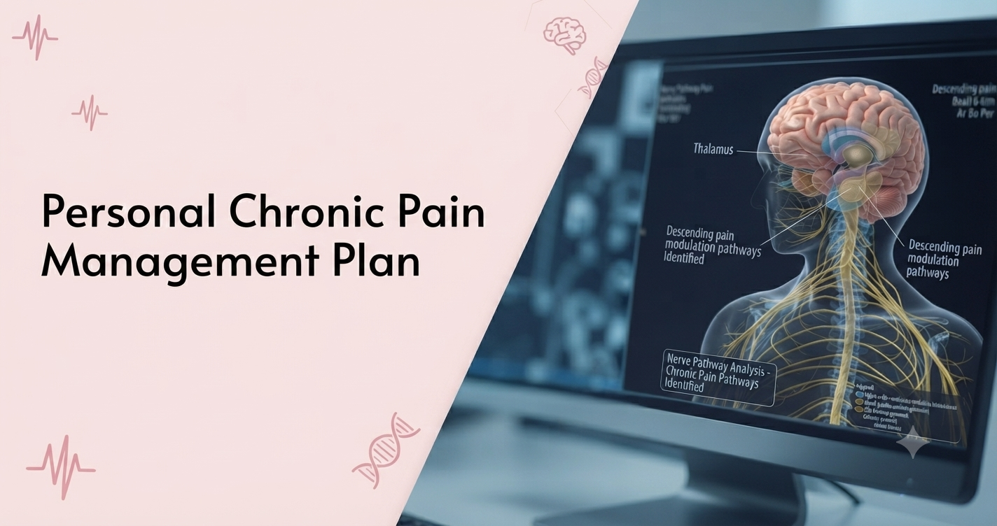
Chronic pain is not the same as the pain you feel after an injury or surgery. Those resolve. Chronic pain stays for months, sometimes years, and it changes how you sleep, how you move, what you eat, and how you think. Most people who live with it spend a long time looking for the one thing that will fix it. That one thing rarely exists. What works is a plan that addresses the pain from several directions at once, adapted to your specific condition, your daily routine, and what you can realistically sustain.
This is how to build that plan.
A pain management plan built on the wrong diagnosis does not work. Many people arrive at a pain specialist after months of partial treatment, physiotherapy for what turned out to be neuropathic pain, or anti-inflammatory medication for what was actually a structural mechanical problem. Before building any plan, confirm what you are dealing with.
Chronic pain broadly falls into three categories. Musculoskeletal pain originates in bones, joints, muscles, tendons, or ligaments. Arthritis, disc disease, and myofascial pain fall here. Neuropathic pain comes from damage or dysfunction in the nerve pathways themselves, producing burning, shooting, or electric-shock sensations. Diabetic neuropathy, post-herpetic neuralgia, and nerve compression injuries are common causes. Cancer pain is driven by tumour pressure, inflammation, or the side effects of treatment and requires a different approach from either of the others.
The type of pain determines what treatments are likely to work. Getting this right first saves months of trial and error. The Pain Management Centre in Gurgaon at CK Birla Hospital uses clinical assessment alongside imaging, nerve conduction studies, and pain mapping to identify the source before any treatment is recommended.
Eliminating pain entirely is rarely achievable with a chronic condition. Setting that as the goal leads to abandoning treatments that were actually working. More useful goals are functional: walking 30 minutes without stopping, sleeping through the night, returning to a specific activity, and reducing pain from a 7 out of 10 to a 4.
Write them down. Be specific about what you want to be able to do, and attach a time frame. Not because chronic pain follows a timetable, but because a time frame forces a review. At 8 weeks, have things improved? If not, what needs to change?
Discuss your goals with your pain specialist at the first appointment. A patient whose priority is returning to work needs something different from one whose priority is managing pain during sleep. The plan should be built around yours, not a default template.
Self-managed approaches, exercise, sleep, diet changes, and support medical treatment. They do not replace it.
For musculoskeletal pain, anti-inflammatory drugs, muscle relaxants, or topical agents are commonly used. For neuropathic pain, the medication class shifts. Antidepressants such as duloxetine and anticonvulsants such as pregabalin work on nerve signalling rather than inflammation, and they take time to reach their effect. Take prescribed medication on schedule rather than only when pain becomes unbearable. Stopping and restarting medications that modulate nerve signalling disrupts the effect and resets the clock.
Injections are used when medication has not provided enough relief, or when the source of pain is specific enough to target directly. Nerve blocks, joint injections, facet injections, and epidural procedures each address different mechanisms. They are not permanent solutions in most cases, but they provide a window of lower pain in which physiotherapy can make more progress than it could against a higher baseline.
Minimally invasive procedures such as radiofrequency ablation, spinal cord stimulation, and intrathecal drug delivery are used for severe or refractory chronic pain where other approaches have not worked. Radiofrequency ablation disrupts the nerve signals carrying pain from specific joints and can provide relief lasting 6 to 18 months. These are specialist-led decisions, not self-referral options.
Physiotherapy is one of the most consistently effective treatments for chronic pain, and one of the most commonly abandoned. The reason is almost always the same: pain increases briefly when movement begins, and patients interpret this as a sign the therapy is making things worse. In most cases it is not.
Deconditioned muscles around a painful joint or spine produce more pain when worked, initially. The improvement comes after weeks of effort, not days. This is not a sign to stop.
Work with a physiotherapist who has experience in chronic pain rehabilitation rather than acute injury recovery. The approach is different. Targets are set in terms of function rather than pain levels. The session itself may feel less intense than expected, because pacing is central to the method and overdoing it early sets the programme back.
Physiotherapy twice a week is not enough on its own. Movement between sessions maintains the gains and breaks the cycle of rest, stiffening, and increased pain that chronic pain patients commonly fall into.
Start with low-impact activity and build gradually. Walking is the simplest entry point: 10 minutes to start, extended by 5 minutes a week until you can walk 30 to 45 minutes continuously. Swimming removes bodyweight load from joints and works well for patients with knee, hip, or spinal pain. Yoga and specific stretching programmes reduce the muscle tension that contributes to pain between flare-ups.
The principle that governs all of this is pacing. Doing a little, resting, doing a little more. Overdoing activity on a good pain day and spending the next three days in bed is the boom-bust pattern that keeps people stuck. Moderate, consistent activity produces better outcomes than occasional intense effort every time.
Pain and sleep have a circular relationship. Pain disrupts sleep; poor sleep lowers the threshold at which pain is felt, making the same stimulus more painful than it would be when rested. Patients with chronic pain who sleep badly rate their pain as more severe and respond less well to treatment.
Keep consistent sleep and wake times seven days a week. Position the body with pillows to support painful areas — a pillow between the knees reduces lumbar and hip pain for side sleepers. Keep the bedroom cool and dark. Avoid screens for an hour before bed.
If pain at night is severe enough to prevent sleep despite these changes, raise it with your pain specialist. Night pain that is worse than daytime pain can indicate a specific mechanism that changes the medication approach, and it is worth distinguishing from general sleep hygiene problems.
Do not use alcohol to manage sleep. It induces sleep but disrupts sleep architecture, reduces restorative sleep stages, and worsens pain sensitivity the following day.
Chronic pain is not psychological, but it has a psychological dimension that cannot be ignored without cost. Pain that persists changes how the nervous system processes signals, called central sensitisation It changes how patients think about their bodies and their future. Anxiety about pain raises pain perception. Depression from months of restricted activity and missed life lowers the threshold at which pain is felt.
This does not mean the pain is imagined. It means treating pain without addressing the psychological dimension leaves part of the problem unaddressed.
Cognitive Behavioural Therapy adapted for chronic pain teaches patients to identify and examine catastrophising thought patterns: the assumption that pain means damage, that activity will make things worse, that improvement is impossible. Patients who complete this alongside medical treatment report lower pain scores and better function than those on medical treatment alone. Mindfulness-based approaches reduce the distress caused by pain, which sounds like a modest benefit until you consider that distress from chronic pain often limits function more than the pain itself does.
If the pain management programme includes psychological support, use it. At CK Birla Hospital, pain specialists work alongside a broader care team rather than in isolation.
Diet does not cure chronic pain. But inflammation makes pain worse, and diet directly affects systemic inflammation. Foods high in refined sugar, refined carbohydrates, and trans fats raise inflammatory markers. A diet built around vegetables, whole grains, oily fish, nuts, and olive oil reduces them.
Hydration matters specifically for musculoskeletal pain. Spinal discs are largely composed of water. Chronic dehydration reduces their height and shock-absorbing capacity over time.
In overweight patients with knee osteoarthritis, weight reduction produces measurable improvement in pain that is not fully explained by reduced mechanical load. The reduction in circulating inflammatory compounds from adipose tissue appears to contribute independently.
If dietary changes feel overwhelming alongside everything else, reduce ultra-processed food first. It is the change with the broadest effect on inflammation and the strongest evidence behind it.
A pain diary helps identify patterns: times of day when pain is worse, activities that trigger it, the relationship between sleep quality and next-day pain levels, how specific medications or treatments affect things. This is useful for your specialist and useful for you in distinguishing between pain that is changing and pain that feels the same because it always feels the same.
The risk is that tracking becomes hypervigilance. Constant monitoring keeps attention on pain and increases the distress it causes. A weekly or fortnightly review is enough. Record date, pain score, activity, sleep quality, and medication taken. Bring it to each appointment. Adjust the plan based on what the patterns show.
A personal plan is not a substitute for specialist care. Some situations require a clinical review:
Pain that is rapidly worsening without a clear reason. New numbness, weakness, or loss of bladder or bowel control alongside back or neck pain. Pain with unexplained weight loss or night sweats. Severe pain uncontrolled despite prescription medication. Pain preventing sleep entirely despite the measures above. Each of these warrants prompt assessment, not further self-management.
The Pain Management team at CK Birla Hospital in Gurgaon and Delhi includes certified pain specialists with experience across musculoskeletal, neuropathic, and cancer pain.
The plan is not a rigid protocol. It is a set of components: medical treatment, physiotherapy, movement, sleep, psychological support, diet, that work together and need regular review. What helps most in month one may need adjustment by month three. A treatment that produces no benefit after an adequate trial should be stopped and replaced.
Bring your goals to each consultation. Be specific about what is limiting you. Ask what a treatment is expected to achieve and over what time frame. Review the plan at every appointment.
Chronic pain rarely resolves in one intervention. It changes over time with attention and consistency sometimes slowly, but enough to matter when it does.
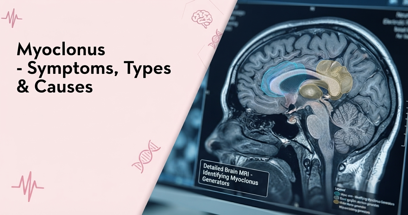
Ever felt a sudden jerk or jump-like sensation just as you were drifting off to sleep? That brief, involuntary movement is called myoclonus. Most people experience it occasionally, especially while falling asleep and it is usually nothing to worry about. But myoclonus can also be linked to epilepsy, other neurological conditions, certain medicines, or metabolic problems.
Myoclonus is not a disease in itself. It is a way of describing a specific type of movement. That is why figuring out what is causing it is such an important part of diagnosis and treatment.
Table of Contents
Myoclonus is a sudden, brief, involuntary jerking or twitching of a muscle or group of muscles. The movement usually lasts only a fraction of a second, although it can be felt repeatedly.
Most myoclonus results from a sudden muscle contraction, known as positive myoclonus. Less commonly, it occurs because an already going muscle contraction suddenly stops. This is called negative myoclonus.
For example, a person with negative myoclonus may briefly lose muscle control when holding a cup and drop it.
The movement can be experienced at rest, during an action, while maintaining a posture or in response to a stimulus such as a sudden sound or touch. When and how it happens can offer doctors important hints about what is causing it.
The main symptom is a sudden, brief muscle jerk that a person cannot voluntarily control.
Depending on its type and severity, myoclonus may cause:
Some people experience an isolated jerk from time to time, whereas others may have frequent movements that interfere with everyday activities.
Neurologists may classify myoclonus according to why it occurs, where it starts in the nervous system or when it happens.
This is common in otherwise healthy people and usually does not need any treatment. For example:
So, not every muscle jerk means there is an underlying neurological disorder.
Here, myoclonus occurs on its own without another identifiable neurological disorder causing it. It is usually stable or progresses very slowly. Some forms may run in families.
Myoclonic jerks can also be experienced as part of epilepsy, a neurological condition that causes seizures. One example is juvenile myoclonic epilepsy, which commonly begins around adolescence and may cause brief jerks involving the shoulders, arms or neck.
A myoclonic jerk does not confirm that a person has epilepsy. However, the pattern and, when appropriate, tests such as an EEG help neurologists determine whether the movements are seizure-related.
This type is usually experienced because of another condition or external factor. It can be associated with neurological diseases, metabolic problems, infections, brain injury, stroke, certain medicines and toxins.
Myoclonus results from abnormal electrical signalling in the nervous system’s movement pathways.
Some possible causes of myoclonus are:
A medication review is particularly useful because some medicines can cause myoclonus. If jerking begins after starting or changing a medicine, tell your doctor.
Diagnosis begins with a detailed medical history and neurological examination. The neurologist may ask:
The evaluation may include:
The investigations are selected according to the movement pattern and suspected underlying cause.
Treatment depends largely on what is causing the myoclonus. When an underlying condition, medicine or metabolic problem can be identified, addressing that cause is usually the first step.
Neurologists or movement disorder specialists may prescribe medicines that reduce abnormal nervous system activity.
For selected focal or segmental forms, botulinum toxin injections (BOTOX) can reduce excessive activity in specific muscles. This approach is particularly useful when jerking is limited to a small, well-defined group of muscles.
If myoclonus results from an electrolyte imbalance, kidney or liver problem, infection, medication or another neurological condition, treating that problem may reduce the jerking.
In some complex cases, other approaches such as immunotherapy, surgery or deep brain stimulation (DBS) may be considered depending on the underlying diagnosis.
You should consider consulting a neurologist if:
A movement disorder specialist or neurologist can determine whether the movements represent myoclonus and investigate the underlying cause.
If sudden jerking movements are becoming frequent or affecting your daily life, an evaluation with a specialist can help identify whether they are harmless or related to an underlying condition. The Neurology and Neurosciences Department at the CK Birla Hospital can help evaluate movement disorders such as myoclonus and determine an appropriate diagnostic and treatment approach. Simply book a consultation to get expert guidance.

Have you ever felt your hand cramp while writing, your foot twist while walking, or your neck suddenly pull to one side?
For people with dystonia, these involuntary muscle contractions are a daily reality. Dystonia is a neurological movement disorder that causes muscles to contract uncontrollably, leading to repetitive movements or abnormal postures.
It affects an estimated 1 in 1,000 people worldwide, yet many people don’t know about it. In this blog, we will talk about what causes dystonia, how to spot its symptoms and the treatment options helping people manage it today.
Table of Contents
Dystonia is a neurological movement disorder that occurs when the brain’s movement-control networks send abnormal signals to muscles. This leads to unwanted muscle activity that can be constant or come and go.
For example, a person with cervical dystonia may find that their neck repeatedly turns or tilts to one side. Someone with focal hand dystonia (also called writer’s cramp) may develop abnormal hand or finger movements specifically when they are writing.
The underlying issue of this disorder involves the nervous system. Unlike some movement disorders that cause weakness or a loss of movement, dystonia is characterised by too much muscle activity. Muscles that should stay relaxed instead tense up, pulling body parts into twisted, repetitive or awkward positions.
Dystonia can affect just one part of the body (focal dystonia), two or more adjacent areas (segmental dystonia), or multiple unrelated body parts at once (generalised dystonia). It can affect a person at any age, though the pattern often differs between children and adults.
Symptoms depend on which muscles are affected and how the condition develops. Some common symptoms include:
Symptoms can also vary during the day and may become more evident with stress, fatigue or particular activities.
Dystonia can be classified according to the parts of the body affected.
Focal dystonia: It affects one body region. For example,
Segmental dystonia: It affects two or more neighbouring areas, such as the neck and an arm.
Multifocal dystonia: It affects two or more unrelated or non-adjacent areas of the body. For example, the eyelids and a foot.
Generalised dystonia: It affects multiple areas of the body, mostly involving the trunk and several limbs.
Hemidystonia: It affects one side of the body.
Sometimes the exact cause of dystonia cannot be identified. In other cases, it may result from a genetic change, another neurological condition, brain injury or certain medicines.
Some possible causes include:
Diagnosis usually begins with a neurological examination, where the neurologist observes the movements, posture and situations in which symptoms occur.
The movement disorder specialist may ask:
Depending on the suspected cause, the specialist may recommend brain imaging tests, blood tests or genetic testing.
The approach of treatment can vary from person to person. Treatment is chosen according to the type of dystonia, affected muscles, severity, underlying cause and how much symptoms interfere with daily life.
The movement disorder specialist may prescribe medicines that reduce abnormal muscle activity or influence chemical signalling in the brain. The choice depends on the individual’s symptoms and response and side effects need to be considered.
Botulinum toxin, commonly known as Botox, can be injected into selected overactive muscles. It temporarily reduces the nerve signals that cause those muscles to contract.
It is particularly useful for many forms of focal dystonia, including cervical dystonia and blepharospasm. Because its effect wears off, injections generally need to be repeated.
Rehabilitation may help maintain movement, improve function and make everyday activities easier. An occupational therapist can also suggest practical adaptations for work or daily tasks.
For some people with severe dystonia that remains disabling despite other treatments, deep brain stimulation (DBS) may be considered.
DBS involves placing electrodes in specific movement-related areas of the brain. A small implanted device then delivers controlled electrical stimulation. DBS can reduce dystonia symptoms and disability in appropriately selected patients.
You should consider seeking medical evaluation if involuntary movements, muscle contractions or abnormal postures:
A movement disorder neurologist can evaluate whether the symptoms are due to dystonia or another condition and determine which investigations and treatments may be appropriate.
If you or a family member is experiencing the symptoms of dystonia, an evaluation can help clarify what is happening and whether treatment is needed. The Neurology and Neurosciences Department at the CK Birla Hospital can provide specialist assessment for movement disorders such as dystonia. Simply book a consultation to get expert guidance.

