Filter :
From lifting a cup to taking a step, our every movement depends on a finely coordinated nervous system. When that coordination is disrupted, the change can be surprisingly visible, sometimes starting with something as simple as a trembling hand.
That disruption is medically called a movement disorder, which is a group of neurological conditions where the brain’s control over the body’s motion does not work the way it should. Read along to know what causes it, what it looks like, and how to treat it.
Table of Contents
Movement disorders are neurological conditions that affect the way a person moves and controls their body. In other words, these disorders affect the nervous system’s ability to control and coordinate body movements. This issue originates in the brain, specifically in the areas that plan, initiate, and coordinate motion, such as the basal ganglia and cerebellum.
Broadly, neurologists split these conditions into two categories:
According to the National Institute of Neurological Disorders and Stroke (NINDS), movement disorders can affect people at different ages, depending on the specific condition. Some, such as dystonia, can begin at any age, whereas others, including certain types of tremor, are more common in middle-aged and older adults. Some disorders are genetic, some develop after injury or illness, and for others, an exact cause cannot be pinned down.
Parkinsonism refers to a cluster of symptoms such as slowness of movement (bradykinesia), muscle stiffness, tremor at rest and balance problems that show up together. Parkinson’s disease is the most common cause of this disorder, but similar symptoms can arise from certain medications, repeated head trauma or other neurodegenerative conditions like progressive supranuclear palsy.
Tremor is an involuntary, rhythmic shaking of a body part. It generally affects the hands first, making everyday tasks like writing, buttoning a shirt, or drinking soup notably harder.
There is a common misconception where people assume any hand tremor means Parkinson’s. It usually doesn’t. Tremor tends to worsen with intentional movement (like reaching for something), while Parkinsonian tremor can also appear at rest and ease during activity.
It causes involuntary muscle contractions that lead to twisting movements or abnormal, sometimes painful postures. It can affect one part of the body like the neck in cervical dystonia or spread more widely. Some people with dystonia experience a phenomenon known as a “sensory trick”, where touching the affected area lightly (a hand to the chin, for instance) temporarily eases the spasm.
Chorea produces brief, irregular movements that seem to flow randomly from one part of the body to another. Huntington’s disease is the commonly known genetic cause, but chorea can also show up with certain autoimmune disorders, during pregnancy (called chorea gravidarum), or as a side effect of some medications.
Because the movements usually look smooth rather than jerky, family members sometimes mistake early chorea for simple restlessness or fidgeting instead of recognising it as a neurological symptom.
Ataxia refers to a loss of coordination that involves unsteady walking, clumsy hand movements, slurred speech. It usually stems from cerebellum dysfunction. It can result from genetic conditions, degenerative conditions, stroke, certain infections or vitamin deficiencies (particularly vitamin E or B12).
Ataxia is sometimes the first visible sign of an underlying nutritional deficiency, which means correcting the deficiency can improve symptoms. It is not true for most other movement disorders on this list.
Tic disorders involve sudden, repetitive movements or sounds that a person feels an urge to make, like blinking, throat-clearing, or shoulder shrugging. Tics are not random; they tend to be the same movement repeated over and over. Many people can briefly hold them back, though this builds up tension that needs to be released.
Tourette syndrome is the most well-known tic disorder, marked by both movement and vocal tics lasting more than a year. Tics start in childhood, can worsen with stress or excitement, and may fade on their own with age. Because kids can suppress tics for short periods, teachers or parents sometimes miss them at first or assume the child is just being fidgety or seeking attention.
This category includes:
Symptoms vary by condition, but common ones are:
Some conditions also bring non-motor symptoms such as:
In Parkinson’s disease specifically, a reduced sense of smell and sleep disturbances can precede visible motor symptoms by years, something even attentive patients rarely anticipate.
Causes fall into a few broad groups and often more than one factor is involved:
Diagnosis usually involves:
Treatment depends mainly on the specific diagnosis, but generally includes:
The goal of treatment is not to erase symptoms completely. It is mainly about helping you maintain your quality of life and independence for as long as possible. Just knowing this tends to ease a lot of the anxiety patients feel after a diagnosis. Also, every movement disorder is different and treatment plans need to be personalised to the individual. It is always best to consult a neurologist for proper diagnosis and an individualised treatment approach.
It is worth having a proper conversation with a neurologist if you notice a tremor that interferes with daily tasks, frequent stiffness or slowness that is getting worse, sudden jerking or twisting movements, changes in handwriting or gait, or tics that persist beyond a few weeks.
Getting evaluated early can help you avoid more serious problems down the line. If you are noticing symptoms of a movement disorder, don’t wait it out. Book an appointment with a neurologist at the CK Birla Hospital today, and take the first step toward a clearer diagnosis and the right treatment plan for you.

Movement disorders like Parkinson’s disease affect more than 8.5 million people worldwide. Many will eventually reach a point where medication no longer provides enough relief. For them, Deep Brain Stimulation (DBS) offers a different path. Its impact can be striking, patients who once struggled with basic tasks like eating or writing often regain a level of control they had not felt in years.
Curious how DBS actually works, who qualifies for it, and what the procedure and recovery really involve? Let’s break it down.
Table of Contents
Your brain holds the main control of your body. It sends electrical signals to tell your body how to move, react, and function. But in certain conditions, some of these signals become disrupted, causing tremors, stiffness or movements you can’t control.
Deep Brain Stimulation (DBS) works by gently correcting these faulty signals. Thin wires, called electrodes, are placed in specific areas of the brain where the problem starts. These electrodes connect to a small device called a neurostimulator, similar to a pacemaker, usually placed under the skin near the collarbone.
Once activated, the neurostimulator sends mild electrical pulses to the targeted brain area. These pulses help calm the abnormal signals, easing symptoms that medication alone could not fully control.
The DBS system has three main parts:
DBS does not remove or destroy the affected part of the brain. It also does not stop the underlying progression of conditions such as Parkinson’s disease. Instead, it is designed to help manage symptoms.
Patient selection is one of the most important parts of DBS treatment.
It is an established treatment for movement disorders such as Parkinson’s disease, essential tremor and dystonia. In some cases, it may also be used for conditions such as epilepsy, Tourette syndrome and obsessive-compulsive disorder, though this depends on the specific case and relevant medical approvals.
For Parkinson’s disease, DBS may be considered when simply medication can no longer keep symptoms under control. In fact, NICE recommends considering deep brain stimulation (DBS) for people with advanced Parkinson’s disease whose symptoms are not adequately controlled with best medical therapy.
A specialist assessment by a neurosurgeon and movement disorder neurologist may consider:
A 2026 expert consensus from the Parkinson’s Research Alliance India also highlights the importance of careful patient selection for DBS, ensuring psychiatric stability, precisely identifying the right brain target, and involving a multidisciplinary team throughout the assessment process.
The potential benefit of DBS depends on the condition being treated, the symptoms present and how carefully the patient is selected.
For appropriate patients, DBS may:
DBS is generally considered safe when performed in carefully selected patients by an experienced, functional neurosurgeon. Serious complications are uncommon, and several precautions are taken to minimise risks.
Potential risks include:
DBS does not end with implantation. The neurology specialists may need several followup appointments to check the device and achieve the best balance between symptom control and side effects.
DBS treatment begins with careful planning before moving on to the actual implantation of the stimulation system. The exact approach can vary from patient to patient, depending on their condition, brain anatomy, surgical plan, and the specific protocols followed by the hospital.
Before surgery, the patient undergoes a detailed neurological evaluation. Brain imaging, such as MRI or CT, may be used to map the brain and identify the precise target for electrode placement. Cognitive and psychological assessments may also form part of the evaluation.
The neurosurgeon uses specialised imaging and stereotactic (3D coordinate system) techniques, which means highly precise methods for locating a specific point inside the brain, to guide the electrodes into the selected target.
In some cases, the patient may remain awake during part of this stage so the team can assess symptoms and responses during electrode placement. In other situations, DBS can be performed using general anaesthesia. The approach is individualised rather than one method being suitable for everyone.
The neurostimulator is placed beneath the skin, usually in the upper chest near the collarbone. Extension wires under the skin connect it to the electrodes in the brain.
The device is generally activated after the initial surgical recovery period. A neurologist or trained DBS specialist then adjusts the stimulation settings according to the patient’s symptoms and response.
This is an important part of treatment. The best settings are not necessarily found during the first programming session. Mayo Clinic notes that optimisation can take several months in some patients.
Recovery has two parts:
During the first few days, patients may experience tiredness, discomfort or swelling around the surgical areas. Before you head home, the healthcare team walks you through everything you need to know, how to care for the incision, which medicines to take, and how much physical activity is safe during recovery.
Over the following weeks, the surgical wounds continue to heal and followup appointments are scheduled. The neurostimulator may then be activated and programmed according to the treatment plan.
It is important to know that your symptoms do not always improve immediately or completely right away. The care team gradually adjusts the stimulation settings over time, based on how symptoms respond and whether any side effects show up along the way.
You should follow your neurosurgical team’s guidance on when to return to work, resume physical work and get back to other daily activities.
For many patients with certain neurological conditions, treatment like DBS can turn symptoms that once felt unmanageable into something livable again. If you would like to know more about this treatment approach or need guidance on managing a neurological condition, start by talking to our neurologist or neurosurgeon. At the CK Birla Hospital, our Neurosciences team is here to guide you through a personalised assessment and walk alongside you at every step. Simply reach out to us to book a consultation!

Do you check the toilet bowl after you poop? If not, you probably should, because your stool says a lot about what is happening inside your body. Many people spot a jelly-like streak of mucus in their stool and immediately start spiraling: “Is this normal?” or “Is my body warning me about something serious?”
Truth is, your gut produces some mucus daily as part of its normal function, so a small amount usually is not concerning. But frequent or excessive mucus, especially with stomach pain, diarrhoea, or blood, should be checked by a specialist.
So how do you know which one you are dealing with? Read along to find the answer!
Table of Contents
Your intestines naturally produce a slippery, jelly-like substance called mucus. It coats the lining of your gut and helps stool move through the colon smoothly, without friction.
In a nutshell, mucus helps by:
Small amounts are usually mixed into stool and may not be visible. You may notice mucus more clearly when the intestine is irritated or inflamed, since mucus production increases as a protective response.
So, spotting a small amount of mucus is not necessarily abnormal. What matters more is the amount, appearance, how frequently it occurs, and whether other symptoms are present.
Mucus deserves more attention when it:
Mucus itself is a symptom rather than a diagnosis. Various digestive conditions can cause it, ranging from relatively common problems such as IBS to inflammatory bowel disease and, less commonly, colorectal cancer.
Intestinal Irritation and Constipation
When stool gets stuck and you strain, it irritates your gut and triggers extra mucus. Diarrhoea can also have the same effect, since intestinal lining gets irritated due to more frequent bowel movements.
Inflammatory Bowel Disease (IBD)
IBD covers two main conditions: ulcerative colitis and Crohn’s disease, both of which can cause long-term inflammation in your digestive tract.
Ulcerative colitis specifically affects the large intestine and produces mucus or pus in stool. Crohn’s disease can affect any part of the digestive tract.
Either way, expect it to come with diarrhoea, abdominal pain, rectal bleeding, fatigue, or unexplained weight loss.
Irritable Bowel Syndrome (IBS)
IBS usually shows up as whitish mucus in stool, along with bloating, cramping, and unpredictable bowel habits, sometimes diarrhoea, sometimes constipation, sometimes both. Since IBS is closely tied to the gut and brain connection, stress can make symptoms worse.
Intestinal Infections
Viral, bacterial and parasitic infections can irritate the intestine and lead to more mucus, which may come along with diarrhoea, abdominal cramps, fever or blood in the stool.
Food Allergies or Intolerances
Certain food allergies and intolerances can cause digestive symptoms like diarrhoea and abdominal discomfort in children.
Tip: A food diary can sometimes help identify a consistent relationship between a food and symptoms, but restrictive diets should not be started without appropriate medical or dietary guidance.
Piles (Haemorrhoids)
These are swollen veins around the anus or rectum, usually caused by straining or constipation. They usually bleed bright red and can come with some mucus but don’t assume mucus is always from piles. If it is persistent, get it checked.
Proctitis
This is inflammation of the rectal lining. It can cause mucus, blood, rectal pain, urgency and a frustrating feeling that you need to pass stool even after having a bowel movement.
Colorectal Cancer
It is a less common cause of mucus in stool, but it should be considered when mucus is coming alongside warning signs such as blood in the stool, persistent changes in bowel habits, abdominal discomfort, sudden weight loss or fatigue.
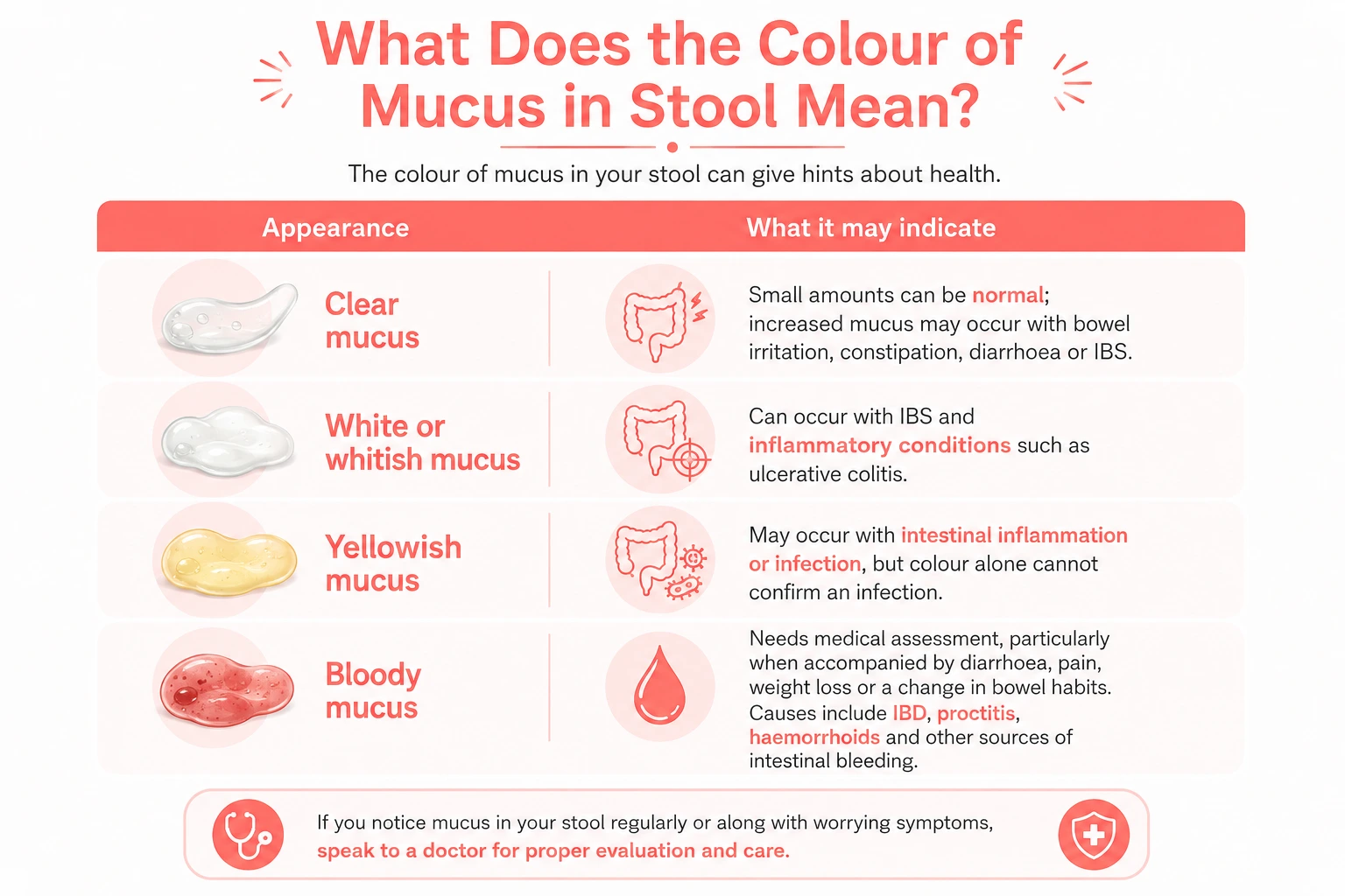
You should speak with a doctor if mucus is persistent, recurring or accompanied by:
Blood in stool or persistent mucus should not simply be assumed to be IBS or piles without getting it properly checked. Initially, you can speak with an internal medicine doctor or general practitioner (GP). If there is anything that needs special assessment, they may refer you to a specialist doctor like a gastroenterologist.
A gastroenterologist usually begins by asking about your bowel habits, diet, medicines, recent infections, family history and other symptoms.
Depending on the situation, your evaluation may involve:
Not everybody with mucus in stool needs to go through all of these tests. The choice of tests depends on your symptoms, age, previous conditions, and other factors.
Once you know what is causing the mucus, the next step is figuring out what actually helps. Let’s see how you can take care of this problem.
Dietary and Lifestyle Measures
Treating the Underlying Condition
Home Remedies
You cannot prevent every cause, but there are various habits that can improve your bowel health.
A small amount of clear mucus in stool can be normal. But persistent, excessive, coloured or bloody mucus needs attention, especially when it comes along with abdominal pain, diarrhoea, weight loss or changes in bowel habits.
If you are unsure about a recurring change in your stool, a consultation with the Gastroenterology Department at the CK Birla Hospital can help identify the cause and determine whether any tests or treatment are needed. Book a consultation with us to get personalised guidance.

Have you noticed pale or discolored patches showing up on your skin, especially after a day in the sun or a sweaty workout? This can be the sign of tinea versicolor, one of the most common fungal skin conditions worldwide, affecting people of all skin types. Worse, many people don’t go for a medical checkup and mistake it for vitiligo, eczema, or simple sun damage, and end up using the wrong creams for months with zero results. So, in this blog, let’s find out what this condition actually is, and how you can manage it and keep it from coming back.
Table of Contents
Tinea versicolor, also called pityriasis versicolor, is a superficial fungal infection. It is caused by an overgrowth of Malassezia which is a type of yeast that normally lives on healthy skin.
The yeast is not something you catch from another person. In certain conditions, it grows excessively and affects the skin’s normal pigmentation, producing patches that may be lighter or darker than the surrounding skin.
Tinea versicolor is not contagious and is not a sign of being unclean.
Tinea versicolor symptoms can vary with skin tone. The patches may be:
They most commonly appear on the upper back, chest, shoulders, neck and upper arms. Several small patches can gradually join together and form larger areas of discolouration.
One easily overlooked clue is what happens after sun exposure. The affected areas do not tan normally, so the contrast between the patches and surrounding skin can become much more evident during summer. This does not necessarily mean the infection has suddenly become worse.
The underlying cause is an overgrowth of Malassezia. Some other factors that can make this more likely are:
Malassezia normally lives on the skin. Problems arise when the yeast multiplies excessively and alters pigmentation and the surface of the skin. Most people who develop tinea versicolor are otherwise healthy.
Warm, humid conditions favour yeast growth. This is why tinea versicolor is particularly common in tropical and subtropical regions and may become more common during hot months.
Malassezia thrives in environments rich in skin oils. Increased oil production, including around puberty, can therefore increase susceptibility.
Changes in hormone levels may alter skin oil production. Pregnancy is one situation in which hormonal changes may increase susceptibility to tinea versicolor.
People with a weakened immune system, or living with some underlying conditions, may be more prone. This does not mean that having tinea versicolor automatically indicates an immune problem.
Patches can understandably raise concern about vitiligo; however the two conditions are different.
| Feature | Tinea Versicolor | Vitiligo |
| Cause | It is caused by the overgrowth of Malassezia yeast. | In this case, the autoimmune condition affects pigment-producing cells. |
| Texture | It usually has fine scaling. | It is smooth, without scaling. |
| Color of patches | May be white, tan, pink, brown, or lighter than surrounding skin | Clearly depigmented, milky-white patches |
| Common areas affected | It commonly affects the chest, back, and shoulders. | Hands, face, and around body openings, among others |
| Treatment | Usually responds to antifungal treatment | Requires different medical management |
These differences can provide clues, but a dermatologist may need to examine the skin to confirm the diagnosis.
A dermatologist (skin specialist doctor) can diagnose tinea versicolor by examining the affected skin.
Depending on the appearance, they may use:
A biopsy is rarely necessary but may be considered when the diagnosis remains uncertain.
Treatment aims to control the yeast overgrowth. The choice depends on how widespread the infection is and whether it keeps returning.
For mild cases, antifungal creams, lotions, washes or shampoos may be sufficient. Some antifungal products are designed to be applied to the skin rather than used only on the scalp. But make sure you are using them as per your dermatologist’s recommendation only.
A dermatologist may prescribe stronger or specific topical antifungal treatment when over-the-counter products are unsuitable or have not worked.
For extensive disease or frequent recurrence, a skin specialist may consider oral antifungal medicines. These medicines are not appropriate for everyone and should not be self-prescribed because they can have drug interactions and other risks.
Note – Killing the yeast does not immediately restore normal skin colour. Pigmentation can take weeks or months to even out, and in some people it can take considerably longer.
Tinea versicolor can return because Malassezia is normally present on the skin. Hygiene cannot permanently remove the yeast, but practical habits can reduce conditions that encourage overgrowth.
There is limited reliable evidence that household remedies such as vinegar, lemon juice or other acidic substances can safely treat tinea versicolor. They may irritate the skin and make discolouration harder to assess. That is why evidence-based antifungal treatment is a safer approach.
You should consider seeing a dermatologist if:
If you have persistent or recurring skin discolouration, a dermatologist at The CK Birla Hospital can help determine whether it is tinea versicolor or another skin condition and guide you towards appropriate treatment. Book a consultation to get expert advice.

Your feet are meant to move. Movement helps push fluid back up through your legs, but stillness lets gravity pull it down instead. That’s why pregnancy, aging, or standing and sitting for too long can cause your feet to swell. But does swelling feet mean something to worry about? Read along to understand why your feet swell, how to get relief and when it is time to see a doctor.
Table of Contents
We colloquially call it “swelling feet,” but the medical term is edema, a condition caused by excess fluid collecting in body tissue, commonly in the feet, ankles, and lower legs. It happens when fluid is not circulating back up through the body efficiently, so it settles in the lower extremities instead.
You have probably noticed the signs like your shoes feeling tighter by evening, or a dent left behind when you press on your ankle. That is called pitting edema. Sometimes the swelling does not leave a mark at all when pressed; this is non-pitting edema, and it is generally linked to conditions like lymphedema.
What should you keep in mind?
Not all foot swelling is edema. Injury, inflammation, and infection can make a foot look swollen too, even when fluid retention is not the main cause.
In edema (swelling feet), the most common signs to watch for are:
Pay attention to what comes with the swelling. Pain after an injury may point towards tissue damage. Redness and warmth may be a sign of some infection or inflammation. If you are suffering with swelling in just one leg, along with sudden pain or tenderness, it can be a sign of DVT (Deep Vein Thrombosis), which is a blood clot in a deep vein.
If your swelling feet are accompanied by any of these signs, it is best to see an internal medicine doctor.
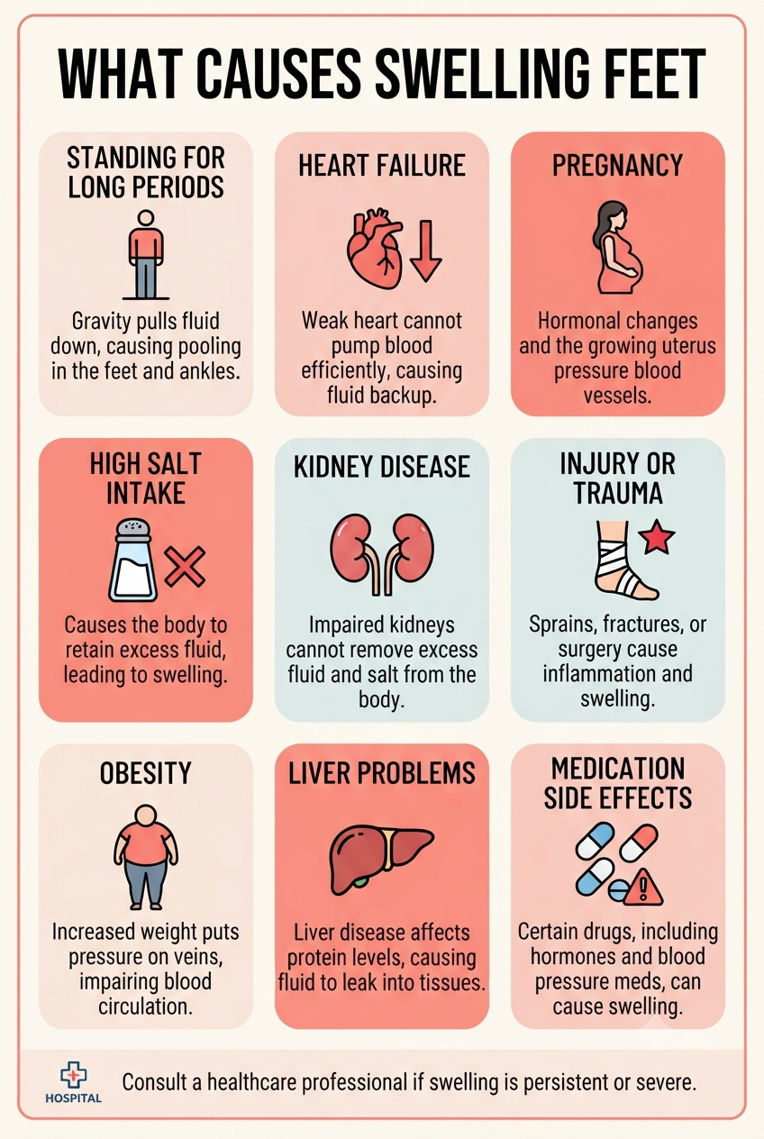
The causes of swelling in the feet range from temporary fluid retention to conditions affecting circulation, the lymphatic system or major organs.
The most common cause is sitting, standing or staying in one position for long periods without much movement, which lets gravity pull fluid down and accumulate in the lower body. This usually happens after a long workday, a road journey or a flight.
As we age, veins can lose some of their elasticity, making it harder for blood to flow efficiently back up the legs. So, mild swelling becomes more common over time.
Gradual swelling of the feet and ankles is common during pregnancy, particularly later in pregnancy and towards the end of the day. During pregnancy, the growing uterus puts extra pressure on the veins carrying blood from the lower body, while hormonal changes cause the body to retain more fluid overall. In both cases, some swelling is considered a normal part of the process.
Sodium causes the body to retain water. A particularly salty meal may therefore make swelling feet and ankles more noticeable in some people who are already prone to fluid retention.
This is also a common cause in some cases as heat can cause blood vessels to widen, which may contribute to fluid moving into surrounding tissues. Swelling can be more evident towards the end of a hot day.
Dehydration is not the most common explanation for persistent foot swelling. However, in some situations, inadequate fluid intake can affect the body’s fluid balance. Recurrent swelling should not simply be attributed to dehydration without considering other causes.
If you previously have any sprain, strain, fracture or other local injury, it can trigger inflammation and swelling in the affected foot or ankle. In this situation, pain and tenderness usually comes with the swelling.
Chronic venous insufficiency is caused when the veins in the legs have difficulty returning blood efficiently to the heart. Fluid can then collect around the ankles and lower legs. Swelling that becomes worse after standing and improves after resting or elevating the legs can fit this pattern.
This is the condition when lymph fluid that normally drains through the lymphatic system, builds up in tissues. It may affect one or both limbs and can cause chronic swelling and changes in the skin.
Certain medications used for high blood pressure, diabetes, pain and inflammation, hormones and steroids, can also cause fluid retention as a side effect. Do not stop a prescribed medicine on your own. A doctor can determine whether the medicine may be contributing to the swelling and whether it should be changed.
An infection or inflammatory condition can cause local swelling that usually comes with pain, redness, warmth or tenderness. An insect bite or sting can also cause temporary swelling.
DVT is a blood clot that develops in a deep vein, usually in the leg. Sudden swelling in one foot or leg, particularly when accompanied by pain, tenderness, warmth or redness, should not be assumed to be ordinary fluid retention. It may require medical assessment.
In some cases, edema can be a sign of an underlying issue with the heart, kidneys, liver, or veins. That is why, if the swelling feet are affecting you more than often, it is always a good idea to initially consult an internal medicine doctor.
Swelling affecting one foot or leg may occur because of:
After a long journey, one-sided swelling deserves particular attention. Long-distance travel, generally involving more than four hours of sitting, can increase the risk of blood clots.
Swelling in both feet may be due to prolonged sitting or standing, pregnancy, venous insufficiency, medication effects or general fluid retention. In some cases, heart, kidney and liver conditions can also cause swelling in both legs.
You may be more likely to develop foot and ankle swelling if you:
Older adults may also experience swelling because several of these factors can occur together.
Medical evaluation is advisable when swelling is:
If you have diabetes, unexplained swelling in the foot, ankle or leg is worth getting checked, particularly if there is a wound, redness, numbness, or any other noticeable change.
There is no specific test for identifying the cause behind swelling feet. The right investigation depends on what the doctor finds during the consultation.
A doctor may ask:
The doctor may examine the skin, veins, pulses, swelling and whether pressing the area produces a pit.
Depending on the suspected cause, investigations may include blood or urine tests, a Doppler ultrasound to assess blood flow and look for a clot, or other imaging and specialised tests. Not everyone with swelling feet needs all of these tests.
If the swelling is mild, familiar and not accompanied by warning signs, a few measures may help.
What to avoid:
Do not rely on massage, herbal drinks, aloe vera or apple cider vinegar soaks as treatments for unexplained swelling. In particular, avoid massaging a suddenly swollen, painful leg before a possible blood clot has been assessed.
If swelling feet are persistent, recurrent or difficult to explain, a medical checkup can help identify the cause rather than simply treating the swelling. The Internal Medicine Department at the CK Birla Hospital can help with the initial evaluation and coordinate care with other medical specialists when needed. To get expert advice, book a consultation.
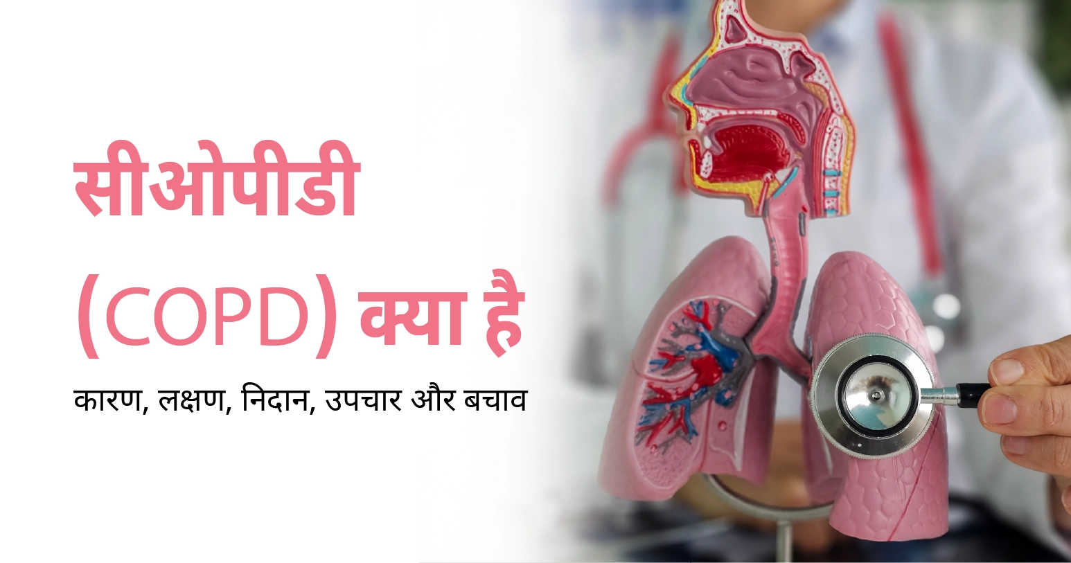
रात में अचानक सांस फूलना या मामूली काम में भी दम घुटना—यह सिर्फ ‘सामान्य थकान’ नहीं, बल्कि COPD (क्रॉनिक ऑब्सट्रक्टिव पल्मोनरी डिजीज) का संकेत हो सकता है। यह बीमारी चुपचाप फेफड़ों की हवा थैलियों को नुकसान पहुंचाती है, जिससे समय के साथ सांस लेने की क्षमता घटने लगती है। WHO के अनुसार यह दुनिया भर में जानलेवा बीमारियों में से एक है, इसलिए शुरुआती लक्षणों को पहचानना ही इससे बचाव की सबसे बड़ी चाबी है।
सही समय पर पहचान और उचित इलाज से फेफड़ों को और नुकसान पहुंचने से रोका जा सकता है और मरीज एक सक्रिय जीवन जी सकते हैं। अगर आपको या आपके किसी प्रियजन को लगातार खांसी, सांस फूलने या सीने में जकड़न की समस्या है, तो इसे नजरअंदाज न करें।
Table of Contents
सीओपीडी बीमारी क्या है, यह समझने के लिए पहले फेफड़ों के काम करने के तरीके को समझना जरूरी है। सामान्य फेफड़ों में हवा आसानी से अंदर-बाहर होती है, लेकिन सीओपीडी में सांस की नलियां सूज जाती हैं, संकरी हो जाती हैं और उनमें अतिरिक्त बलगम बनने लगता है। इससे हवा फेफड़ों में फंस जाती है और मरीज को सांस लेने और छोड़ने दोनों में तकलीफ होती है।
यह कोई अचानक होने वाली बीमारी नहीं है, यह वर्षों में धीरे-धीरे विकसित होती है, अक्सर 40 साल की उम्र के बाद ज्यादा दिखाई देती है। एक बार फेफड़ों को नुकसान हो जाने पर उसे पूरी तरह पलटा नहीं जा सकता, इसलिए जल्दी पहचान जरूरी है।
इस स्थिति के जोखिम कारक एवं कारण एक ही हैं, जिन्हें नीचे लिखा गया है –
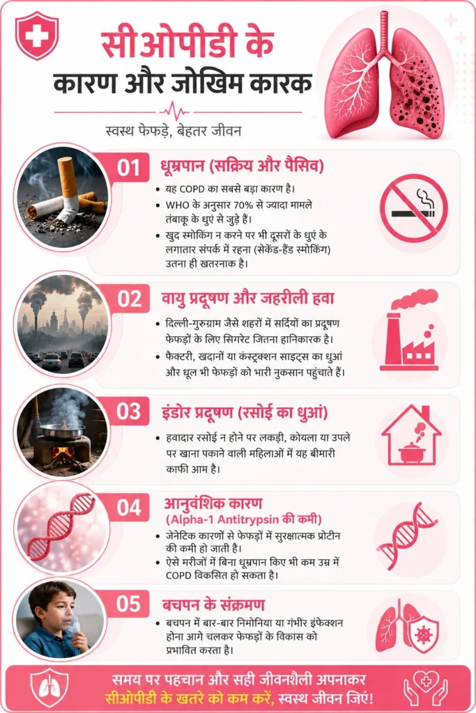
हमारे पल्मोनोलॉजी विभाग में अक्सर मरीज सालों पुरानी “सामान्य खांसी” समझकर बीमारी को टालते रहते हैं। जब तक जांच होती है, फेफड़ों की क्षमता 30 से 40% तक घट चुकी होती है। समय पर कराया गया एक ‘स्पायरोमेट्री टेस्ट’ बीमारी को शुरुआती चरण में ही पकड़ सकता है।
सीओपीडी के इलाज में कई विकल्प उपलब्ध हैं, और डॉक्टर निदान के आधार पर ही इलाज के विकल्प का चयन करते हैं –
सीओपीडी एक गंभीर बीमारी जरूर है, लेकिन सही समय पर पहचान, दवाएं और जीवनशैली में सुधार करके मरीज एक सामान्य और सक्रिय जीवन जी सकते हैं। सांस की तकलीफ या लगातार खांसी को बढ़ती उम्र का असर समझकर नजरअंदाज न करें। दिल्ली और गुरुग्राम स्थित हमारे सेंटर्स पर अनुभवी पल्मोनोलॉजिस्ट और आधुनिक स्पायरोमेट्री टेस्ट की सुविधा उपलब्ध है। अपने फेफड़ों को सुरक्षित रखने के लिए आज ही अपना अपॉइंटमेंट बुक करें।
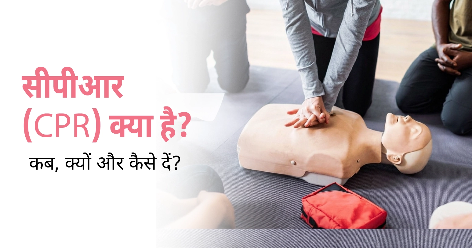
एक पल के लिए सोचिए। आप अपने घर के किसी बड़े-बुजुर्ग के साथ बैठे हैं, बातें हो रही हैं, और अचानक वो बेहोश होकर गिर जाते हैं। सांस थम जाती है, दिल की धड़कन महसूस नहीं होती। ऐसे में अगले दो-तीन मिनट तय करते हैं कि सामने वाला व्यक्ति जिंदगी की तरफ लौटेगा या नहीं। यही वो पल है, जहां सीपीआर (CPR) किसी की जान बचाने वाला सबसे बड़ा हथियार बन जाता है। भारत में आज भी बहुत कम लोगों को यह बुनियादी तकनीक आती है, और यही अनजान होना कई बार किसी अपने को समय रहते खोने की वजह बन जाता है। इस ब्लॉग में हम आसान भाषा में समझेंगे कि सीपीआर क्या है, कब देनी चाहिए और सही तरीका क्या है, ताकि इमरजेंसी में आप घबराएं नहीं, बल्कि किसी की जान बचाने वाले हीरो बन सकें।
सीके बिरला अस्पताल की इमरजेंसी और कार्डियक टीम अक्सर ऐसे मामले देखती है, जहां परिवार के किसी सदस्य ने समय पर सीपीआर दे दी और मरीज को नई जिंदगी मिल गई। दिल्ली और गुरुग्राम, दोनों सेंटर पर हमारे अनुभवी कार्डियोलॉजिस्ट और इमरजेंसी स्पेशलिस्ट से मिलकर सही जानकारी लें। हृदय संबंधी जोखिम हो तो देर न करें, आज ही अपॉइंटमेंट बुक करें।
Table of Contents
सीपीआर का पूरा नाम है Cardiopulmonary Resuscitation, यानी कार्डियोपल्मोनरी रिससिटेशन। यह एक इमरजेंसी फर्स्ट-एड तकनीक है, जिसमें छाती को बार-बार दबाकर और जरूरत पड़ने पर मुंह से सांस देकर, दिल और फेफड़ों का काम अस्थायी रूप से खुद अपने हाथों से किया जाता है, ताकि मस्तिष्क और शरीर के अंगों तक ऑक्सीजन-युक्त रक्त पहुंचता रहे, जब तक दिल दोबारा खुद से धड़कना शुरू न कर दे या मेडिकल टीम मौके पर न पहुंच जाए।
सीपीआर हर बेहोशी में नहीं, बल्कि सिर्फ उन स्थितियों में देनी चाहिए जहां सांस या धड़कन रुक चुकी हो या रुकने की कगार पर हो।
जब दिल धड़कना बंद कर देता है, तो शरीर में रक्त का प्रवाह रुक जाता है और मस्तिष्क को ऑक्सीजन मिलनी बंद हो जाती है। सिर्फ चार से छह मिनट के भीतर मस्तिष्क की कोशिकाएं स्थायी रूप से क्षतिग्रस्त होना शुरू कर सकती हैं। सीपीआर के दौरान छाती को बार-बार दबाने से दिल पर दबाव पड़ता है, जिससे खून शरीर में थोड़ा-बहुत घूमता रहता है और मस्तिष्क तक ऑक्सीजन पहुंचती रहती है। यही वजह है कि सीपीआर को “टाइम-सेंसिटिव” इमरजेंसी माना जाता है, जहां हर एक मिनट की देरी सर्वाइवल चांस को करीब 7 से 10 प्रतिशत तक कम कर सकती है।
भारत में यह चुनौती और भी गंभीर है। कई रिसर्च बताते हैं कि यहां बाईस्टैंडर सीपीआर (आम लोगों द्वारा दी गई सीपीआर) की दर बेहद कम है, कुछ अध्ययनों में यह 10 प्रतिशत से भी कम पाई गई है, जबकि अमेरिका और यूरोप जैसे देशों में यह आंकड़ा कहीं ज़्यादा है। यही अंतर भारत में कार्डियक अरेस्ट के बाद जीवित बचने की दर को प्रभावित करता है।
सीपीआर शुरू करने से पहले कुछ जरूरी कदम उठाना बहुत मायने रखता है –
व्यक्ति को सख्त सतह पर पीठ के बल लिटाएं। एक हथेली छाती के बीचों-बीच, दूसरी उसके ऊपर रखकर उंगलियां जोड़ लें। सीधी बाहों से छाती को 5–6 सेंटीमीटर गहराई तक, 100–120 प्रति मिनट की रफ्तार से दबाएं। हर 30 संपीड़न के बाद, प्रशिक्षित हों तो 2 बचाव सांस दें। यही 30:2 अनुपात है, जिसे सामान्य सांस लौटने या मेडिकल टीम के आने तक दोहराएं।
1 से 8 साल के बच्चों में एक हाथ से हल्का दबाव, गहराई छाती की करीब एक तिहाई तक। अनुपात वही 30:2 रहता है।
एक साल से कम उम्र में सिर्फ दो उंगलियों से हल्का पर नियमित दबाव दिया जाता है। बचाव सांस देते समय मुंह और नाक दोनों एक साथ कवर करने होते हैं।
अगर आप प्रशिक्षित नहीं हैं, तो सिर्फ लगातार छाती संपीड़न देकर भी जान बचाई जा सकती है। यह Hands-Only CPR आम लोगों के लिए सीखना और लागू करना सबसे आसान तरीका माना जाता है।
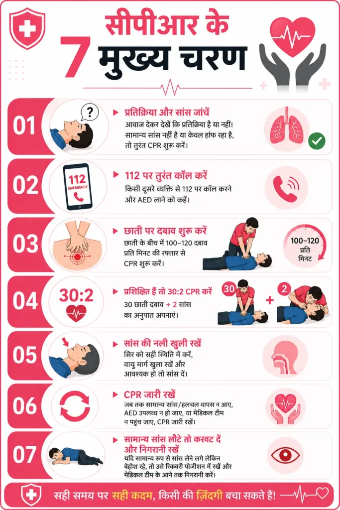
सीपीआर को आप मुख्य रूप से 7 चरणों में बांटों और इमरजेंसी स्थिति में उन्हें एक-एक करके फॉलो करें –
सीपीआर एक जान बचाने वाली प्रक्रिया है, लेकिन इसके दौरान कुछ जटिलताएं भी हो सकती हैं, जैसे छाती पर ज्यादा दबाव पड़ने से पसली टूटना, या मुंह से सांस देते समय पेट में हवा भर जाना (गैस्ट्रिक इन्सफ्लेशन)। इन जोखिमों के बावजूद, विशेषज्ञ मानते हैं कि सीपीआर न देने का जोखिम, इन संभावित जटिलताओं से कहीं ज़्यादा गंभीर होता है। इसलिए घबराए बिना, सही तकनीक के साथ सीपीआर देना ही सबसे सुरक्षित रास्ता है।
सीपीआर सिर्फ एक मेडिकल शब्द नहीं, बल्कि किसी की जिंदगी और मौत के बीच का फासला मिटाने वाला हुनर है। भारत में आज भी बहुत कम लोग इसे सही तरीके से जानते हैं, और यही वजह है कि हृदयाघात के कई मामलों में समय रहते मदद नहीं मिल पाती। अगर आप, आपका परिवार या आपके ऑफिस के साथी सीपीआर सीख लें, तो आप किसी अनजान पल में किसी की जान बचाने वाले हीरो बन सकते हैं। दिल्ली और गुरुग्राम दोनों शहरों में हमारे अस्पताल है, जो आपके दिल का ख्याल अच्छे से रख सकते हैं। अपने दिल की सेहत को नजरअंदाज न करें, आज ही हमारे कार्डियोलॉजी और इमरजेंसी विशेषज्ञों से अपॉइंटमेंट बुक करें और अपने परिवार को सुरक्षित भविष्य दें।
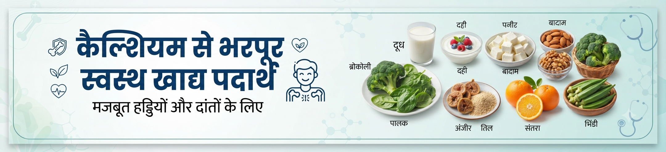
कैल्शियम हड्डियों की “साइलेंट पावर” और आपकी सेहत का असली फाउंडेशन है। इसे आप इस तरह से समझ सकते हैं कि जैसे हड्डियां हमारे शरीर का ढांचा बनाने में मदद करता है, उसी प्रकार से कैल्शियम हमारे शरीर की हड्डियों को मजबूत करने का कार्य करता है। अक्सर लोग थकान (tiredness in hindi) , हल्की दर्द या कमजोरी जैसे लक्षण (kamjori ke lakshan) को नजरअंदाज कर देते हैं, लेकिन जब वह अचानक हल्की चोट के कारण फ्रैक्चर के साथ हमारे पास आते हैं, तब पता चलता है कि उनकी हड्डियां लो कैल्शियम के कारण बहुत कमजोर हो गई है। नेशनल इंडियन ऑर्थोपेडिक एसोसिएशन के 2024 की रिपोर्ट के अनुसार आज भारत में 8 करोड़ से ज्यादा लोग हड्डियों की बीमारियों से पीड़ित हैं, जिसमें ऑस्टियोपोरोसिस (osteoporosis in hindi) का खतरा सबसे ज्यादा होता है।
यदि आप या आपकी मां, आपकी दादी, या आपके बच्चे कभी उठने-बैठने में दर्द महसूस करते हैं, तो आज से ही अपने घर के खान-पान में कैल्शियम को जोडें। यही छोटी शुरुआत आपको अस्पताल के बिस्तर से दूर रख सकती है। इसके अतिरिक्त हड्डियों की बीमारी से बचने या फिर किसी भी समस्या के इलाज के लिए सबसे पहला कदम आपका एक अनुभवी ऑर्थोपेडिक सर्जन या फिर हड्डी रोग विशेषज्ञ से परामर्श होना चाहिए।
Table of Contents
सिर्फ हड्डियां या दांत ही नहीं, आपका दिल, आपकी मांसपेशियां, तंत्रिका तंत्र, और रक्त का थक्का बनने की प्रक्रिया, सबके लिए कैल्शियम जरूरी है। शरीर की हर कोशिका कैल्शियम की मदद से ही अपने आप को रिकवर और दुसरे काम करती है। बचपन, युवावस्था, प्रेग्नेंसी या बुढ़ापे, उम्र के हर पड़ाव में कैल्शियम की आवश्यकता अलग-अलग होती है।
यदि शरीर में कैल्शियम की मात्रा बनी रहती है, तो इसके कारण न ही हड्डी टूटती है और दिल की धड़कन भी सही चलती है। नैशनल इन्स्टिट्यूट ऑफ हेल्थ के एक रिसर्च के अनुसार, कैल्शियम की कमी से बच्चों में रिकेट्स और बुजुर्गों में फ्रैक्चर का खतरा दोगुना हो जाता है।
अगर आप या आपके घर के किसी सदस्य में ये लक्षण दिखें, तो बिना देर किए डॉक्टर से मिलें और इलाज के सभी विकल्पों पर बात करें –
यह सारे लक्षण शरीर में कैल्शियम की कमी के लक्षण हैं। आजकल महिलाओं में ऑस्टियोपोरोसिस और बच्चों में रिकेट्स तेजी से बढ़ रहा है, विशेषकर गलत खानपान और धूप से दूरी इसका मुख्य कारण हैं।
बाजार में सैकड़ों सप्लीमेंट्स हैं, लेकिन घर पर ही कुछ ऐसे पदार्थ होते हैं, जो इन सभी सप्लीमेंट्स को मात दे सकते हैं। प्राकृतिक पदार्थों की बात जब आती है, तो ये हैं सबसे ज्यादा कैल्शियम के स्रोत, जो सस्ता भी हैं और आसानी से उपलब्ध हैं –
इन सभी चीजों को रोज की रूटीन में शामिल करें। सुबह का नाश्ता, लंच या डिनर, इन सभी खाद्य पदार्थों को अपने आहार में शामिल करें और शरीर में कैल्शियम की लय को बनाए रखें।
हर व्यक्ति की उम्र और लाइफस्टाइल के अनुसार शरीर में कैल्शियम की आवश्यकता अलग-अलग होती है। जैसे कि –
| आयु वर्ग | अनुशंसित कैल्शियम मात्रा (प्रतिदिन) |
| शिशु (1–3 वर्ष) | 700mg |
| बच्चे (4–8 वर्ष) | 1,000mg |
| किशोर (9–18 वर्ष) | 1,300mg |
| वयस्क (19–50 वर्ष) | 1,000mg |
| महिलाएं (51+), पुरुष (71+) | 1,200mg |
गर्भवती या स्तनपान कराने वाली महिलाओं को अतिरिक्त कैल्शियम चाहिए। इस स्थिति में महिलाओं को संतुलित खानपान दें और नियमित डॉक्टर से सलाह आवश्यक है।
यदि आप रोजाना सही मात्रा में कैल्शियम का सेवन करते हैं, तो इससे आपको निम्न लाभ मिल सकते हैं –
एक हालिया रिपोर्ट में बताया गया है कि नियमित कैल्शियम युक्त आहार लेने से 30% तक हड्डी टूटने की संभावना घट जाती है। यह तथ्य 2024 में प्रकाशित हावर्ड हेल्थ पब्लिशिंग की रिपोर्ट पर आधारित है।
यदि लंबे समय तक कैल्शियम की कमी रह जाए, तो आपके शरीर में निम्न समस्याएं उत्पन्न हो सकती हैं –
इंडियन जर्नल ऑफ एंडोक्रिनोलॉजी के अनुसार, देश में 30% वयस्कों में ऑस्टियोपोरोसिस की आशंका है। इन सभी लोगों में से महिलाएं और बुजुर्ग कैल्शियम और विटामिन डी की कमी से सबसे ज्यादा प्रभावित होते हैं।
आज शायद आप सोच रहे होंगे कि प्लेट में सिर्फ एक केला, एक गिलास दूध या थोड़ी दही जोड़कर क्या फर्क पड़ेगा? लेकिन यह छोटा कदम आपके, आपके बच्चों और परिजनों के जीवन में अमूल्य सुधार ला सकती है। कैल्शियम को नजरअंदाज न करें। यह आपके शरीर की नींव है। हालांकि हड्डियों की समस्या जैसे कि ऑस्टियोपोरोसिस को आप घर पर ठीक नहीं कर सकते हैं। इसके लिए आपको एक अनुभवी हड्डी रोग विशेषज्ञ की आवश्यकता पड़ेगी। इसलिए प्रयास करें कि आप परामर्श लें और इलाज के सभी विकल्पों के बारे में बात करें।
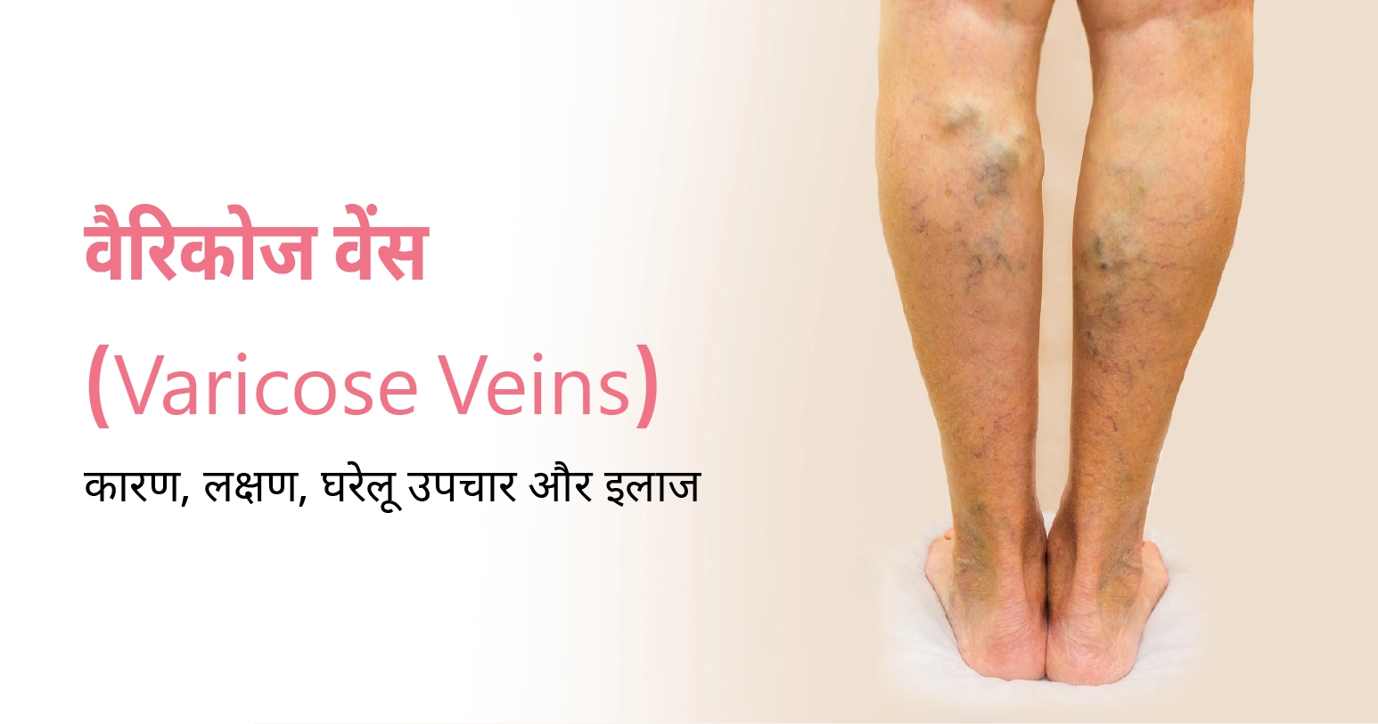
शाम को ऑफिस से घर लौटते वक्त क्या आपके पैर भी भारी-भारी से लगने लगते हैं? क्या टांगों में हल्की सूजन या नसों का उभार आपको आईने के सामने खड़े होकर छिपाना पड़ता है? अगर हां, तो आप अकेले नहीं हैं। भारत में हर चौथा व्यक्ति किसी न किसी स्तर पर वैरिकोज वेंस से जूझ रहा है, और ज्यादातर लोग इसे सिर्फ “थकान” समझ कर टाल देते हैं। यही सबसे बड़ी गलती है।
वैरिकोज वेंस सिर्फ दिखने की समस्या नहीं है, यह आपके रक्त संचार (blood circulation) से जुड़ा एक चिकित्सीय संकेत है, जिसे समय पर पहचानना और सही ढंग से मैनेज करना जरूरी है। इस ब्लॉग में हम इस समस्या को जड़ से समझेंगे, इसके कारण, लक्षण, घरेलू उपाय और आधुनिक इलाज के हर पहलू को विस्तार से जानेंगे। अगर आपको या आपके परिवार में किसी को पैरों की नसें उभरी हुई महसूस हो रही हैं, तो देर न करें, हमारे अनुभवी वैस्कुलर स्पेशलिस्ट से अपॉइंटमेंट बुक करें और सही समय पर सही सलाह पाएं।
Table of Contents
वैरिकोज वेंस उन नसों को कहा जाता है जो त्वचा के ठीक नीचे सूजी हुई, मुड़ी हुई और उभरी हुई दिखाई देते हैं, अक्सर नीले या बैंगनी रंग की। ये ज्यादातर पैरों, टखनों और पिंडलियों में देखने को मिलती है, क्योंकि खड़े होने और चलने से इन हिस्सों की नसों पर दबाव सबसे ज्यादा पड़ता है। हमारी नसों के अंदर छोटे-छोटे शिरा वाल्व (venous valves) होते हैं, जिनका काम खून को एक ही दिशा में, यानी वापस दिल की तरफ, धकेलना है। जब ये वाल्व कमजोर या क्षतिग्रस्त हो जाते हैं, तो खून सही तरीके से आगे नहीं बढ़ पाता और नस में ही जमा होने लगता है। इसी वजह से नस फूल जाती है और वैरिकोज वेंस का रूप ले लेती है। कई बार इसके साथ-साथ स्पाइडर वेंस (spider veins) भी दिखाई देते हैं, जो छोटी, लाल या नीली महीन रेखाओं जैसी होती हैं और आमतौर पर कम गंभीर मानी जाती हैं, लेकिन इन्हें भी नजरअंदाज नहीं करना चाहिए।
यह समस्या रातों-रात नहीं आती, बल्कि धीरे-धीरे विकसित होती है। शुरुआत में नस की दीवार थोड़ी कमजोर पड़ती है, फिर शिरा वाल्व अपना काम ठीक से नहीं कर पाते। नतीजतन, खून नस में पीछे की ओर बहने लगता है, जिसे मेडिकल भाषा में “रिफ्लक्स” कहा जाता है। समय के साथ यह दबाव नस की दीवारों को और कमजोर करता जाता है, जिससे नस और ज्यादा फैलती और मुड़ती जाती है। अगर इस प्रक्रिया को शुरुआत में ही नहीं रोका गया, तो यह धीरे-धीरे क्रॉनिक वेनस इन्सफिशिएंसी (chronic venous insufficiency) जैसी गंभीर स्थिति में बदल सकती है, जिसमें पैरों की त्वचा का रंग बदलना और वेनस अल्सर (venous ulcer) जैसी जटिलताएं भी सामने आ सकती हैं।
हर वैरिकोज वेंस एक जैसी नहीं होती। डॉक्टर इन्हें आमतौर पर तीन श्रेणियों में बांटते हैं। पहली, ट्रंक वैरिकोज वेंस, जो बड़ी और साफ दिखने वाली, उभरी हुई नसें होती हैं। दूसरी, रेटिकुलर वेंस, जो थोड़ी छोटी होती हैं लेकिन त्वचा के नीचे जालनुमा पैटर्न बनाती हैं। तीसरी, स्पाइडर वेंस, जो सबसे महीन होती हैं और अक्सर सिर्फ कॉस्मेटिक समस्या मानी जाती हैं। इसके अलावा कुछ मामलों में पेल्विक क्षेत्र में भी वैरिकोज वेंस विकसित हो सकती हैं, खासकर उन महिलाओं में जिन्होंने एक से ज्यादा बार प्रसव कराया हो। सही इलाज तय करने के लिए यह जानना जरूरी है कि आपकी नस किस श्रेणी में आती है, इसलिए स्व-निदान की बजाय डॉक्टरी जांच पर भरोसा करें।
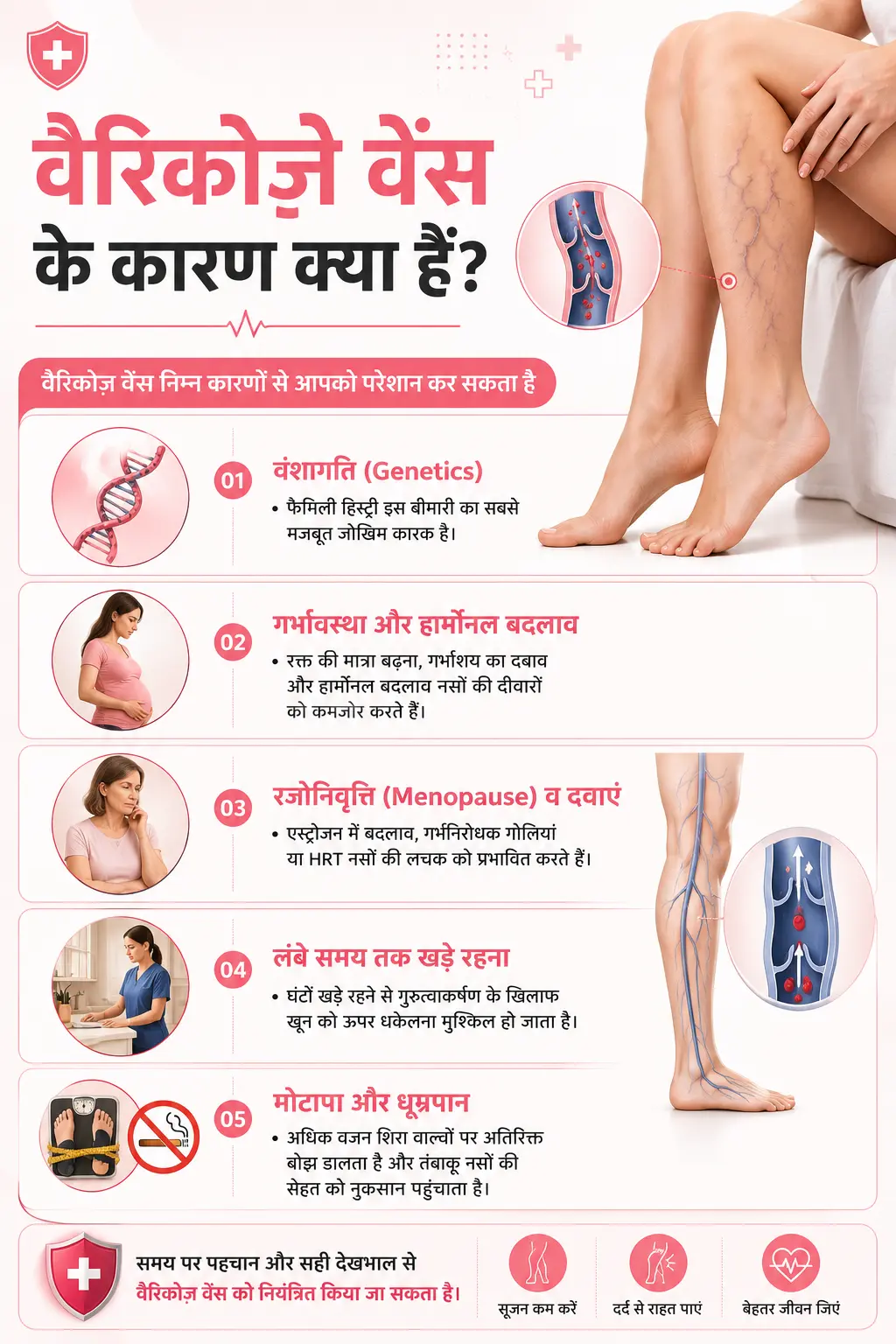
वैरिकोज वेंस निम्न कारणों से आपको परेशान कर सकती है –
उत्तर भारत में हुए एक अध्ययन के अनुसार, महिलाओं में यह समस्या पुरुषों की तुलना में लगभग दोगुनी पाई गई, जिसमें करीब आधी महिलाएं वैरिकोज वेंस से प्रभावित मिलीं। यह आंकड़ा साफ बताता है कि हार्मोनल बदलाव इस बीमारी में कितनी बड़ी भूमिका निभाते हैं।
शुरुआत में लक्षण बहुत मामूली लगते हैं, इसलिए ज्यादातर लोग इन्हें गंभीरता से नहीं लेते:
विशेषज्ञ सलाह: हमारे पास ऐसे कई मामले आते हैं जहां मरीज शुरुआत में सिर्फ “पैर भारी लगते हैं” कहकर आते हैं, लेकिन जांच में पता चलता है कि उनकी सैफेनस नस (Saphenous Vein) में रिफ्लक्स शुरू हो चुका है। अगर इस स्तर पर सही सलाह मिल जाए, तो आगे की जटिलताओं से आसानी से बचा जा सकता है। इसलिए किसी भी लक्षण को “सामान्य थकान” समझकर टालना सही रणनीति नहीं है।
वैरिकोज वेंस की स्थिति में कुछ घरेलू उपचार आपके लिए बहुत लाभकारी साबित हो सकते हैं –
ध्यान दें: घरेलू नुस्खे सिर्फ सहायक उपाय हैं, इन्हें मुख्य मेडिकल इलाज का विकल्प न मानें।
जब नसें काफी फैल चुकी हों या लक्षण बढ़ने लगें, तो चिकित्सकीय इलाज जरूरी हो जाता है। आधुनिक विकल्प कम-इनवेसिव (Minimally Invasive) और डे-केयर आधारित है, जिनमें अस्पताल में भर्ती होने की ज़रूरत नहीं पड़ती।
जेनेटिक कारणों में पूरी रोकथाम संभव नहीं है, लेकिन जोखिम काफी हद तक कम किया जा सकता है:
अगर आपको पैरों में लगातार दर्द, सूजन जो आराम करने पर भी कम न हो, त्वचा के रंग में बदलाव, नस के आसपास घाव या जख्म, या नस से खून बहना जैसे लक्षण दिखें, तो तुरंत डॉक्टर से संपर्क करें। इसी तरह अगर घरेलू उपाय अपनाने के बावजूद कुछ हफ्तों में कोई राहत नहीं मिल रही, तो यह जांच करवाने का सही समय है, क्योंकि लंबे समय तक अनदेखी करने पर यह डीप वेन थ्रोम्बोसिस (DVT) जैसी गंभीर स्थिति का खतरा भी बढ़ सकता है।
वैरिकोज वेंस को नज़रअंदाज़ करना आसान लगता है, लेकिन समय रहते सही कदम उठाना आपकी जिंदगी को कहीं ज्यादा आरामदायक बना सकता है। सीके बिरला अस्पताल में दिल्ली और गुरुग्राम, दोनों केंद्रों पर अनुभवी वैस्कुलर सर्जन आपकी जांच से लेकर स्क्लेरोथेरेपी, EVLA जैसी आधुनिक प्रक्रियाओं तक, हर कदम पर आपके साथ हैं। अपने नजदीकी सेंटर पर आज ही अपॉइंटमेंट बुक करें और अपने पैरों को वह देखभाल दें, जिसके वे हकदार हैं।

