Filter :
ऑफिस की डेडलाइन, बच्चों की पढ़ाई, घर की जिम्मेदारियां, सब कुछ मिलकर किसी की भी नींद छीन सकते हैं। यदि आपके साथ भी ये सब है, तो इसमें आप अकेले नहीं हैं। यहीं पर एक पुरानी आयुर्वेदिक जड़ी-बूटी का काम आता है, जिसके बारे में आपने कभी न कभी सुना होगा, अश्वगंधा।
यह कोई नया ट्रेंड नहीं है, बल्कि हजारों साल पुराना आयुर्वेदिक ज्ञान है जिसे अब आधुनिक विज्ञान भी परख रहा है। इस लेख में हम अश्वगंधा के फायदे, इसकी सही खुराक, सेवन का सही तरीका और किन लोगों को इससे बचना चाहिए, यह सब विस्तार से समझेंगे। अगर आप भी तनाव, नींद की समस्या या कमजोरी से परेशान हैं और सही मार्गदर्शन चाहते हैं, तो सीके बिरला अस्पताल के अनुभवी विशेषज्ञों से अपॉइंटमेंट बुक करें और अपनी सेहत का सही आकलन कराएं।
Table of Contents
अश्वगंधा का साइंटिफिक नाम विथानिया सोमनिफेरा (Withania Somnifera) है। इसे आयुर्वेद में “रसायन” श्रेणी की जड़ी बूटी माना गया है, यानी ऐसी औषधि जो शरीर की समग्र ताकत और जीवन शक्ति बढ़ाने में मदद करती है। इसकी जड़ों की गंध घोड़े जैसी होने के कारण इसे “अश्व” (घोड़ा) और “गंध” से मिलाकर यह नाम दिया गया। आधुनिक विज्ञान की भाषा में इसे एडाप्टोजेन (adaptogen) कहा जाता है, यानी एक ऐसा प्राकृतिक तत्व जो शरीर को शारीरिक और मानसिक तनाव के प्रति बेहतर तालमेल बिठाने में मदद करता है। इसकी जड़ों में विथानोलाइड्स नाम के सक्रिय यौगिक पाए जाते हैं, जिन्हें इसके अधिकतम स्वास्थ्य लाभों के पीछे की वजह माना जाता है।
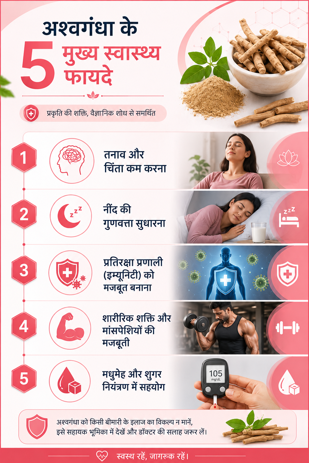
अश्वगंधा एक आयुर्वेदिक जड़ी बूटी है, जिसके फायदे भी भरपूर हैं –
यहां यह समझना जरूरी है कि अश्वगंधा पर हुए सभी शोध एक जैसे मजबूत नहीं हैं। तनाव और कॉर्टिसोल पर इसका असर सबसे ज्यादा और सबसे भरोसेमंद रिसर्च से समर्थित है। बाकी फायदों (इम्यूनिटी, शुगर, मांसपेशियां) पर शोध शुरुआती स्तर के हैं, और छोटे सैंपल साइज़ या सीमित अवधि के कारण इन्हें “संभावित” कहना ज्यादा सही होगा, न कि “साबित” फायदा। हमारे डॉक्टर मरीजों को यही सलाह देते हैं, इसे किसी बीमारी के इलाज का विकल्प न मानें, बल्कि सहायक भूमिका में देखें।
अश्वगंधा की खुराक व्यक्ति की उम्र, स्वास्थ्य स्थिति और उद्देश्य के अनुसार अलग-अलग हो सकती है। सामान्यतः वयस्कों के लिए रोजाना 250 मिलीग्राम से 600 मिलीग्राम तक की खुराक सुरक्षित मानी जाती है, और ज्यादातर क्लिनिकल अध्ययन इसी सीमा में किए गए हैं। पाउडर के रूप में लेने पर आमतौर पर आधा से एक चम्मच (लगभग 3 से 6 ग्राम) प्रतिदिन की सलाह दी जाती है। लेकिन यह सामान्य जानकारी है, हर व्यक्ति की जरूरत अलग होती है, इसलिए खुराक तय करने से पहले किसी योग्य आयुर्वेदिक चिकित्सक या डॉक्टर से जरूर सलाह लें। खासकर अगर आप पहले से कोई दवा ले रहे हैं या किसी पुरानी बीमारी से जूझ रहे हैं।
यह भी ध्यान रखें कि ज्यादातर डॉक्टर अश्वगंधा को खुद से शुरू करने की सलाह नहीं देते, बल्कि अगर मरीज पहले से इसे ले रहा है और कोई समस्या नहीं है, तो उसे जारी रखने की छूट देते हैं और सिर्फ मौजूदा दवाओं के साथ इंटरैक्शन की जांच करते हैं।
अश्वगंधा को लेकर समाज में एक बड़ा मिथक फैला हुआ है कि यह सिर्फ पुरुषों की यौन शक्ति बढ़ाने के लिए इस्तेमाल की जाने वाली जड़ी बूटी है। यह पूरी तरह सही नहीं है। हां, यह टेस्टोस्टेरोन स्तर और प्रजनन स्वास्थ्य को सहारा देने में मदद कर सकती है, इसमें कोई शक नहीं। लेकिन इसका दायरा इससे कहीं ज्यादा बड़ा है। यह तनाव प्रबंधन, नींद, इम्यूनिटी, मांसपेशियों की मजबूती और मानसिक स्पष्टता, यानी शरीर और दिमाग दोनों के समग्र स्वास्थ्य के लिए काम करती है। महिलाएं भी इसे हार्मोनल संतुलन, ऊर्जा स्तर और तनाव प्रबंधन के लिए उतनी ही प्रभावी ढंग से इस्तेमाल कर सकती हैं। इसे सिर्फ एक “मर्दाना” जड़ी बूटी के दायरे में सीमित कर देना इसके असली आयुर्वेदिक महत्व को कम आंकना है।
हर प्राकृतिक चीज हर किसी के लिए सुरक्षित हो, यह जरूरी नहीं। कुछ स्थितियों में अश्वगंधा से परहेज करने की सलाह दी जाती है –
अश्वगंधा अकेले कोई जादुई इलाज नहीं है, यह एक सहायक की भूमिका निभाती है। बेहतर परिणाम के लिए इसे सही जीवन शैली के साथ जोड़ना जरूरी है। यदि आपको भी संदेह है कि आपको अश्वगंधा लेना चाहिए या नहीं, तो बिना देर किए हमारे अनुभवी विशेषज्ञों से परामर्श लें। वे आपका मार्गदर्शन सही से करेंगे।
अगर पिछले कुछ हफ्तों से आपके आसपास भी लोग बुखार, खांसी और बदन दर्द की शिकायत लेकर घर बैठे हैं, तो आप अकेले नहीं हैं। दिल्ली और आसपास के इलाकों में इन दिनों स्वाइन फ्लू यानी H1N1 के मामलों में तेजी देखी जा रही है, और सोशल मीडिया से लेकर पड़ोस के व्हाट्सएप ग्रुप तक, हर जगह यही चर्चा है।
सच कहें तो एक हल्का सा बुखार भी अब चिंता की वजह बन जाता है, खासकर जब घर में बच्चे, बुजुर्ग या गर्भवती महिलाएं हों। यही वजह है कि सही जानकारी होना बेहद जरूरी हो जाता है, ताकि आप घबराने की बजाय समझदारी से कदम उठा सकें। इस ब्लॉग में हम स्वाइन फ्लू से जुड़ी हर जरूरी बात, लक्षणों से लेकर इलाज और बचाव तक, आसान भाषा में समझाएंगे। अगर आपको या आपके परिवार में किसी को लगातार बुखार या सांस लेने में तकलीफ महसूस हो रही है, तो देर न करें, हमारे अनुभवी विशेषज्ञों से अभी अपॉइंटमेंट बुक करें और सही समय पर सही इलाज पाएं।
Table of Contents
स्वाइन फ्लू, इन्फ्लूएंजा-A वायरस की एक किस्म H1N1 के कारण होने वाला संक्रामक रोग है। इसे “स्वाइन” इसलिए कहा जाता है क्योंकि यह वायरस मूल रूप से सूअरों में पाए जाने वाले फ्लू वायरस से समान पाया गया था, हालांकि अब यह इंसानों से इंसानों में ही फैलता है और इसका सूअर के संपर्क से कोई सीधा संबंध जरूरी नहीं है।
भारतीय आयुर्विज्ञान अनुसंधान परिषद (ICMR) के अनुसार, यह कोई नया वायरस नहीं बल्कि हर साल लौटने वाला एक मौसमी इन्फ्लूएंजा स्ट्रेन है। दिल्ली स्वास्थ्य विभाग के हालिया आंकड़ों की मानें तो अकेले दिल्ली में इस साल अब तक इन्फ्लूएंजा-A के लगभग तीन हज़ार मामले दर्ज हो चुके हैं, जिनमें बड़ा हिस्सा H1N1 का है। मानसून के बाद नमी और भीड़ भाड़ वाले माहौल में यह वायरस तेजी से सक्रिय हो जाता है, इसलिए इस मौसम में सतर्क रहना और भी ज़रूरी हो जाता है।
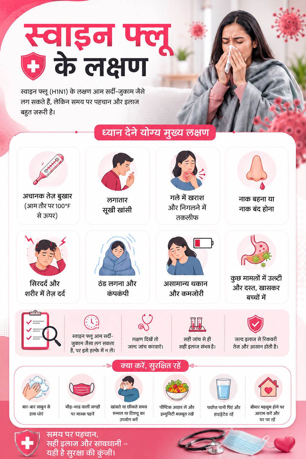
स्वाइन फ्लू के लक्षण आम मौसमी फ्लू से काफी मिलते-जुलते हैं, इसलिए शुरुआत में इसे पहचानना थोड़ा मुश्किल हो सकता है। ध्यान रखने योग्य मुख्य लक्षण इस प्रकार हैं –
हमारे OPD में अक्सर ऐसे मरीज आते हैं, जो शुरुआत में इसे सामान्य सर्दी-जुकाम समझकर घरेलू नुस्खों से इलाज करते रहते हैं, और जब बुखार तीन-चार दिन तक नहीं उतरता तब जांच के लिए आते हैं। ऐसे में देखा गया है कि जितनी जल्दी सही जांच और इलाज शुरू होता है, रिकवरी उतनी ही आसान और तेज होती है। इसलिए लक्षण हल्के भी लगे तो उन्हें नज़रअंदाज़ करना सही नहीं है।
वायरस के संपर्क में आने के बाद आमतौर पर 1 से 4 दिनों के भीतर लक्षण दिखना शुरू हो जाते हैं। कुछ लोगों में यह अवधि थोड़ी लंबी भी हो सकती है, खासकर अगर उनकी रोग प्रतिरोधक क्षमता (immunity) कमजोर हो। यह भी ध्यान देने वाली बात है कि लक्षण दिखने से एक दिन पहले से लेकर बीमारी के 5 से 7 दिन बाद तक व्यक्ति दूसरों को संक्रमित कर सकता है, इसलिए इस दौरान सावधानी बरतना और भी ज़रूरी हो जाता है।
स्वाइन फ्लू मुख्य रूप से एक श्वसन संक्रमण (respiratory infection) है, जो निम्न तरीकों से फैलता है –
विशेषज्ञों का मानना है कि मानसून के बाद बदलता तापमान, नमी और घर के अंदर भीड़ भाड़ इस मौसमी फ्लू वायरस के फैलाव के लिए अनुकूल माहौल बनाते हैं, यही वजह है कि हर साल इसी समय के आसपास मामलों में उछाल देखा जाता है।
अगर लक्षण दो-तीन दिन से ज़्यादा बने रहें, तो डॉक्टर आमतौर पर एक विस्तृत जांच के बाद निम्न परीक्षणों की सलाह देते हैं –
समय पर सही जांच होने से न सिर्फ इलाज की दिशा तय करने में मदद मिलती है, बल्कि गंभीर जटिलताओं से बचाव भी संभव हो पाता है।
अधिकतर स्वस्थ लोगों में स्वाइन फ्लू अपने आप 5 से 7 दिनों में ठीक हो जाता है, बशर्ते सही देखभाल की जाए। इलाज में आमतौर पर शामिल होता है –
एक बात हमेशा याद रखें, बिना डॉक्टर की सलाह के खुद से एंटीबायोटिक या एंटीवायरल दवा लेना नुकसानदायक हो सकता है। हाल ही में इंडियन मेडिकल एसोसिएशन (IMA) ने भी अपनी एडवाइजरी में लोगों को सेल्फ-मेडिकेशन से बचने की सलाह दी है।
अगर लक्षण हल्के हैं और डॉक्टर ने घर पर आराम करने की सलाह दी है, तो इन बातों का ध्यान रखें –
स्वाइन फ्लू से बचाव, इलाज से कहीं आसान है। ये उपाय अपनाकर आप खुद को और अपने परिवार को सुरक्षित रख सकते हैं –
कुछ लक्षण ऐसे हैं जिन्हें बिल्कुल नजरअंदाज नहीं करना चाहिए। अगर इनमें से कोई भी संकेत दिखे तो तुरंत नज़दीकी अस्पताल जाएं –
बच्चों, गर्भवती महिलाओं, 65 वर्ष से ऊपर के बुजुर्गों और डायबिटीज, अस्थमा या हृदय रोग जैसी पहले से मौजूद बीमारियों वाले लोगों को हल्के लक्षण होने पर भी देर किए बिना डॉक्टर से सलाह लेनी चाहिए, क्योंकि इन लोगों में जटिलताओं का खतरा अपेक्षाकृत अधिक होता है। सीके बिरला अस्पताल की इंटरनल मेडिसिन और पल्मोनोलॉजी की अनुभवी टीम मौजूद है, जो हर उम्र के मरीजों के लिए 24×7 आपातकालीन सेवाएं भी उपलब्ध कराती है। अगर आपको या आपके परिवार में किसी को स्वाइन फ्लू के लक्षण महसूस हो रहे हैं, तो जांच में देरी न करें, आज ही हमारे विशेषज्ञ से अपॉइंटमेंट बुक करें।
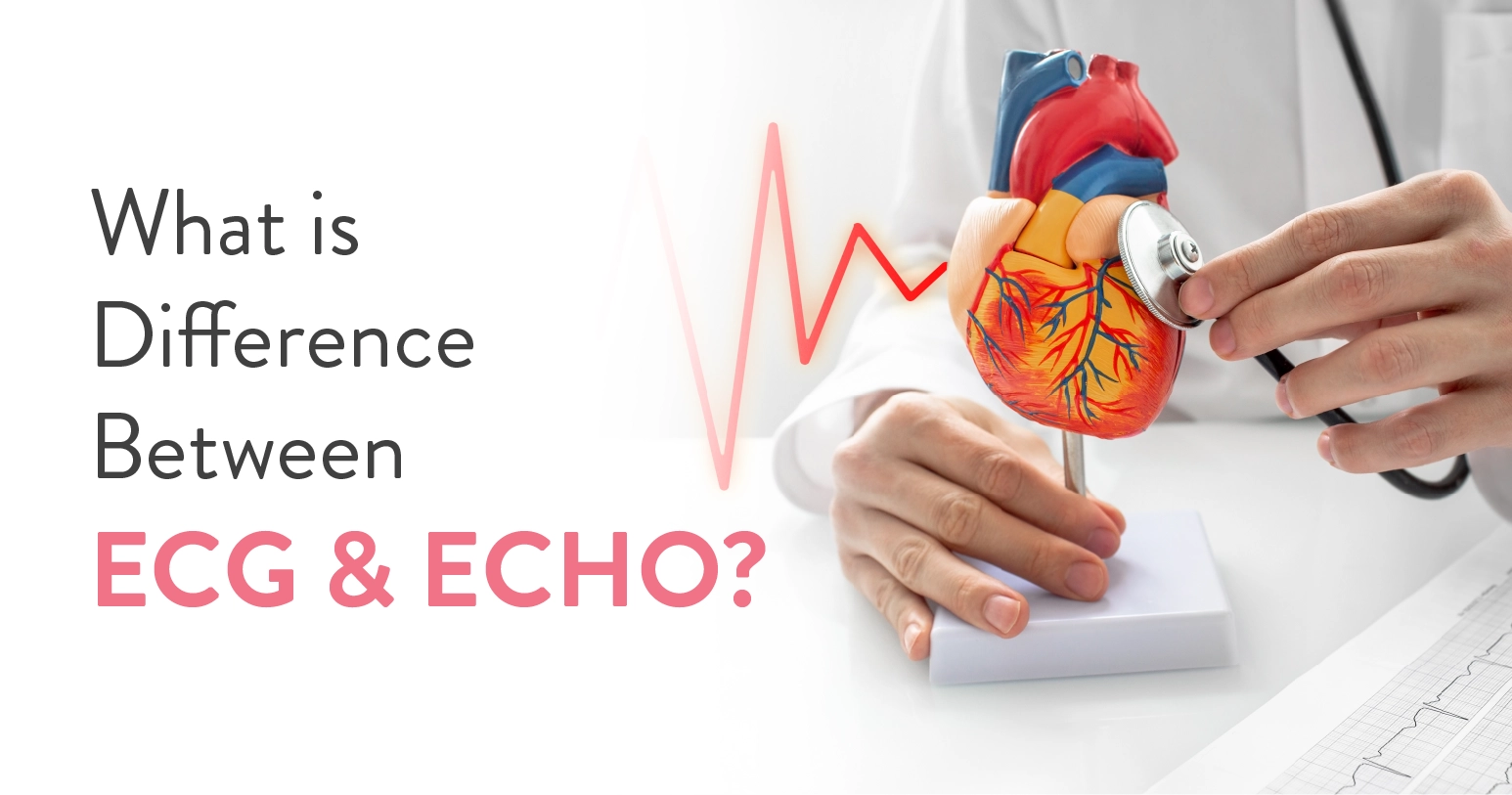
If your cardiologist has mentioned an ECG and an Echo test in the same breath, and you are left wondering, “Aren’t these two tests the same?” We get you! Many people confuse the two because both are used to examine the same organ, the heart. But they serve different purposes and provide different information about how your heart is functioning. That is why doctors often recommend them together to get an accurate picture of your heart health. So, how are they actually different? Let’s find out.
Table of Contents
An ECG measures the electrical signals that make your heart beat, whereas an Echo uses ultrasound to create moving images of your heart’s structure and pumping action.
An ECG (electrocardiogram) tells you about the heart’s rhythm and timing.
An Echo (echocardiogram) tells you about the heart’s shape, size, valves and pumping strength.
Neither test replaces the other. They answer different questions about the same organ.
You might also come across the term “electrocardiograph,” which simply refers to the machine that records the ECG, not the test itself. This connection between electrocardiogram vs electrocardiograph confuses many patients, so it is worth understanding the difference. The electrocardiogram is the test and its output, and the electrocardiograph is the device that produces it.
| Feature | ECG (Electrocardiogram) | Echo (Echocardiogram) |
| What it checks | Your heart’s electrical timing that is the signal that tells it when to beat | The physical heart itself, including the structure, size, shape and how well it is pumping |
| Technology used | Small electrode patches on your skin, connected to a monitor | A handheld ultrasound probe moved gently over your chest |
| Duration | Usually takes 5 to 10 minutes | Closer to 30 to 45 minutes, since the technician needs multiple angles |
| Can it catch an irregular heartbeat (heart rhythm)? | Yes | Limited, since it is not designed for this. |
| Can it see your heart valves working? | No | Yes |
| Shows chamber size/pumping strength | No | Yes |
| Detects arrhythmias | Yes | Sometimes |
| Detects structural defects | No | Yes |
| Any Pain or discomfort involved | None | None |
| When it is usually recommended | Chest pain, palpitations, routine health checkups | Suspected heart failure, murmurs, valve disease |
An ECG, or electrocardiogram, records the electrical impulses that travel through your heart with every beat. These impulses tell your heart muscle when to contract and relax. It is completely painless, non-invasive and does not send any electricity to your body. Small sticky electrodes placed on your chest, arms, and legs pick up this activity and translate it into wavy lines on a screen or paper.
An ECG can help detect:
An ECG records your heart’s electrical activity, but it does not show the heart’s structure or how well it pumps blood. For that, cardiologists usually recommend an echocardiogram (Echo), which creates real-time images of the heart using ultrasound.
An echocardiogram, commonly called Echo, uses high-frequency sound waves (ultrasound) to create a live, moving image of your heart. This test lets doctors know your heart’s size, how thick its walls are, how well each chamber is pumping and whether the four valves are opening and closing properly.
Echo is particularly useful for diagnosing heart failure, valve disease, congenital heart defects and fluid around the heart. It is also the go-to test for measuring ejection fraction, which is a number that shows how much blood your heart pumps out with each beat.
It is a non-invasive test and does not hurt; however, you may feel a cold gel or mild pressure on your chest from the ultrasound tool.
An Echo test helps doctors evaluate:
What Can’t an Echo Show?
Although an Echo provides detailed images of the heart’s structure and pumping function, it does not record the heart’s electrical activity or detect abnormal heart rhythms as effectively as an ECG.
This decision totally depends on your symptoms. If you are experiencing palpitations, irregular pulse or frequent fainting spells, consult a cardiologist. They will likely start with an ECG since it captures rhythm problems almost instantly.
If you have breathlessness, swelling in your legs, a known heart murmur or a family history of structural heart disease, an Echo gives a complete and detailed picture of your heart health.
No test is better than the other. Both tests are simply designed to answer different questions. Choosing the right test mainly depends on your symptoms.
Note: Do not undergo either test without a doctor’s prescription or recommendation. Speak with a doctor first. After assessing your symptoms and understanding your condition, they will recommend the most appropriate test for you.
In many cases, cardiologists suggest both ECG and Echo because your heart’s electrical system and its physical structure are deeply connected, even though they are measured differently.
An abnormal ECG might raise a red flag that only an Echo can fully explain. For instance, your ECG shows a pattern that looks like a past heart attack. Your cardiologist will likely want an Echo next, just to see how much of the heart muscle was affected and how well it is still pumping blood.
ECG and Echo may sound similar, but they look at your heart through two completely different lenses.
An ECG records your heart’s electrical activity, whereas an Echo shows its structure and how well it pumps blood. Depending on your symptoms and medical history, your cardiologist may recommend one or both tests to arrive at an accurate diagnosis.
Rather than trying to decide which test you need, speak with a cardiologist who can recommend the most appropriate evaluation for your condition. At the CK Birla Hospital, our experienced cardiac specialists can help identify the underlying cause of your symptoms, assess your heart health and guide you towards the right diagnostic test and treatment plan.

सुबह आंख खुलते ही पूरे शरीर में भारीपन, खिंचाव और लगातार थकान महसूस होना सामान्य नहीं है। अगर यह दर्द कई दिनों तक बना रहे, तो यह सिर्फ थकान नहीं, बल्कि आपका शरीर आपको कोई गंभीर चेतावनी दे रहा है। ICMR की एक रिपोर्ट के अनुसार, भारत में हर चार में से एक व्यक्ति मस्कुलोस्केलेटल (हड्डी और मांसपेशी) की समस्याओं से जूझ रहा है।
अगर आप भी पूरे शरीर में दर्द और सूजन के कारणों को लेकर परेशान हैं, तो यह ब्लॉग आपके लिए है। यहाँ हम इसके मुख्य कारण, सही उपचार और प्रभावी घरेलू उपाय जानेंगे। लंबे समय के दर्द को नजरअंदाज न करें; सही जांच के लिए आज ही हमारे अनुभवी विशेषज्ञों से मिलें और अपने स्वास्थ्य समस्या के बारे में विस्तार से बात करें।
Table of Contents
हर दर्द खतरनाक नहीं होता। कई बार शरीर में दर्द का होना बिल्कुल सामान्य होता है और कुछ दिनों में अपने आप ठीक भी हो जाता है। चलिए इसे उदाहरण की मदद से समझते हैं –
जब आपने बहुत दिनों बाद जिम जाना शुरू किया हो और मांसपेशियों पर अचानक जोर पड़े। जब सर्दी-जुकाम या फ्लू हो और पूरे बदन में दर्द और टूटन हो। जब लंबी यात्रा के बाद शरीर अकड़ा हुआ हो। इन सभी स्थितियों में “तीव्र दर्द” (Acute Pain) होता है। यह आमतौर पर 2 से 5 दिनों में ठीक हो जाता है।
लेकिन जब पूरे शरीर में दर्द नियमित हो जाए, यानी हफ्तों या महीनों तक बना रहे – तो यह “दीर्घकालिक दर्द” (Chronic Pain) की श्रेणी में आता है। और यही वह स्थिति है जब डॉक्टर की सलाह जरूरी हो जाती है।
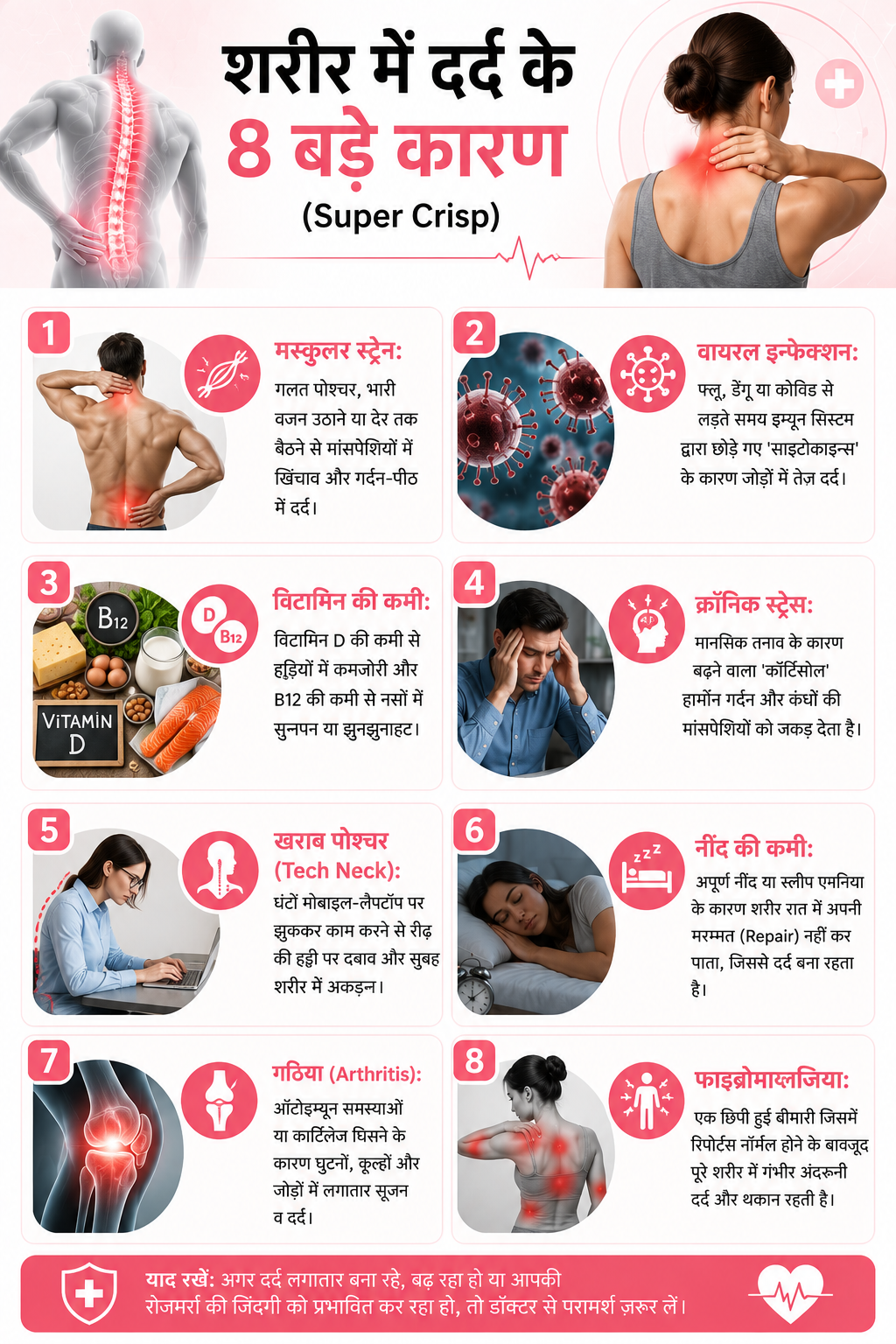
बिना तैयारी के भारी काम करना, गलत तरीके से वजन उठाना, या बहुत देर तक एक ही पोजीशन में बैठे रहना – इन सबसे मांसपेशियों में सूक्ष्म चोटें आ जाती हैं। नतीजा – अगले दिन पूरे शरीर में दर्द और सूजन। यह दर्द खासकर कमर, पीठ, गर्दन और कंधों में महसूस होता है।
जब शरीर किसी वायरस से लड़ रहा होता है, तब प्रतिरक्षा प्रणाली (Immune System) कुछ रसायन (Cytokines) छोड़ती है जो मांसपेशियों और जोड़ों में दर्द पैदा करते हैं। इसलिए फ्लू में “पूरे शरीर में दर्द की दवा” की जरूरत पड़ती है। COVID-19 के बाद भी कई मरीजों में हफ्तों तक शरीर में दर्द और थकान रहती है, जिसे “Long COVID” कहा जाता है।
यह आधुनिक भारत की सबसे बड़ी समस्या है। AIIMS दिल्ली के एक रिसर्च के अनुसार, भारत में लगभग 70-80% शहरी आबादी विटामिन D की कमी का सामना कर रहे हैं। घरों के भीतर काम करना, धूप से बचना और खराब खानपान इसके मुख्य कारण हैं।
यह सुनने में अजीब लग सकता है, लेकिन मानसिक तनाव शारीरिक दर्द में सीधे तौर पर बदल सकता है। जब हम लंबे समय तक तनाव में रहते हैं, तो शरीर “कोर्टिसोल” (Cortisol) हार्मोन रिलीज करता है। इससे गर्दन और कंधों की मांसपेशियां कस जाती हैं और पुराना दर्द बन जाता है। बहुत से लोगों में डिप्रेशन और चिंता (Anxiety) भी शरीर में व्यापक दर्द के रूप में सामने आती हैं।
आज आप मेट्रो में जाएं और देखें कि हर व्यक्ति अपने फोन में घुसा बैठा है। इसे “Tech Neck” कहा जाता है। मोबाइल और लैपटॉप पर झुककर काम करने से गर्दन, कंधे और पीठ पर भारी दबाव पड़ता है। WHO की एक रिपोर्ट के अनुसार, कमर दर्द (Back Pain) दुनिया में विकलांगता का नंबर एक कारण बन चुका है।
नींद वह समय है जब शरीर खुद को ठीक करता है। अगर यह रिपेयर का समय न मिले, तो दर्द और थकान बनी रहती हैं। Sleep Apnea (नींद में सांस रुकना) और अनिद्रा (Insomnia) जैसी स्थितियां इस चक्र को और खराब कर देते हैं।
रूमेटाइड गठिया (Rheumatoid Arthritis) एक स्वप्रतिरक्षित विकार या ऑटोइम्यून विकार है, जिसमें शरीर अपने ही जोड़ों पर हमला करता है। इससे पूरे शरीर में दर्द और सूजन होती है, खासकर सुबह के समय। ऑस्टियोआर्थराइटिस (Osteoarthritis) में जोड़ों की उपास्थि (Cartilage) घिसने लगती है, जिससे घुटने, कूल्हे और रीढ़ में दर्द होता है।
फाइब्रोमायल्जिया एक ऐसी स्थिति है, जिसमें पूरे शरीर में अंदरूनी दर्द होना लंबे समय तक बना रहता है, लेकिन खून की जांच में कुछ नहीं दिखता। इसमें थकान, नींद की गड़बड़ी और याददाश्त की समस्याएं भी होती हैं। भारत में यह बीमारी अक्सर देर से पहचानी जाती है।
पूरे शरीर में दर्द की दवा लेने से पहले सही निदान जरूरी है। डॉक्टर जांच से दर्द के मूल कारण का पता लगाते हैं और उसी दर्द के कारण के आधार पर इलाज की योजना बनाते हैं –
नोट: बिना डॉक्टर की लिखित पर्ची (Prescription) के किसी भी दर्दनिवारक दवा का नियमित सेवन लिवर और किडनी को नुकसान पहुंचा सकता है।
हल्के और अस्थायी शरीर दर्द के लिए ये घरेलू उपाय आपको तुरंत राहत दे सकते हैं –
घरेलू उपाय और आराम तब तक ठीक हैं, जब तक दर्द हल्का और अस्थायी हो। लेकिन निम्न स्थितियों में बिना देर किए डॉक्टर से मिलें –
शरीर में दर्द 2 हफ्ते से ज्यादा समय से बना हुआ हो। दर्द के साथ तेज बुखार, ठंड लगना या वजन अचानक कम होना हो। जोड़ों में सूजन और लाली हो, खासकर सुबह के समय। पूरे शरीर में अंदरूनी दर्द होना, जिसके कोई कारण समझ न आएं। सांस लेने में तकलीफ या सीने में दर्द के साथ शरीर दर्द हो। दर्द की वजह से रोजमर्रा के काम करना मुश्किल हो रहा हो।
इन स्थितियों में खुद से पूरे शरीर में दर्द की दवा लेना खतरनाक हो सकता है। सही जांच ही सही इलाज की नींव है।
पूरे शरीर में दर्द सिर्फ एक तकलीफ नहीं, बल्कि आपके शरीर का संकेत है। चाहे वह थकान हो, विटामिन की कमी या कोई बीमारी, इन संकेतों को हल्के में न लें। इस ब्लॉग में आपने जाना कि शरीर में दर्द क्यों होता है, इसके कारण क्या हैं और इसका डॉक्टरी व घरेलू उपचार कैसे किया जा सकता है।
सबसे जरूरी बात यह है कि अपने दर्द को नजरअंदाज न करें। समय पर सही जांच और इलाज से आप जल्द स्वस्थ हो सकते हैं। सीके बिरला अस्पताल दिल्ली और गुरुग्राम के अनुभवी विशेषज्ञ डॉक्टर आपकी समस्या के सही उपचार के लिए तैयार हैं। आज ही अपॉइंटमेंट बुक करें और दर्द-मुक्त जीवन की शुरुआत करें।
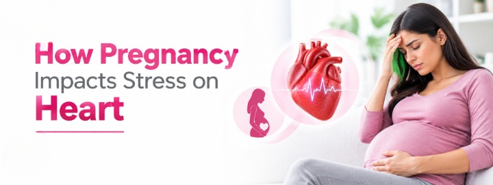
Pregnancy is full of joy and excitement, but it also brings changes you may not expect. As your baby grows, your heart pumps more blood and works harder to meet the needs of both you and your baby. For most healthy women, this is a normal part of pregnancy.
Still, a faster heartbeat, unusual tiredness or breathlessness can leave you wondering, “Is this normal?” Most of the time, these are normal pregnancy changes. Occasionally, though, pregnancy can reveal an underlying heart condition or make an existing one harder to manage. Knowing what to look out for can help you know when it is time to speak with your doctor.
Table of Contents
One of the most important effects of pregnancy on the cardiovascular system is the increased amount of blood circulating through the body.
The placenta, uterus and growing baby all need a steady blood supply. To meet this demand, the cardiovascular system of a mother undergoes several adaptations:
Cardiac function can rise by around 30% to 50% during pregnancy, with changes beginning early and becoming more evident as pregnancy progresses. This increase results from a combination of a faster heart rate and a greater amount of blood being pumped with each heartbeat.
Your blood volume also increases during pregnancy. This helps deliver enough blood and oxygen to your baby while keeping your own organs well supplied.
An interesting point that is usually overlooked is that the heart’s workload does not simply return to its normal level immediately after childbirth. Labour and the period immediately after delivery involve further rapid changes in circulation. This is one reason women with significant heart disease may require careful monitoring around delivery and after birth.
A faster heartbeat can be one of the first things a pregnant woman notices.
As pregnancy progresses, the resting heart rate commonly increases because the heart needs to circulate more blood. You may also notice your heartbeat more clearly after going upstairs, walking fast or changing position.
Mild palpitations can occur during pregnancy and are not usually a sign of heart disease.
Still, if palpitations are persistent or severe, especially with dizziness, fainting, chest pain or significant breathlessness, talk to your doctor.
Some symptoms associated with increased cardiac function can overlap with ordinary pregnancy symptoms.
These may include:
However, you should pay attention to how severe, frequent or unusual your symptoms feel.
For example, feeling mildly breathless after climbing stairs can be normal during pregnancy. But feeling breathless while resting or suddenly struggling to lie flat is different and should be checked by a doctor.
This is especially important because some heart conditions, such as peripartum cardiomyopathy, can cause symptoms that may initially feel like normal pregnancy changes.
Pregnancy generally places a greater cardiovascular demand on the body, but most healthy hearts adapt successfully.
The stress on the heart during pregnancy may become more significant when a woman already has a cardiovascular condition, such as:
Pregnancy can sometimes reveal a previously unrecognised heart problem because the cardiovascular system is working more than usual.
Women with known heart disease should ideally discuss pregnancy with their cardiologist and obstetrician before conception. The level of risk varies considerably depending on the specific heart condition and how well the heart is functioning.
High blood pressure during pregnancy needs timely monitoring. Preeclampsia is a pregnancy complication that involves high blood pressure, usually after 20 weeks, along with signs that other organs may be affected.
It requires medical attention because it can affect both maternal and fetal health.
Preeclampsia and other hypertensive disorders of pregnancy should also be considered beyond childbirth. They are associated with an increased risk of cardiovascular disease later in life.
Peripartum cardiomyopathy is an uncommon form of heart failure. It develops towards the end of pregnancy or during the months following delivery. It can even affect women who have never previously been diagnosed with heart disease.
Its symptoms can resemble ordinary pregnancy or postpartum changes, including tiredness, breathlessness and swelling. New or worsening breathlessness, particularly at rest or when lying down, marked swelling, chest discomfort or a racing heartbeat should therefore not simply be attributed to pregnancy.
An echocardiogram or ultrasound scan of the heart, can help doctors evaluate how well the heart is pumping when this condition is suspected.
You should contact your doctor promptly if you experience:
If you have severe chest pain, trouble breathing or fainting, please seek urgent medical care. And after delivery, don’t simply brush off new or worsening symptoms as tiredness or sleepless nights. Your body is recovering, but some heart conditions can also develop during this time, so it is always better to get checked.
Taking care of your heart during pregnancy starts with regular antenatal care. Depending on your health and pregnancy, your doctor may recommend:
Exercise should always be individualised. Women with uncomplicated pregnancies are generally encouraged to remain physically active, whereas those with certain medical or pregnancy complications may need specific restrictions or guidance.
The connection between pregnancy and heart health does not end after your baby is born. Having conditions such as preeclampsia, high blood pressure or gestational diabetes during pregnancy can increase your risk of heart disease later in life.
These complications are a reason to pay a little more attention to your heart health after pregnancy. Keeping an eye on your blood pressure and blood sugar, eating well, staying active and maintaining a healthy weight can all help lower your future risk.
Women who maintain better cardiovascular health after pregnancy have a lower risk of developing heart disease later on.
If you already have a heart condition, have had heart surgery, or are experiencing symptoms such as frequent palpitations, fainting or ongoing breathlessness, it is a good idea to speak with a cardiologist.
If you have heart disease and are planning a pregnancy, consider getting checked before you conceive. Your cardiologist and obstetrician can review your heart health, medications and pregnancy plan, so you know what to expect during pregnancy, delivery and the postpartum period.
Pregnancy naturally makes your heart work a little harder. Your blood volume, heart rate and cardiac output increase to give you and your baby the blood and oxygen you need. For most healthy women, these changes are a normal part of pregnancy.
If you have an existing heart condition or notice symptoms that feel unusual or are getting worse, don’t ignore them. Conditions such as high blood pressure, preeclampsia and peripartum cardiomyopathy may need closer monitoring.
If you are pregnant, planning a pregnancy or simply worried about a new heart-related symptom, talking to a specialist can help you. The cardiology and obstetric department at the CK Birla Hospital work with a multi-disciplinary team-based approach to help you understand your heart health and plan the right care through pregnancy and after delivery.

Many people start their day with a glass of lemon water, soaked almonds, fruit or another ‘fat burning’ food hoping it will speed up their weight loss. But does eating a particular food on an empty stomach actually help you lose weight?
Actually, just one food cannot burn your body fat simply because you eat it first thing in the morning. Sustainable weight loss depends on your overall balanced diet, energy intake, physical activity, sleep and other factors. However, the quality of your first meal can still matter because choosing foods rich in protein and fibre may help you feel fuller and make it easier to manage your food intake through the day.
So, what are some of the best foods to eat for weight loss when your stomach is empty? Let’s look at nutritious options that can help you start your day with a filling and balanced meal.
Eating a particular food on an empty stomach does not have any special fat burning powers. Weight management is influenced by the balance between the energy you consume and the energy your body uses.
A healthy diet should provide enough nutrition while maintaining balance, moderation and variety. WHO recommends a diet based largely on minimally processed foods that include vegetables, fruits, whole grains, pulses, nuts and other nutritious sources of protein.
So, if you are trying to lose weight and feel hungry in the morning, you can give the following food options a try:
Eggs are a good source of high quality protein and also provide important nutrients such as vitamin B12, vitamin D, choline, selenium and phosphorus. The protein in eggs can help you feel full and may make it easier to manage hunger through the morning.
You can try boiled eggs, a vegetable omelette or egg bhurji prepared with a moderate amount of oil. Adding vegetables such as spinach, tomato, onion, capsicum or mushrooms can increase the fibre, vitamins and minerals in your meal.
For a more balanced breakfast, you can pair eggs with vegetables and a whole grain food such as chapati or toast instead of relying on eggs only. If you are watching your overall calorie intake, limit the amount of oil, butter, cheese or other high calorie ingredients added during preparation.
Oats are a healthy breakfast option because they provide carbohydrates and dietary fibre, including beta-glucan.
You can prepare oats with milk or plain curd and add fruit, chia seeds or a small portion of nuts. Avoid turning a nutritious bowl of oats into a dessert by adding large amounts of sugar, syrups or sweetened toppings.
Unsweetened curd or Greek yoghurt can give you protein and make breakfast more satisfying. They also provide important nutrients such as calcium, phosphorus and vitamin B12, while curd contains beneficial bacteria that are good for your gut health. Greek yoghurt is generally higher in protein than regular curd, which can help you feel fuller for longer. You can try it with fruit, oats, seeds or a small portion of nuts for a more balanced breakfast. If you are buying packaged yoghurt, don’t forget to check the label for added sugars.
Apple, guava, orange, pear, papaya, berries and other seasonal fruits can all fit into a weight loss friendly diet. Whole fruit is generally a better everyday choice than fruit juice because it retains its fibre. WHO recommends regular consumption of fruits and vegetables as part of a healthy diet and advises limiting free sugars, including those found in many fruit juices. There is also no need to avoid fruit simply because you are eating it on an empty stomach.
Different nuts and seeds offer different nutrients. Almonds provide vitamin E, magnesium, fibre and protein, whereas walnuts are a source of alpha-linolenic acid (ALA), a plant-based omega-3 fatty acid. Chia and flaxseeds provide fibre and ALA, along with other minerals.
You can add a small portion of nuts or seeds to oats, plain curd, fruit or a smoothie. If you prefer eating them on their own, a small handful is usually enough. Remember, nuts and seeds are nutrient-dense but also relatively high in calories, so eating large handfuls can add more calories to your diet. For a weight loss friendly option, choose plain or dry roasted nuts without added sugar or excess salt.
Sprouts is a convenient Indian breakfast ingredient that provides plant-based protein, fibre, folate, vitamin C, vitamin K and other micronutrients. Their fibre and protein content can help make a meal more filling, which may be useful when you are trying to manage your overall food intake.
You can have a bowl of mixed sprouts with vegetables, lemon and spices, which can be combined with curd, paneer, eggs or another suitable protein source to make the meal more balanced.
If you have a sensitive digestive system, start with a small portion, as raw sprouts may cause bloating or discomfort in some people.
Besan chilla, made from gram flour, can be a practical Indian breakfast choice. Besan provides plant-based protein, fibre, iron, folate and other micronutrients. Adding vegetables such as onion, tomato, spinach, carrot or capsicum can further increase the fibre, vitamins and minerals in the meal.
You can pair a besan chilla with plain curd to add more protein and calcium. Use oil in moderation when cooking, as the amount of oil can significantly increase the calorie content of the meal.
For vegetarians looking for a protein-rich breakfast, paneer or tofu can be combined with vegetables and a suitable whole grain food.
Paneer provides protein, calcium and phosphorus, on the other hand, tofu is a plant-based food made by coagulating soy milk and curd that provides protein and minerals such as calcium and iron, depending on how it is prepared and processed. Tofu can be particularly useful for people who are lactose intolerant and looking to include more plant-based foods in their diet.
Morning drinks like lemon water, cumin water, green tea and more are surrounded by some of the biggest weight loss claims online. Some of these can be nutritious or useful for hydration, but none should be considered a substitute for a balanced diet or a fat removal treatment.
Warm water can help you meet your daily fluid needs, improve digestion and help you feel fuller, but there is no strong, scientific evidence that drinking water at a particular temperature causes major fat loss.
You can drink warm, room-temperature or cool water according to your preference. The important thing is maintaining adequate hydration throughout the day.
It can be a simple way to increase your fluid intake if you enjoy its taste. Lemon also provides a small amount of vitamin C, however the amount you get depends on how much lemon you add.
What lemon water does not do is directly burn belly fat or cause major weight loss. Its main benefit is hydration, not fat burning. So, we can say that it indirectly helps in weight loss by replacing high calorie drinks and keeping you hydrated.
Green tea originates from the plant compounds called catechins and contains caffeine. It may have some effect on body weight, but it should not be viewed as a powerful fat burning drink. Any potential effect is modest and does not replace a balanced diet, physical activity or other healthy lifestyle habits. People who are sensitive to caffeine may also want to limit their intake, particularly later in the day.
Apple cider vinegar is commonly promoted online as a weight loss drink. Vinegar has been studied for its possible effects on blood sugar and appetite, but there is still not enough evidence to say that it can reliably help with weight loss. So, you do not need to add apple cider vinegar to your morning routine just because it is often promoted as a weight-loss drink.
Jeera (Cumin) water, made by soaking or boiling cumin seeds in water, is a popular traditional drink. Cumin contains small amounts of compounds and nutrients such as iron and antioxidants. But, the amount obtained from a typical cup of jeera water is unlikely to be nutritionally significant.
You can have jeera water if you enjoy it, but do not treat it as a magical drink for weight loss.
You do not have to completely eliminate any one food to lose weight. Still, there are some breakfast choices that can make it harder to manage overall energy intake when consumed frequently or in large portions.
These may include:
The issue is not that these foods are eaten on an empty stomach. The bigger concern is their overall nutritional profile, portion size and place in your total daily diet.
A simple way to approach breakfast is to combine:
Protein + fibre-rich foods + vegetables or fruit + a moderate amount of healthy fat
The best breakfast is not the one with the fewest calories. It is one that provides enough nutrition, suits your preferences and helps you maintain a sustainable eating pattern. If you would like a ready-made plan to follow, see our diet chart for weight loss for day-wise meal ideas.
Also, if you are not hungry, don’t force yourself to eat a heavy breakfast. At the same time, skipping breakfast should not be used as a weight loss strategy if it leaves you excessively hungry and leads to overeating later.
General healthy eating advice may work well for many healthy adults, but the right approach to weight loss can be different for each person.
You should consider speaking with a dietician before adding or eliminating foods or making major dietary changes if you:
Obesity can be due to several factors, including genetics, how the body regulates appetite and energy, eating habits, the environment, certain health conditions and some medicines. It is a complex, long-term health condition.
If you are living with obesity, experiencing sudden weight changes, or have an underlying health condition, avoid making major dietary changes based only on random advice. A personalised plan may be more appropriate for your nutritional and medical needs. You can consult a dietician at the CK Birla Hospital for personalised nutrition advice, or book an appointment directly, and discuss whether further medical evaluation is needed.
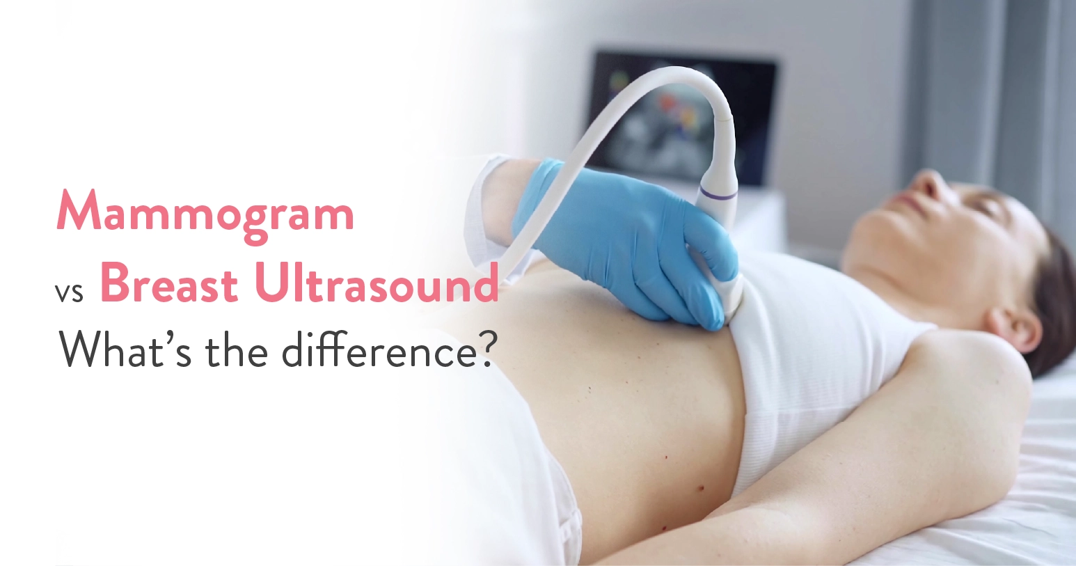
Breast cancer is one of the most common cancers among women, which is why regular breast screening is very important. Being advised to get a mammogram or breast ultrasound comes with questions, mainly, what is the difference, and do you really need one over the other?
Mammograms and breast ultrasounds are not interchangeable. They look at breast tissue in different ways and are sometimes used together to give doctors a clearer understanding of your breast health.
Whether you are going for your first breast screening, checking a new lump or simply want to understand your options, knowing the difference between a mammogram and a breast ultrasound can help you feel more informed. In this blog, we will explain how each test works, when it is recommended and what you can expect during the procedure.
Table of Contents
A mammogram is one of the most effective ways to check for breast cancer before it causes any noticeable symptoms. It uses low-dose X-rays to capture detailed images of the breast to spot tiny changes that may not be felt during a self-exam or physical examination.
Generally, routine mammogram screening begins around the age of 40, although the right age and screening schedule depend on your personal risk factors and your breast specialist’s advice.
A mammogram is used for:
Many women assume mammograms only detect cancer. In reality, they also identify non-cancerous (benign) breast conditions such as cysts, fibroadenomas and calcifications that simply need monitoring rather than treatment.
During the examination:
The test is usually completed within 15 to 20 minutes.
Many women delay screening because they worry the compression will be painful. Most describe the sensation as temporary pressure rather than severe pain. Scheduling the test a few days after your menstrual period, when breasts are less tender, may make the experience more comfortable.
A breast ultrasound is an imaging test that uses high-frequency sound waves instead of radiation to produce pictures of breast tissue.
Unlike mammograms, ultrasounds do not routinely screen for breast cancer. Instead, they help doctors investigate specific concerns found during a physical examination or after a mammogram.
Breast ultrasound is particularly useful for:
Breast ultrasounds are usually performed immediately after an abnormal mammogram. This combination frequently prevents unnecessary biopsies by confirming that a suspicious looking area is simply a harmless fluid-filled cyst.
The procedure of USG is simple and painless.
A healthcare professional:
The examination usually takes 15 to 30 minutes.
Since ultrasound does not use radiation, it is considered safe during pregnancy.
Ultrasound images are captured in real time, allowing doctors to observe blood flow and tissue movement, which can sometimes provide additional clues about a breast abnormality.
A mammogram is generally recommended if:
Your doctor may recommend an ultrasound if:
Age plays an important role in selecting the most appropriate imaging test.
The best screening schedule depends on your personal risk factors, family history and your doctor’s recommendations.
Dense breast tissue can reduce the visibility of some cancers on mammograms. In these cases, ultrasound acts as a complementary test rather than a replacement.
The accuracy of the test depends on what doctors are looking for.
Mammograms are generally better for:
Ultrasounds are generally better for:
Some people worry that needing an ultrasound after a mammogram means cancer has been found. However, additional imaging is common and usually performed simply to obtain clearer information before making any diagnosis.
| Misconception | Reality |
| “An ultrasound is better than a mammogram.” | It is not necessary. An ultrasound cannot reliably detect certain early breast cancers that mammography can identify. |
| “Mammograms expose me to dangerous radiation.” | The radiation dose used in mammography is very low. The benefits of early cancer detection greatly outweigh the small radiation exposure. |
| “Young women never need mammograms.” | Women at increased risk due to genetic mutations or a strong family history should begin screening earlier under specialist guidance. |
A mammogram remains an essential test of routine breast cancer screening, while a breast ultrasound provides valuable additional information when investigating lumps, dense breast tissue or unclear mammogram findings.
The right test depends on your age, symptoms, personal risk factors, and your doctor’s advice. Rather than choosing one over the other, remember that these imaging techniques work together to ensure the most accurate diagnosis and timely care.
If you have concerns about breast health, have noticed a new breast change, or are due for routine screening, the dedicated ‘Breast Care’ unit and Radiology Department at the CK Birla Hospital can guide you on the most appropriate imaging approach. Reach out to us to book a consultation with an experienced breast specialist.
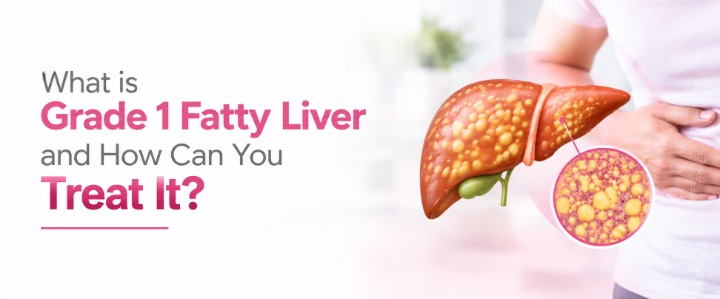
Nearly one in three adults in India today has some degree of fatty liver; many without even knowing it, until a routine scan flags Grade 1 fatty liver. It has become almost an unspoken side effect of modern day sedentary life: long hours at a desk, junk meals grabbed on the go, screens replacing sleep, stress replacing peace of mind. It is surprising to know, but your body, especially the liver, absorbs all of this and shows the consequences gradually.
But when the problem is detected early, at the Grade 1 level, it can alert you before it progresses further.
So, what exactly does Grade 1 fatty liver mean, how serious is it really and what can you do about it starting today? Keep reading to find out.
Table of Contents
Your body naturally stores fat in different places and some fat is also found around internal organs, where it helps cushion and protect them. The liver can normally contain a small amount of fat too. The concern begins when excess fat starts accumulating inside liver cells.
When this buildup is mild, an ultrasound may describe it as Grade 1 fatty liver, also called mild hepatic steatosis.
But what does “Grade 1” actually mean?
Consider grading as a way of describing how much fat is visible in the liver on imaging:
| Grade | What it Generally Means |
| Grade 1 | Mild fat accumulation in the liver |
| Grade 2 | Moderate fat accumulation in the liver |
| Grade 3 | Severe fat accumulation in the liver |
There is one important detail that often gets missed. The grade of fatty liver seen on an ultrasound is not the same as the stage of liver damage.
An ultrasound can suggest that excess fat is present, but it cannot reliably determine whether the liver has inflammation or how much fibrosis (scarring) has developed. If you have conditions such as diabetes, obesity or abnormal liver tests, your liver specialist (Hepatologist or Gastroenterologist) may recommend additional tests to evaluate your risk of liver fibrosis.
So, if your report mentions ‘Grade 1 fatty liver,’ it does not mean that your liver is severely damaged or that you have cirrhosis. It means that mild fat accumulation has been detected and it is a good time to understand why it happened and take steps to improve your liver health.
These days, Grade 1 fatty liver has become very common, with unhealthy diets, lack of physical activity and metabolic conditions such as obesity, insulin resistance and type 2 diabetes contributing to its increasing prevalence. In some people, persistent liver fat can progress to inflammation and fibrosis over time.
In most cases, Grade 1 fatty liver is not an emergency. It is usually an early and manageable finding. However, it should still not be taken lightly. Working on it as soon as you find it can help improve your liver health and reduce the risk of it progressing further.
The right approach is therefore:
Don’t panic. Don’t ignore it. Find out why it happened and address the underlying risk factors.
Fat accumulation in the liver does not happen overnight. It can be the result of small, everyday habits and changes in metabolic health that build up over the years. What you eat, how active you are and how your body handles blood sugar and fat can all influence how much fat gets stored in the liver.
●Being overweight or having obesity, particularly when excess fat is stored around the abdomen
● Insulin resistance, where the body does not respond to insulin effectively
● Type 2 diabetes or high blood sugar
● High triglycerides or abnormal cholesterol levels
● Long periods of physical inactivity
● A diet high in added sugars, refined carbohydrates and highly processed foods
● Excessive alcohol intake, which can contribute to alcohol-related liver disease
But fatty liver is not always a result of being overweight or lifestyle only. It can also occur due to certain medicines, rapid weight loss, malnutrition, genetic factors or other liver conditions.
If you have been reading about fatty liver, you may have come across the term nonalcoholic fatty liver disease (NAFLD). Now, often called metabolic dysfunction-associated steatotic liver disease (MASLD).
MASLD refers to fat accumulation in the liver in people who have one or more metabolic risk factors, such as obesity, insulin resistance, type 2 diabetes, high blood pressure or abnormal cholesterol and triglyceride levels, after other causes of liver fat have been considered.
So, if your older report mentions NAFLD, there is no need to get confused by the different names. NAFLD is the older term, whereas MASLD is the newer terminology used for metabolic fatty liver disease.
Fatty liver can be difficult to notice in the beginning because even when fat starts building up in the liver, many people continue to feel normal and overlook mild symptoms. This is why fatty liver is usually discovered during a routine health checkup or ultrasound.
When symptoms do occur, they include:
These symptoms are not specific to fatty liver and can also affect a person with many other health conditions. So, having these symptoms does not necessarily mean that you have fatty liver. But, to be sure, it is always better to get a proper diagnosis from a liver specialist.
In more advanced liver disease, a person may develop symptoms such as:
These are signs that need thorough medical evaluation and should not be assumed normal.
Key takeaway: Don’t wait for severe symptoms to take fatty liver seriously. If your medical reports already mention Grade 1 fatty liver, it is worth finding out what may be causing it and what steps you can take to manage it and improve your liver health.
Depending on your health history and risk factors, a Hepatologist or Gastroenterologist may check:
Your doctor may also want to check whether there is any fibrosis (scarring) in the liver, especially if you have certain risk factors or your blood test results are abnormal. This helps understand your liver health beyond the amount of fat seen on an ultrasound.
One of the first ways to assess this is the FIB-4 score.
It uses your age and results from routine blood tests to estimate your risk of advanced liver fibrosis. If further assessment is needed, your doctor may recommend FibroScan or another non-invasive test to look more closely at liver stiffness.
An ultrasound showing Grade 1 fatty liver tells you that there is a mild amount of fat in the liver. It does not indicate whether the liver has significant scarring. The need for further testing depends on your individual risk.
So, don’t be alarmed if your doctor recommends additional tests. These tests are usually done to understand your liver health better and decide what treatment or followup may be right for you.
Years of unhealthy eating habits, long sitting hours and lack of physical activity have made Grade 1 fatty liver increasingly common today. But finding it early also gives you a chance to do something about it. Paying attention to your liver and metabolic health, understanding what may be causing the fat accumulation and making the right lifestyle changes can help you take better care of your liver in the long run.
If you have recently been diagnosed with fatty liver and are unsure what your report means or what you should do next, speaking to a specialist can help. Book a consultation with the Gastroenterology & Hepatology team at the CK Birla Hospital to get personalised guidance for managing fatty liver and taking better care of your liver health.
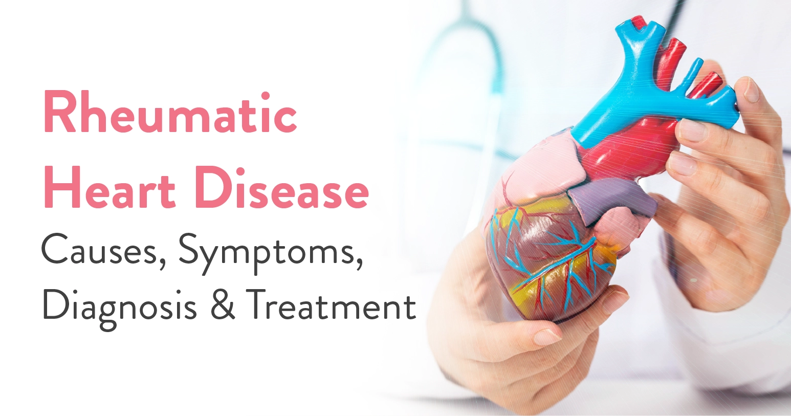
Can a throat infection damage your heart? In certain cases, yes.
An infection caused by group A Streptococcus bacteria can trigger rheumatic fever, an inflammatory reaction that may affect the heart and its valves. If the valves are repeatedly inflamed, permanent scarring can develop, leading to rheumatic heart disease (RHD).
Recognising the early signs, understanding how RHD is diagnosed and knowing how recurrent rheumatic fever can be prevented can help protect the heart from further damage. Read along to understand the causes, symptoms, diagnosis and treatment of rheumatic heart disease.
Table of Contents
Rheumatic heart disease (RHD) is long term damage to one or more heart valves caused by rheumatic fever. Rheumatic fever is an inflammatory illness that can develop after an infection with group A Streptococcus, commonly known as strep. In some people, the immune response to the infection triggers rheumatic fever, which can cause inflammation in the heart.
When the heart valves become inflamed, they can develop scarring and structural changes. Over time, a damaged valve may become narrowed, making it difficult for blood to pass through, or may fail to close properly, causing blood to leak backwards.
This lasting valve damage is called RHD (Rheumatic Heart Disease). RHD usually begins in childhood, although symptoms of valve disease may not appear until many years later. The World Health Organization estimates that around 55 million people worldwide are living with RHD.
The chain is usually:
Group A strep infection → rheumatic fever → inflammation of the heart → valve scarring → rheumatic heart disease
Strep throat is a common cause, although group A Streptococcus can also cause skin infections such as impetigo. Rheumatic fever does not develop after every strep infection. It affects you when the body’s immune response becomes misdirected.
Factors that increase the risk of RHD are:
In children, an echocardiogram can sometimes detect heart valve changes that may not be found during a routine examination.
A process called molecular mimicry plays a major role in this reaction.
After a group A strep infection, the immune system produces antibodies to fight the bacteria. In some people, these antibodies may mistakenly react with certain tissues in the body because they resemble parts of the bacteria.
This can cause inflammation in the heart, particularly the valves. Repeated episodes of rheumatic fever may gradually produce permanent scarring and structural changes.
Rheumatic fever does not usually show up right after a strep infection. It generally develops within 1 to 5 weeks.
Rheumatic heart disease symptoms depend on whether a person is experiencing acute rheumatic fever or has developed chronic valve damage.
Rheumatic fever may cause:
Heart inflammation, called carditis, may produce a new heart murmur. Importantly, some heart involvement can be detected only through echocardiography and may not produce obvious symptoms.
Rheumatic heart disease does not always cause symptoms in the early stages. They may develop gradually as the damaged valve starts affecting how the heart functions. The long term symptoms include:
A person may therefore develop RHD symptoms many years after the original rheumatic fever episode. The absence of symptoms immediately after childhood rheumatic fever does not necessarily mean there is no valve damage.
Diagnosis combines the person’s medical history, examination and heart investigations.
Untreated or advanced RHD can lead to:
The treatment approach depends on the severity of the condition, the valves affected and whether further episodes of rheumatic fever need to be prevented.
Catching and treating a group A strep infection early is one of the most effective ways to prevent rheumatic fever from developing in the first place.
For people who have already had rheumatic fever or been diagnosed with RHD, ongoing antibiotics are usually necessary to prevent another attack. This is known as secondary prophylaxis. How long the medication continues varies from person to person, depending on their age, whether they have had rheumatic fever before and the extent of any heart or valve involvement.
If the disease has led to complications like fluid buildup, heart failure or irregular heart rhythms, cardiologists may prescribe additional medications to manage these issues and reduce the risk of blood clots.
When valve damage is very serious, surgery to repair or replace the affected valve may be needed. The right approach depends on which valve is involved, how severe the damage is and factors like the patient’s age and overall health.
Rheumatic heart disease (RHD) usually begins with something as ordinary as a sore throat, which makes it easy to overlook until real damage has already been done. However, much of this damage is preventable. Treating strep infections timely, staying consistent with your medications and knowing the warning signs early can go a long way in protecting the heart valves from lasting harm.
So don’t ignore a sore throat, and don’t wait out breathlessness or unexplained fatigue. When it comes to rheumatic heart disease, early action is what helps you in the long run. If you have any concerns, whether about a past infection or new symptoms, a conversation with an internal medicine doctor or cardiologist can help clear the confusion. Book a consultation with the Cardiology Department at the CK Birla Hospital to get the right advice from a specialist.

