Filter :

Congratulations! You have entered the third trimester of your pregnancy! It is amazing how you have been able to overcome some of the most testing phases of your pregnancy journey. Now, all we do is make sure we welcome the baby in the most healthy and happy way. Your pregnant belly is booming and you are glowing in anticipation with the happy pregnancy hormones doing their job.
This is also a time when you and your family start planning for the actual delivery. How to choose a birthing plan? How to remain agile during this phase? What are the best sleeping positions? How to ensure breastfeeding is properly done? For that here’s a brief guide on how to prep for your delivery.
But in the third trimester, what are the problems pregnant women need to deal with? What should you eat, what should you avoid, there are a whole lot of questions you need answers to. Don’t worry, just read on…
Table of Contents
Pregnancy happens in 3 stages of 3 months each called trimester. When you reach week 27 of your pregnancy tenure, it means you’re now in your third trimester. In an ideal scenario, this trimester ends at week 40, however, depending on your condition or complications it ends when your baby is delivered. A full-term pregnancy ranges between weeks 37 to 42 of pregnancy. Anything before week 37 is considered premature, and if your baby has not arrived by week 42, your OB-GYN will induce labour to avoid complications or opt for a c-section if there are complications.
Currently, around 49.4% of pregnancies in India are under the high-risk pregnancy bracket as per research data. This is why it is very important to be extra careful in the final trimester to avoid unwanted complications.
Source: https://www.ncbi.nlm.nih.gov/pmc/articles/PMC10502764/
The third trimester is more tiring and uncomfortable than the other two trimesters primarily because the embryo grows to its full form. The discomfort is mainly attributed to the baby’s position and size, and with the due date approaching soon, anxiety also reaches its peak, making this trimester emotionally challenging as well. It is very important that at this stage you remain positive and try to adapt to the upcoming changes. Here is what you can expect during this stage:
It is essential that you take all sorts of health and medical precautions for some of the most commonly seen problems during the third trimester of pregnancy. These are:
Apart from this adequate prenatal care should be taken w.r.t. Your babies, such as checking the baby’s progress in terms of size, heart rate, position and movements. Make sure you and your gynaecologist have chalked out a proper birthing plan and made your choices.
During the third trimester, you will need frequent checkups, especially during week 32 to week 36. The doctors will check your weight and blood pressure and thoroughly examine you for any signs or symptoms you experience. Take your vaccines and more importantly the doses of tetanus, diphtheria and acellular pertussis (Tdap) vaccines. These are recommended for each pregnancy ideally taken between weeks 27 and 36 of the third trimester. This will build immunity for the baby at the time of birth.
There are ample emotional changes in expectant mothers. Some are good and some not so good, but it must be ensured that the mother is always in a positive frame of mind.
Make a checklist of all the things you will need during your third trimester. Start with charting out a birthing plan with your OB-GYN and healthcare provider. This will include monitoring your health pre-labour, any concerns that may indicate a c-section and consultations post-delivery.
At the CK Birla Hospital®, we take pride in planning things out for our expectant families for a seamless and memorable experience of welcoming a child into this world. For this, we don’t leave any stones unturned.
Everyone who sees you now is extra careful around you and more attentive to your needs. This is by far the second-best feeling during a pregnancy trumped only by the news of the baby’s arrival. But like they say, “with great power comes great responsibility”. This is why, our hands-on team of professional caregivers make sure you are well supported and looked after in a homely atmosphere, under compassionate care. Check out our comprehensive maternity package offer here (link to offer blog)
There is a virus ready to affect your body the moment your immunity shows the slightest dip. The onset of the recent pandemic has increased our general awareness about our health and possible diseases incredibly. One of the silent pandemics the world is still suffering from is cancer. This has been there for a long time mostly without a cure. Cervical cancer is also becoming a growing concern worldwide affecting a few men and mostly women. Its main cause is HPV or human papillomavirus.
What are the ways this virus attacks? Is it contagious? Is there any cure?
Table of Contents
The widely popular Human papillomavirus (HPV), can affect different parts of your body. There are over 100 types, including strains that cause warts on different parts of your body.
Roughly 30 HPV strains can affect your genitals, this includes your vulva, vagina, cervix, penis, scrotum, rectum and anus. Most of these are sexually transmitted infections (STIs) that pass on through skin-to-skin contact. However, the majority of genital HPV strains are harmless, including those responsible for genital warts. But others can be potentially high-risk variants, which may lead to cervical cancer. Early detection can lead to better results and prevent cancer from becoming severe. This leads us to the next question: Are all warts HPV?
Yes. All warts happen due to HPV, but all forms of HPV do not form warts. But whether or not these are fatal is the main concern. The HPV strains which lead to genital warts are nuisances.
The other types of HPV warts are:
| Flat Warts | smooth, flat-headed, fleshy or brownish-coloured bumps. |
| Plantar Warts | Small rough growths around the balls of the feet. |
| Common Warts | Small, grainy skin growths on your fingers or hands. These warts are transmitted by touch. |
| Periungual and Subungual Warts | These happen around your nails and fingers and disappear on their own. |
The type of HPV that can progress to cancer doesn’t cause warts. Which brings us to the next question: How is HPV related to cervical cancer?
HPV type 16 and type 18 can cause cervical dysplasia. Simply put this changes the cells of your cervix, the opening between your vagina and your uterus in women. When it is left untreated, it tends to advance to cervical cancer. Up to the age of 30, most HPV strains clear up by themselves. If found during a pap smear, it is advisable to perform regular screening to ensure the risk is eliminated at the onset. This then brings up the next question: Who is at a higher risk of getting HPV?
Irrespective of your reproductive anatomy, it’s vital to prevent HPV by either getting vaccinated and/or practising safe sex.
You Can Also Read: All you need to know about cervical cancer | Know your risk
First and foremost one must understand how one can get an HPV infection.
HPV is highly contagious since it spreads easily via skin-to-skin contact. No body fluids have to be exchanged either, so it will not spread through ejaculation (cum).
Genital HPV doesn’t usually come with any symptoms. The only visible symptom is a wart in your genital area. These are rough, cauliflower-like lumps that suddenly start growing on your skin. The timeline for the infection and the wart is not defined. It may appear weeks, months or even years after you’ve been infected. The only saving grace is that these are harmless.
The rest of the variants don’t have any symptoms. The only time the symptoms surface is when they progress to cancer. With cervical cancer, we must keep in mind that having HPV does not guarantee developing cancers.
The first step to any treatment is diagnosis. The doctor/ specialist will advise the following tests:
When the virus’s presence is confirmed, treatments will be prescribed. However, please note that once you get HPV it will never leave your body. So your healthcare partner will provide means to manage the situation by:
You Can Also Read: 10 Best Gynaecologists to Visit in Delhi-Gurgaon
Since the only way to prevent HPV is by abstaining from sex, here are some realistic ways in which you can protect yourself and your loved ones:
To conclude, HPV is a cause of concern but it can be prevented from escalating into cancer. As per WHO, the Human papillomavirus (HPV) is the name of a group of 200 known viruses, which can cause genital warts or cancer. Our body is able to control up to 90% of the existing HPV in our bodies, but a persistent infection can lead to Gynae, Uro and Cervical cancers. While HPV contributes to only 5% of all cancer cases worldwide, women are more likely to get it. Therefore a regular screening is essential.
Source: https://www.who.int/news-room/fact-sheets/detail/human-papilloma-virus-and-cancer
At the CK Birla Hospital, we understand the importance of being a reliable healthcare provider, especially in cases that need extra care. Our Gynae and Cancer specialists will not only help you prevent these morbid diseases but also guide you towards early detection, protection and treatment. Our judgement-free approach ensures we give the best possible treatment for your problem in the most compassionate and caring manner. To reach out to our sexual health experts book your appointment here.

Pregnancy is one of the most precious moments in a woman’s life. But it is also a time when women are most delicate, in terms of their bodies, mind and overall well-being. The main factor responsible for this is the changes that happen in her body, while the baby grows inside the womb. Additionally, some women also experience high blood pressure that might suddenly spike up if not monitored during pregnancy. This can put both the mother and her baby at risk during and after delivery.
High blood pressure or hypertensive disorders are preventable. But can these be prevented? Is there a way to properly manage the condition based on the stage of pregnancy? Yes. The good news is that it is both preventable and treatable.
Table of Contents
High blood pressure is a fairly common condition observed in most adults these days. Also called hypertension, it is observed among pregnant women between the ages of 20 to 44. However, when there is good control of blood pressure throughout the pregnancy, both the mother and her baby are likely to stay stable and healthy. This is why it is essential to get your medication sorted before, during and after pregnancy.
Monitoring high blood pressure is essential to avoid unwanted complications during pregnancy. This can impact both the mother and her baby.
Complications for the mother due to high BP:
Complications for the baby due to high BP:
| Pattern Delivery | The baby is born in less than 37 weeks of pregnancy. |
| Low Birth Weight | Baby weighs less than 5 pounds at birth |
| Malnourishment at birth | Lack of enough oxygen and nutrients in the womb causes this and triggers premature delivery. |
So given the extremity of the complications, if you are expecting or planning, you must discuss the problems with your healthcare partner before, during, and after pregnancy.
To understand the symptoms we need to understand the types of hypertensive disorders that might happen to a woman during her pregnancy.
The following profile of people are more likely to get preeclampsia:
You Can Also Read: A Guide to Increasing Baby Weight When 9 Months Pregnant
Depending on the stage of the pregnancy, the treatment approach will differ before, during and after pregnancy.
| Stage of pregnancy | Treatment and management of Hypertension |
| Before Pregnancy: When planning for pregnancy, set up a discussion with your healthcare partner or gynaecologist. | Make sure you discuss the following with your obstetrician:
|
| During Pregnancy | Once the pregnancy is confirmed, the following needs to be done in consultation with your Obstetrician:
|
| After Pregnancy | Once the baby is delivered monitor the following under the guidance of your doctor:
|
In conclusion, there can be multiple problems that can occur during a pregnancy, hypertension or high blood pressure is one of them. But this can be managed with proper planning and consultation with an experienced obstetrician. In case you are planning your pregnancy, it is always advisable to discuss your concerns with an expert who will give you the real picture based on your health. At the CK Birla Hospital, our expert OB GYNs provide holistic counselling and support to all our patients to prepare them physically and mentally towards a smooth pregnancy despite all concerns. Reach out to us to book an appointment.
With the flick of the wrist, a cricketer plays masterstrokes that make the team win matches. The same wrist is as work when you sweep and dust to keep your house clean. Our wrists do much more than just exist as a part of our bodies from where we can feel our pulse. Every time you want to know the time you look at your wrist, every time you hold something it involves your wrist. From typing to writing, cooking and even playing sports, our wrists make sure we are able to seamlessly move our hands to our needs. So when the wrists start to hurt, that’s when we realise exactly how much it contributes to a good quality of life for us.
So what can cause wrist pain? And what are the ways in which you can treat them?
Table of Contents
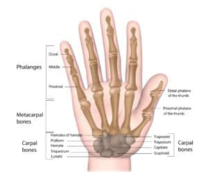
The wrist is a joint, connecting your hand and forearm. The wrist joint is made up of many small bones, which enable you to bend, straighten and rotate your hand. If you experience pain, numbness or tingling sensation in this region, then you are suffering from wrist pain. However, there is no need to be concerned about this pain as it is completely treatable. So now let us understand what are the reasons behind this pain in the next section.
As per the research carried out on risk factors associated with wrist pain by experts at the BMC Musculoskeletal Disorders, it was observed that women (14.7%) have a higher risk of getting wrist pain than men (5.6%). The study also indicated that those employed in manual labour and those who work out with weights are at a higher risk of getting wrist pain.
Source: https://d-nb.info/1206413336/34
Primarily any trauma impact on any part of the body will make it painful. So, if you have experienced a fall, and you have landed on your wrist, then it is bound to pain, due to the stress and trauma exposed to it. An X-ray will reveal whether this impact resulted in a sprain, fracture, or partial ligament disruption.
Otherwise, sometimes aches and pains may tend to appear out of the blue, with symptoms that may come and go. When the symptoms start getting worse or do not seem to subside for days then this will be termed chronic pain in the wrist. Get a healthcare provider to carefully examine the pain and obtain appropriate imaging studies to isolate the cause of the pain.
Some of the most identified causes of wrist pain are:
Depending on the cause of the pain the treatment approach will differ.
At home, you may do the following to help manage the pain:
If the orthopaedic identifies that the pain is chronic, then they might recommend
These days orthopaedic surgeons prefer using arthroscopic (minimally invasive) techniques to ensure a speedy recovery and complete treatment.
It is always said by medical practitioners that prevention is always better than cure. In the case of wrist pain, repetitive movements can tend to make it hurt more. For example, we all spend a lot of time typing. So you can avoid wrist pain from it by:
In conclusion, the underlying cause of pain once identified can help locate the cause and enable your healthcare partner to apply the appropriate treatment for it. When in doubt regarding pain that refuses to go away on its own, always reach out to a hand and wrist expert who will help resolve your problem. At the CK Birla Hospital, our award-winning team of experts and orthopaedic surgeons will provide the best solution with the fastest chance of recovery.
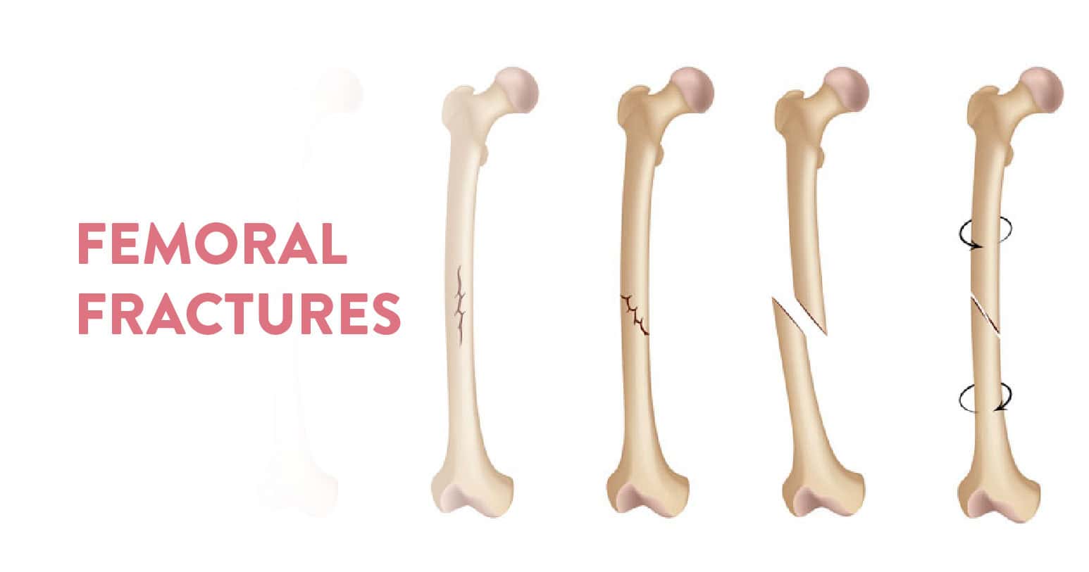
Table of Contents
The femur bone is the biggest and most significant bone in our body. It is found in our legs and it mainly connects the hip and the knee. A broken femur is therefore a serious injury that calls for immediate medical care. The only possible treatments are surgery and physical therapy. And these fractures are very painful, so a lot of pain medication is also prescribed.
Since the bone is the biggest and heaviest in our body, a femoral fracture often takes months to heal. Since the bone is big, the impact for fracture also has to be great. So one usually suffers from this during a car crash, an accident or an assault. Otherwise, elderly people who have weaker bones and are prone to injuries face this problem.
Since it is the biggest bone in your body, a femoral fracture will impact the quality of life of the person suffering from it. The possible consequences of a broken femur are:
When the injury happens it will show up the usual signs of pain, and swelling and in the worst cases the broken bone will pierce out of the skin of the leg. Rush to an orthopaedic, who will be able to physically examine the problem and suggest imaging tests to assess the extent of the injury.
Based on the results of X-rays and computed tomography scans (CT), the orthopaedic will diagnose the type of fracture and possible treatment options. The following types of fractures are usually diagnosed in a femoral fracture:
First and foremost the fracture must be cleaned and covered to avoid infection of any kind. Mostly, healthcare providers use a splint that will cover the whole leg. This splint goes up towards the hip. This is a temporary measure to help manage the pain, keep the bone in place and ensure the best outcomes.
Next, your healthcare provider will use traction to treat the fracture. These are broadly categorised into two types based on the placement of the traction:
If the fracture has happened, surgery will be performed within 24 to 48 hours to ensure the timely recovery of your injury. In this case, there are two types of surgical options available:
A fractured femur will mean that the internal skeletal framework of your leg has been compromised. So it will be extremely difficult to put any kind of weight or pressure on your leg. The bipedal movement of our legs needs the entire leg to function properly. So at any point, while walking we transfer our body weight on either of our legs to move. Since the fracture does not allow for any weight to be put on it, it will be very difficult to walk in this case. Support will be necessary for any kind of movement.
A fractured femur bone is an indication that the body will undergo a lot of pain and trauma due to the sheer magnitude of impact necessary to cause the break. The quality of life of the patient will be compromised and mobility will be substantially restricted. Since the events that lead to such painful and severe fractures are big, it will leave you with a long and tedious recovery tenure.
Even after the femur heals a significant amount of physiotherapy and rehab is required to reinstate the natural mobility of the person.
Recovery is very long for femoral fractures. It usually takes anywhere between 4 to 6 months. The length of your recovery also depends on the severity of your fracture, associated wounds and impact on the nerves and blood vessels around it. So even beyond the time when the trauma impact wears off, the pain sustains and involves a lot of pain medication to stabilise the condition. So the pain stays till the time the associated complications and the internal issues are completely healed.
A femoral neck fracture is a type of intracapsular hip fracture. This is because the femoral neck connects the femoral shaft with the femoral head. The femoral head connects with the acetabulum to form the hip joint. Therefore the conjunctional location of the bones makes the femoral neck prone to fracture. If the upper end of the femur bone has fracture this will therefore lead to a hip fracture, especially in patients with vitamin D and calcium deficiency.
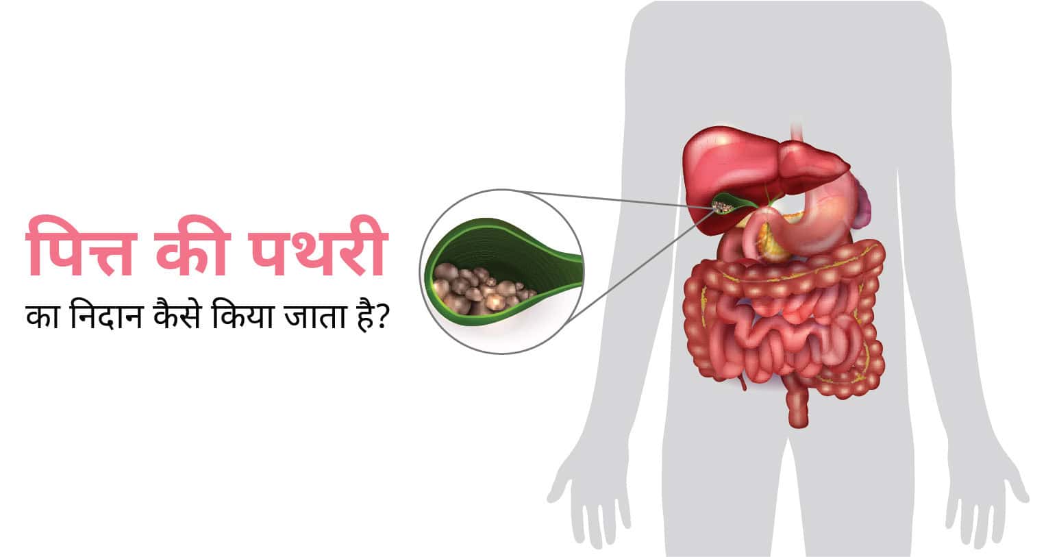
अस्वस्थ जीवनशैली, गलत पान-पान, फ़ास्ट फूड्स, कोल्ड ड्रिंक्स, निष्क्रियता आदि हमारे जीवन का एक अहम् हिस्सा बन चुके हैं। इन सबकी वजह से हमें कई तरह के प्रॉब्लम्स का सामना करना पड़ता है जिसमें से एक है पित्त की पथरी। वैसे तो यह समस्या किसी भी उम्र के व्यक्ति को हो सकती है।
लेकिन अधिकतर मामलों में यह महिलाओं और अधिक उम्र के लोगों में देखने को मिलता है। पित्त की पथरी एक गंभीर समस्या है जिसका समय पर उचित निदान और उपचार कराना आवश्यक है, क्योंकि एक समय के बाद यह असहनीय दर्द, सूजन, संक्रमण और दुर्लभ मामलों में कैंसर का कारण भी बन सकता है।
पित्त की पथरी के दो प्रकार होते हैं जिसमें पहला कोलेस्ट्रॉल स्टोन और दूसरा पिगमेंट स्टोन है।
पित्त की पथरी होने पर कभी-कभी मरीज को कई महीनों या 1-2 सालों तक लक्षण अनुभव नहीं होते हैं। लेकिन जैसे-जैसे पथरी का आकार बढ़ता है, इसके कारण पित्ताशय में सूजन और दूसरी समस्याएं शुरू होती हैं – मरीज खुद में अनेक लक्षणों को अनुभव करना शुरू कर देते हैं।
पित्त की पथरी के मुख्य लक्षणों में निम्न शामिल हो सकते हैं:
इन सबके अलावा, आप अन्य लक्षण भी अनुभव कर सकते हैं। अगर आप ऊपर दिए गए लक्षणों को खुद में अनुभव करते हैं तो आपको बिना देरी किए विशेषज्ञ डॉक्टर से परामर्श करना चाहिए। आमतौर पर डॉक्टर पित्त की पथरी का निदान करने के बाद उपचार की प्रक्रिया शुरू करते हैं।
पित्त की पथरी का निदान करने के लिए डॉक्टर सबसे पहले मरीज से उनके लक्षणों के बारे में कुछ प्रश्न पूछते हैं, मरीज का शारीरिक परीक्षण करते हैं और फिर कुछ विशेष जांच करने का सुझाव देते हैं। पित्त की पथरी का निदान करने के लिए निम्न जांच किए जाते हैं:
इस जांच के दौरान, डॉक्टर मरीज का ब्लड सैम्पल लेकर उसे लैब में जांच करने के लिए भेजते हैं। खून जांच के दौरान, पित्त की पथरी के कारण संक्रमण, पीलिया, अग्नाशयशोध (पैंक्रियाटाइटिस) या दूसरी जटिलताओं के बारे में पता लगाया जाता है।
इस दौरान, डॉक्टर पित्त की पथरी की तस्वीर प्राप्त करते हैं जिनकी मदद से पित्ताशय के मोटापा और सूजन की पुष्टि करने में मदद मिलती है।
जब अल्ट्रासाउंड के दौरान छोटी पथरी की पुष्टि नहीं हो पाती है तो उसके बारे में पता लगाने के लिए डॉक्टर एंडोस्कोपिक अल्ट्रासाउंड का सुझाव देते हैं। इस जाँच के दौरान, डॉक्टर एंडोस्कोप नामक उपकरण का इस्तेमाल करके छोटी आकार की पथरी को आसानी से देख पाते हैं।
इमेजिंग परीक्षण के दौरान डॉक्टर सिटी स्कैन, ओरल कोलेसिस्टोग्राफी, हेपेटोबिलरी एमिनो डायसोटिक एसिड स्कैन, मैग्नेटिक रेजोनेंस कोलेजनोपैंटोग्राफी या एंडोस्कोपिक रेट्रोग्रेड कोलेजनोपैनोग्राफी आदि जांच प्रक्रियाएं करते हैं।
आवश्यकता होने पर, ऊपर दिए गए जांचों के आलावा डॉक्टर अन्य भी जांच करने का सुझाव दे सकते हैं। सभी जांचों के परिणामों के आधार पर डॉक्टर उपचार करते हैं। पित्त की पथरी के उपचार में घरेलू नुस्खे, आयुर्वेदिक जड़ी-बूटियां, होम्योपैथिक दवाएं, एलोपैथिक दवाएं और सर्जरी आदि शामिल हैं।
सर्जरी ही पित्त की पथरी का एकमात्र सफल और परमानेंट इलाज है। उपचार के दूसरे माध्यमों से पित्त की पथरी के लक्षणों को कम किया जा सकता है। अगर आप खुद में पित्त की पथरी के लक्षणों को अनुभव करते है या इससे पीड़ित हैं तो हमारे डॉक्टर के साथ परामर्श कर उचित उपचार प्राप्त कर सकते हैं।
प्रश्न 1. क्या पित्ताशय की पथरी अपने आप घूल सकती है?
पित्त पथरी अपने आप दूर हो सकती है, लेकिन आमतौर पर ऐसा नहीं होता है और उपचार की आवश्यकता हो सकती है। पित्ताशय की पथरी हमेशा लक्षणों का कारण नहीं बन सकती है, और उन मामलों में, जटिलताओं को रोकने के लिए केवल आहार परिवर्तन ही आवश्यक हो सकते हैं। लोग पित्ताशय की थैली के बिना सामान्य जीवन जी सकते हैं।
प्रश्न 2. पित्त की पथरी के क्या कारण हैं?
पित्ताशय में पथरी बनने के अनेक कारण होते हैं। इसमें मुख्य रूप से अस्वस्थ जीवनशैली, गलत खान-पान, मोटापा, कम मात्रा में पानी पीना, तेजी से अजान कम करना, जंक फूड्स और कोल्ड ड्रिंक्स आदि का सेवन आदि।
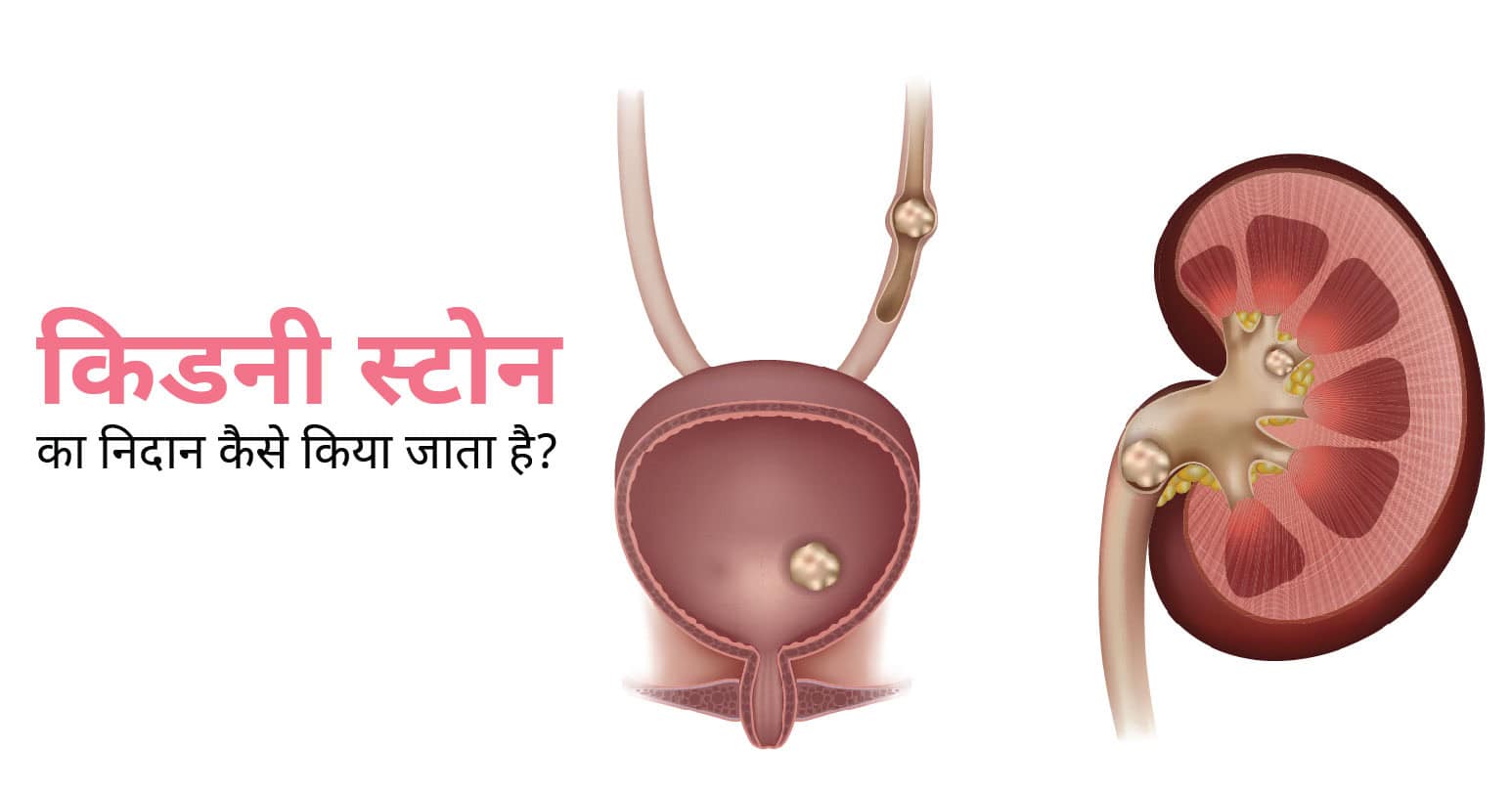
गुर्दे की पथरी को अंग्रेजी में किडनी स्टोन कहते हैं। यह पेशाब में पाए जाने वाले सॉल्ट और मिनरल्स जैसे रसायनों से बानी होती है। आज के समय में किडनी स्टोन होना एक सामान्य समस्या का रूप ले चूका है। पूरी दुनिया में लाखों की संख्या में मरीज इससे पीड़ित हैं।
गुर्दा यानी किडनी का मुख्य काम अपशिष्ट और तरल पदार्थों की अधिक मात्रा को शरीर से बाहर निकालना है। किडनी का काम पेशाब के जरिए सोडियम, पोटाशियम आदि अपशिष्ठ पदार्थों को शरीर से बाहर निकालकर खून का शुद्धिकरण करना है।
हालांकि, जब आपकी डाइट में कैल्शियम, पोटेशियम आदि खनिजों की अधिक मात्रा होती है तो इनकी मात्रा अधिक होने के कारण कई बार यह किडनी से पूरी तरह बाहर नहीं निकल पाते हैं। अंतत यही अपशिष्ट पदार्थ धीरे-धीरे एक जगह जमा होकर पत्थर यानी स्टोन जैसी आकृति की संरचना कर लेते हैं जिसे मेडिकल भाषा में किडनी स्टोन या गुर्दे की पथरी के नाम से जाना जाता है।
आमतौर पर किडनी स्टोन की शुरुआती स्टेज में इसके लक्षण अनुभव नहीं होते हैं। लेकिन जैसे-जैसे इसकी संख्या बढ़ती है या इसका आकार बड़ा होता है – लक्षण अनुभव होने शुरू हो जाते हैं। किडनी स्टोन के लक्षणों में पेशाब से खून आना, बार-बार पेशाब लगना, पेशाब से बदबू आना, पेशाब के रंग में बदलाव आना, बुखार और उल्टी होना, यूरिन मार्ग में संक्रमण होना, पेशाब के साथ खून आना, कमर के निचले हिस्से में तेज और असहनीय दर्द होना आदि शामिल हैं।
किडनी स्टोन के लक्षण अनुभव होने पर मरीज को तुरंत डॉक्टर के पास जाना चाहिए। मरीज से उसकी परेशानियों के बारे में पूछने के बाद, डॉक्टर मरीज का शारीरिक परीक्षण करते हैं और फिर लक्षणों के आधार पर किडनी स्टोन का निदान करने के लिए कुछ जांच का सुझाव देते हैं जिनमें निम्न शामिल हो सकते हैं:
इस दौरान डॉक्टर मरीज के खून का सैम्पल लेकर लैब में जांच करते हैं। खून जांच के दौरान, खून में मौजूद मिनरल्स के बारे में पता लगाया जाता है। जाँच के दौरान, खून में मिनरल्स या यूरिक एसिड की अधिक मात्रा की पुष्टि होना किडनी हेल्थ में किसी प्रकार की गड़बड़ी को दर्शाता है। खून जाँच के परिणाम के आधार पर निदान की प्रक्रिया को आगे बढ़ाया जाता है।
मूत्र परीक्षण यानी यूरिन टेस्ट जिसके दौरान डॉक्टर मरीज के पेशाब का सैंपल लेकर लैब में जांच के लिए भेजते हैं। अगर टेस्ट के दौरान, मूत्र में पथरी बनाने वाले पदार्थों की मात्रा सामान्य से अधिक पायी जाती है तो यह किडनी स्टोन की ओर इशारा करते हैं।
जब ऊपर दिए गए जांचों से किडनी स्टोन की पुष्टि हो जाती है तो डॉक्टर आगे इमेजिंग टेस्ट यानी इमेजिंग परीक्षण करने का सुझाव देते हैं। किडनी स्टोन को विस्तृत रूप से देखने और समझने के लिए डॉक्टर सिटी स्कैन और अल्ट्रासाउंड करते हैं।
इस जाँच के दौरान एक्स-रेज का इस्तेमाल करके डॉक्टर कंप्यूटर स्क्रीन पर किडनी को देखते हैं।
इस जांच के दौरान हाई फ्रीक्वेंसी रेडियो वेव्स का इस्तेमाल करके किडनी स्टोन की तस्वीर प्राप्त की जाती है जिससे उसके आकार एवं प्रकार को समझने में मदद मिलती है।
इन सबके अलावा, कुछ मामलों में कुछ मरीजों को पेशाब के साथ स्टोन बाहर आ जाता है। ऐसे में डॉक्टर, बाहर निकले हुए स्टोन का आकलन करते हैं। जांच के बाद, डॉक्टर स्टोन के प्रकार, आकार और मरीज की उम्र एवं समग्र स्वास्थ्य को ध्यान में रखते हुए उपचार के प्रकार और पद्धति का चयन करते हैं।
अगर आप खुद में किडनी स्टोन के लक्षणों का अनुभव करते हैं या आपको इस बात की आशंका है कि आपको किडनी स्टोन है तो आप हमारे विशेषज्ञ डॉक्टर के साथ परामर्श कर सकते हैं। हमारे डॉक्टर के साथ अपॉइंटमेंट बुक करने के लिए आप इस पेज के ऊपर दिए गए फोन नंबर या बुक अपॉइंटमेंट फॉर्म का इस्तेमाल कर सकते हैं।
प्रश्न 1. कितने एमएम की पथरी अपने आप निकल जाती है?
विशेषज्ञ डॉक्टर का कहना है कि अगर किडनी स्टोन का आकर 5-6 मिलीमीटर है तो वह बिना ऑपरेशन के अपने आप ही पेशाब के रास्ते शरीर से बाहर निकल सकती है। हालाँकि, अगर ऐसा नहीं होता है और लक्षण गंभीर रूप लेने लगते हैं तो मेडिकल उपचार की आवश्यकता पड़ सकती है।
प्रश्न 2. क्या किडनी स्टोन अपने आप घुल सकती है?
किडनी की छोटी पथरी अक्सर शरीर से अपने आप बाहर निकल जाती है। जब तक वे गंभीर दर्द या जटिलताओं का कारण नहीं बनते, उपचार आवश्यक नहीं है। किडनी स्टोन का आकार बड़ा होने पर आमतौर पर इलाज करने की आवश्यकता होती है।
प्रश्न 3. पथरी में चाय पीनी चाहिए क्या?
आमतौर पर किडनी स्टोन से पीड़ित मरीज को डॉक्टर चाय के सेवन से मना करते हैं। काली चाय में ऑक्सालेट भरपूर मात्रा में मौजूद होता है। ऑक्सालेट एक यौगिक है जो अनेक खान-पान की चीजों में पाया जाता है। शरीर में इसकी मात्रा अधिक होने पर किडनी स्टोन का खतरा बढ़ जाता है।
प्रश्न 4. क्या पथरी में दूध पीना चाहिए?
आपको उन खाद्य पदार्थों का सेवन सीमित करना चाहिए जो कैल्शियम ऑक्सालेट स्टोन बनाते हैं जो किडनी स्टोन का प्रमुख प्रकार है। भोजन के दौरान कैल्शियम युक्त खाद्य पदार्थ जैसे दूध, दही, और कुछ पनीर और ऑक्सलेट युक्त खाद्य पदार्थ एक साथ खाएं और पिएं।
गाय के दूध में ऑक्सालेट नहीं होता है और इसमें आपके लिए आवश्यक कैल्शियम होता है, इसलिए यह आपके लिए एक अच्छा विकल्प है। कैल्शियम ऑक्सालेट से बनी गुर्दे की पथरी तब बनती है जब मूत्र में इन पदार्थों की मात्रा अधिक होती है।
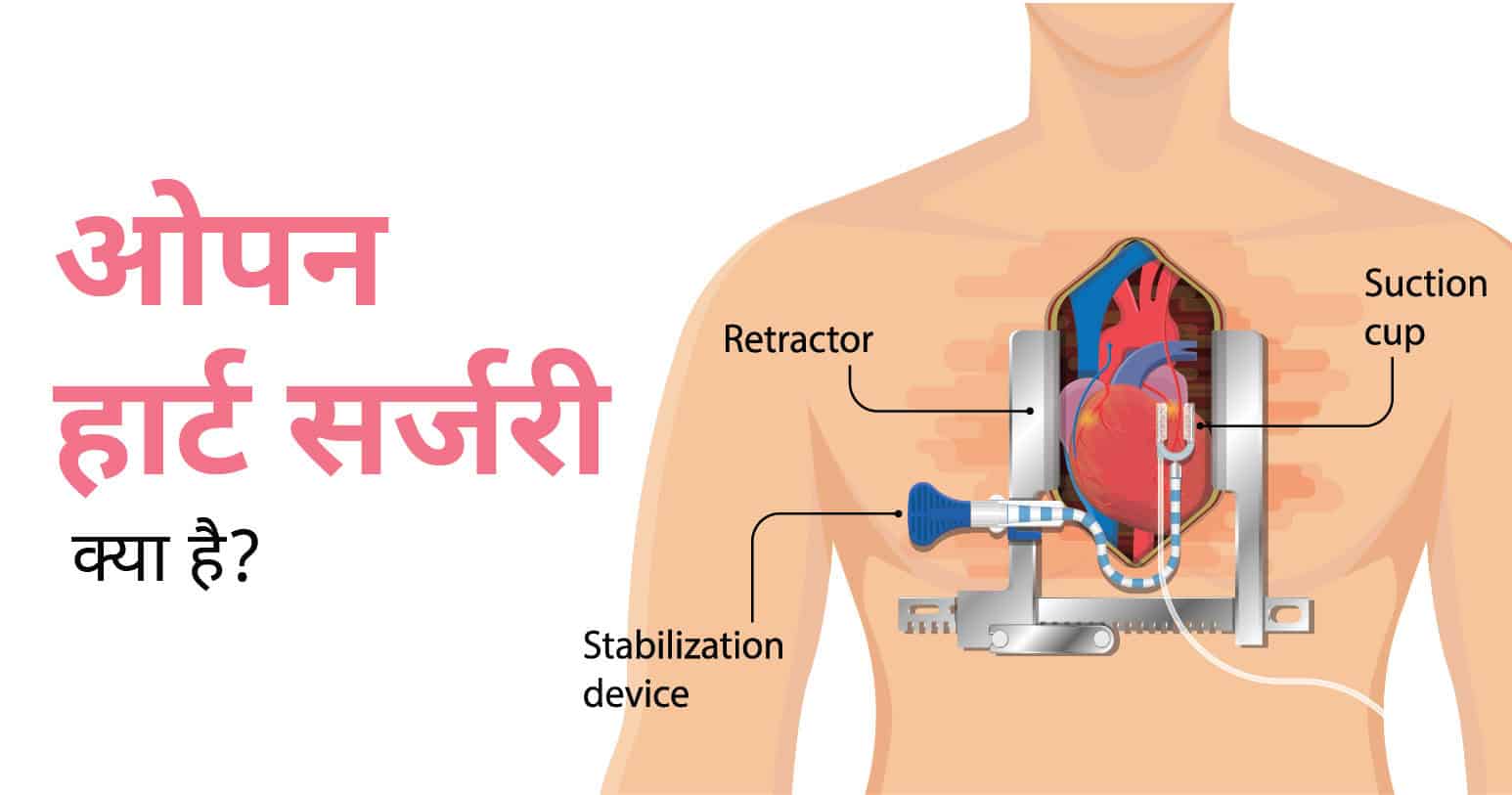
अस्वस्थ जीवनशैली, सिगरेट, शराब और दूसरी नशीली चीजों का सेवन, निष्क्रियता, तनाव, मोटापा आदि हमारे जीवन का हिस्सा बन चुके हैं। इन सबके कारण हमें अनेक प्रकार की बीमारियों का सामना करना पड़ता है जिसमें मुख्य रूप से दिल से संबंधित बीमारियां शामिल हैं। दिल से संबंधित अनेक बीमारियों का इलाज करने के लिए ओपन हार्ट सर्जरी का उपयोग किया जाता है।
Table of Contents
ओपन हार्ट सर्जरी को कई बार हार्ट सर्जरी भी कहा जाता है। इस प्रक्रिया के दौरान, डॉक्टर मरीज की छाती में चीरा लगाकर उसे खोलते हैं और दिल यानी हृदय की मांसपेशियों, वाल्वों या धमनियों की सर्जरी करते हैं।
ओपन हार्ट सर्जरी के दौरान, अवरुद्ध धमनियां (Blocked Arteries) में सुधार करने के लिए डॉक्टर उन्हें नई आर्टरीज से जोड़ते हैं। इससे अवरुद्ध धमनियों को बायपास करके फ्रेश यानी ताजा खून को दिल तक पहुँचाया जाता है।
नेशनल हार्ट, लंग एंड ब्लड इंस्टीट्यूट के अनुसार, ओपन हार्ट सर्जरी का सबसे सामान्य प्रकार है कोरोनरी आर्टरी बाईपास ग्राफ्टिंग। चिकित्सा विज्ञान में विकास होने के कारण आज दिल से संबंधित अनेक बीमारियों को छोटे चीरे की मदद से ठीक किया जा सकता है। इसलिए काफी लोग ओपन हार्ट सर्जरी करवाने से कतराते हैं।
सीएबीजी यानी कोरोनरी आर्टरी बायपास ग्राफ्टिंग करने के लिए ओपन हार्ट सर्जरी की जाती है। हार्ट डिजीज यानी दिल की बीमारियों से पीड़ित मरीजों का उपचार करने के लिए कोरोनरी आर्टरी बायपास ग्राफ्ट का इस्तेमाल किया जाता है।
कोरोनरी हार्ट डिजीज से पीड़ित मरीज के दिल की मांसपेशियां ठोस और संकुचित (solid and compact) हो जाती हैं जिसके कारण वे रक्त कोशिकाओं में ऑक्सीजन और खून नहीं पहुंचा पाती हैं। जब दिल तक खून सही ठंग से नहीं पहुंचता है तो हार्ट अटैक का खतरा होता है।
निम्न स्थितियों में डॉक्टर ओपन हार्ट सर्जरी की आवश्यकता पड़ सकती है:
विशेषज्ञ के अनुसार, ओपन हार्ट सर्जरी को पूरा होने में आमतौर पर 3-6 घंटे का समय लगता है। ओपन हार्ट सर्जरी करने से पहले डॉक्टर मरीज को अनेक जांच करने का सुझाव देते हैं ताकि इस बात की पुष्टि की जा सके की मरीज इस सर्जरी के लिए पूर्ण रूप से तैयार है।
जांच के परिणामों के बाद जब डॉक्टर हर तरह से संतुष्ट हो जाते हैं तो सर्जरी की एक तारीख तय करते हैं, मरीज को सर्जरी से पहले की कुछ दिशा-निर्देश देते हैं और अंतत चयनित दिन पर सर्जरी को पूरा किया जाता है।
ओपन हार्ट सर्जरी में निम्न चरण शामिल हैं:
ओपन हार्ट सर्जरी खत्म होने के बाद मरीज की छाती में 2-3 ट्यूब लगे होते हैं जिससे फ्लूइड बाहर निकलता है। इसके अलावा, मरीज को इंट्रावेनस ट्यूब भी लगी होती है जिससे उन्हें आवश्यक पोषक पहुंचाए जाते हैं। साथ ही, कैथिटर भी लगा होता है जिससे मरीज आसानी से यूरिन पास कर पाता है।
ओपन हार्ट सर्जरी खत्म होने के बाद, मरीज को कुछ 5-7 दिनों तक आईसीयू में रखा जाता है। उसके बाद, उन्हें रेगुलर केयर में और फिर बाद में सभी रिपोर्ट्स नार्मल होने के बाद दवाएं और दिशा-निर्देशों के साथ हॉस्पिटल से डिस्चार्ज कर दिया जाता है।
प्रश्न 1. क्या ओपन हार्ट सर्जरी दर्दनाक है?
ओपन हार्ट सर्जरी को जेनेरल एनेस्थीसिया के प्रभाव में किया जाता है। इसलिए मरीज को इस प्रक्रिया के दौरान दर्द नहीं होता है, लेकिन एनेस्थीसिया के बाद सीने में दर्द हो सकता है। चूँकि यह एक बड़ी सर्जरी, इसलिए रिकवरी के दौरान जोखिम का खतरा भी होते है। सर्जरी के बाद किसी भी तरह की परेशानी होने पर तुरंत अपने डॉक्टर से इस बारे में बात करनी चाहिए ताकि वे उचित उपाय से आपकी समस्या को दूर कर सकें।
प्रश्न 2. ओपन हार्ट सर्जरी के बाद क्या खाना चाहिए?
आमतौर पर ओपन हार्ट सर्जरी के बाद डॉक्टर मरीज को हरी सब्जियां, ताजे फल, साबुत अनाज, रोटी, बिना चर्बी वाले मांस, मछली और कम वसा वाले डेयरी उत्पादों के सेवन का सुझाव देते हैं। लेकिन ध्यान देने वाली बात यह है कि मरीज की उम्र, स्वास्थ्य, पहले से चल रहे इलाज, शुगर, ब्लड प्रेशर और दूसरी बातों को ध्यान में रखते हुए डॉक्टर मरीज के लिए डाइट का सुझाव देते हैं।
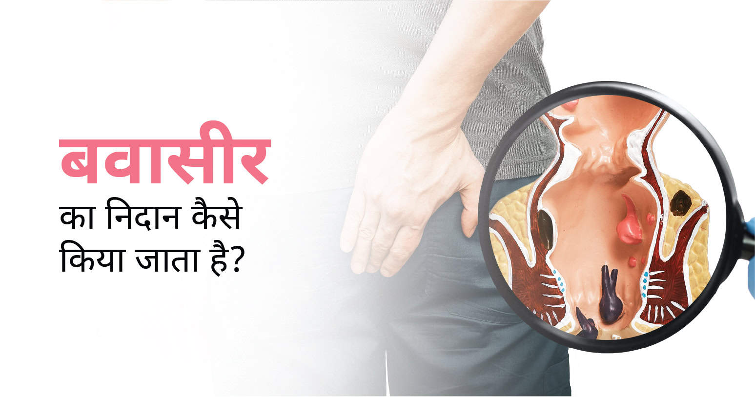
गुदा क्षेत्र की नसों में सूजन आना और उसके कारण मस्सों का निर्माण होना मेडिकल भाषा में बवासीर कहलाता है। बवासीर को अंग्रेजी में पाइल्स कहते हैं। समय पर बवासीर का निदान होने पर सटीक उपचार की मदद से इसे बढ़ने यानी गंभीर रूप लेने से रोका एवं हमेशा के लिए ख़त्म किया जा सकता है।
बवासीर के दो प्रकार होते हैं जिसमें आंतरिक बवासीर और बाहरी बवासीर शामिल हैं। साथ ही, बवासीर के चार अलग-अलग चरण होते हैं जिसमें ग्रेड 1, ग्रेड 2, ग्रेड 3 और ग्रेड 4 शामिल हैं।
बवासीर कई कारणों से होता है जिसमें मुख्य रूप से लंबे समय तक एक ही जगह बैठना, पुरानी कब्ज की शिकायत होना, कम पानी का सेवन करना, गुदा क्षेत्र में चोट लगना, रेक्टल प्रोलैप्स होना, फाइबर-युक्त भोजन नहीं करना, सिगरेट और शराब का सत्याधिक सेवन करना, अत्याधिक तैलीय और मसालेदार चीजों का सेवन करना, फ़ास्ट फूड्स और ड्रिंक्स का सेवन करना आदि शामिल हैं।
बवासीर होने पर आप खुद में अनेक लक्षणों का अनुभव कर सकते हैं जैसे कि बैठते समय गुदा क्षेत्र में दर्द होना, गुदा क्षेत्र में सूजन, जलन और दर्द होना, मल त्याग के समय मल के साथ खून आना और गुदा क्षेत्र में खुजली होना आदि। अगर आप इन लक्षणों को खुद में अनुभव करते हैं या आपको इस बात का संदेह है कि आपको बवासीर है तो जल्द से जल्द विशेषज्ञ डॉक्टर से परामर्श करना चाहिए।
आपके लक्षणों के आधार पर डॉक्टर कुछ जांच करके लक्षणों के पीछे के सटीक कारण का पता लगाते हैं। बवासीर का निदान करने के लिए डॉक्टर निम्न का सुझाव दे सकते हैं:
शारीरिक परीक्षण बवासीर की नैदानिक प्रक्रिया का सबसे शुरुआती चरण है। शारीरिक परीक्षण के दौरान, डॉक्टर मरीज से उसकी मेडिकल हिस्ट्री से संबंधित कुछ प्रश्न पूछते हैं। साथ ही, मरीज के गुदा क्षेत्र के बाहरी हिस्से का परीक्षण भी करते हैं।
यह आमतौर पर आंतरिक बवासीर का निदान करने के लिए किया जाता है। डिजिटल रेक्टल एग्जाम के दौरान डॉक्टर ग्लव्स एवं लुब्रिकेटेड उंगली से गुदा के क्षेत्र की जांच करते हैं। साथ ही, वे उसे गुदा मार्ग के अंदर डालकर वहां के टिशू के असामान्य विकास एवं आंतरिक बवासीर की जांच करते हैं।
इस परीक्षण के दौरान डॉक्टर मरीज के गुदा मार्ग के अंदर एनोस्कोप नामक उपकरण को डालकर कंप्यूटर स्क्रीन पर गुदा के भीतरी हिस्सों को अच्छी तरह से देखते हैं। एनोस्कोप एक पतली ट्यूब की तरह होता है जिसका एक छोर पर कैमरा और लाइट लगा होता है जिसकी मदद से डॉक्टर को गुदा के आंतरिक हिस्सों को कंप्यूटर स्क्रीन पर देखने में मदद मिलती है।
यह प्रक्रिया भी बिलकुल एनोस्कोपी की तरह है, लेकिन इसमें मलाशय के अस्तर और कोलोन(colon) की जांच करने के लिए प्रॉक्टोस्कोप नामक मेडिकल उपकरण का इस्तेमाल किया जाता है।
इस प्रक्रिया के दौरान डॉक्टर कोलोनोस्कोप नामक मेडिकल उपकरण का इस्तेमाल करके मलाशय और कोलोन के आंतरिक हिस्सों को देखते हैं। कोलोनोस्कोप एक पतली ट्यूब की तरह है जिसका एक छोर पर एक छोटी सी लाइट और कैमरा लगा होता है।
जब डॉक्टर को इस बात का संदेह होता है कि मरीज को पाचन संबंधित कोई अन्य बीमारी है, कोलोरेक्टल कैंसर होने का खतरा है या फिर मरीज मध्य आयु वर्ग का है और उसने हाल में कोई कोलोनोस्कोपी नहीं कराई है। इन सभी जांच के परिणाम के आधार पर यह तय किया जाता है बवासीर का कारण क्या है, वह किस चरण में हैं और आगे किस प्रकार की जटिलताओं का खतरा है आदि। बवासीर का निदान करने के बाद डॉक्टर को इसके लिए प्रभावशाली उपचार और पद्धति का चयन करने में मदद मिलती है।
अगर आप खुद में बवासीर के लक्षणों का अनुभव करते हैं या आपको इस बात का संदेह है कि आपको बवासीर है तो आप हमारे डॉक्टर के साथ अपॉइंटमेंट बुक करके अपनी बीमारी उचित निदान और सटीक उपचार करवा सकते हैं। हमारे विशेषज्ञ के साथ अपॉइंटमेंट बुक करने के लिए आप इस पेज के ऊपर दिए गए फोन नंबर या अपॉइंटमेंट बुक फ्रॉम का इस्तेमाल कर सकते हैं।