Filter :
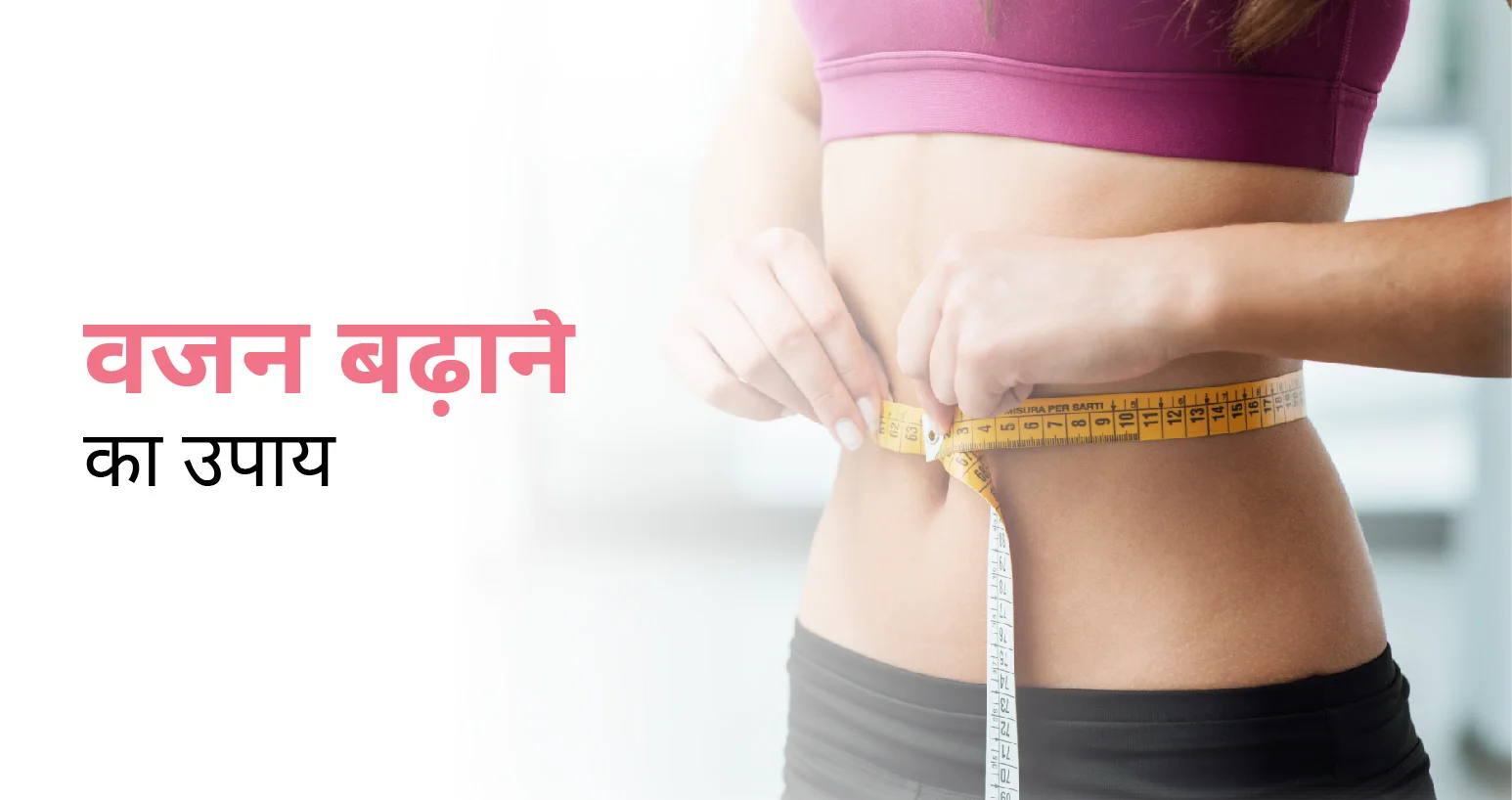
Table of Contents
ये भी पढ़े: हड्डी का कैंसर: प्रकार, कारण, लक्षण और उपचार
कितना भी खाए मोटा नहीं होता हूं! बहुत ट्राई किया, लेकिन वजन नहीं बढ़ रहा है! क्या आप भी इन वाक्यों से वाकिफ हैं? यदि हां, तो आज हम इस ब्लॉग में वजन बढ़ाने के बारे में बात करने वाले हैं। जिस तरह हमारा मोटापा हमारे स्वास्थ्य के लिए एक खतरनाक कारक बन गया है, उसी प्रकार कम वजन भी हमारे जीवन में कई समस्याओं का मुख्य कारण बनता जा रहा है।
कई बार ऐसा होता है कि लोग प्रमुख रूप से कम वजन के कारण एक मजाक का पात्र बन जाते हैं। लेकिन अभ आपको घबराने की आवश्यकता नहीं है। इस ब्लॉग में वह सारी जानकारी आपको मिल जाएगी, जिससे आपका वजन प्राकृतिक रूप से बढ़ने लग जाएगा।
कम वजन कई स्वास्थ्य समस्याओं का संकेत देता है, जैसे कमजोर प्रतिरक्षा प्रणाली, थकान, और कुपोषण। यदि व्यक्ति स्वस्थ वजन बनाए रखने में सफल होता हैं, तो इससे व्यक्ति के शरीर में ऊर्जा के स्तर में भी वृद्धि होती है और मांसपेशियों की ताकत में भी सुधार होता है। इसके कारण समग्र स्वास्थ्य में भी लाभ देखने को मिलता है। चलिए सबसे पहले वजन बढ़ाने से संबंधित मूल बातों के बारे में जानते हैं।
वजन बढ़ाने की युक्तियों को जानने से पहले आपको कुछ बातों को ज़रूर जान लेना चाहिए। यह सारी मूल बातें वजन बढ़ाने की नींव है, जो आपकी मदद कर सकते हैं। निम्नलिखित बातों का विशेष ध्यान रखने की आवश्यकता होती है –
स्वस्थ वजन बनाए रखना बहुत ज्यादा आवश्यक है क्योंकि –
कुछ भी खाने से स्वस्थ वजन नहीं बढ़ता है। यह सत्य है कि बाहर का खाने-पीने से वजन बढ़ता है, लेकिन वह अस्वस्थ वजन होता है, जो कई सारी बीमारियों के साथ आता है। वजन बढ़ाने के लिए तीन मुख्य बातों पर ध्यान देना चाहिए – कैलोरी अधिशेष, मैक्रोन्यूट्रिएंट्स, और सूक्ष्म पोषक तत्वों का सेवन। चलिए तीनों को एक-एक करके समझते हैं।
वजन कम करने के लिए कम कैलोरी का सेवन करना चाहिए, उसी प्रकार वजन को बढ़ाने के लिए कैलोरी के सेवन को बढ़ाने की आवश्यकता होती है। कैलोरी का मुख्य कार्य शरीर में ऊर्जा और ऊतकों का निर्माण करना है, जिससे शरीर को आवश्यक ईंधन भी मिलता है। एक बात का और खास ख्याल रखना होगा कि कैलोरी के लिए अस्वस्थ आहार से आपको दूरी बनानी चाहिए।
मैक्रोन्यूट्रिएंट्स वजन बढ़ाने में बहुत अहम भूमिका निभाते हैं। इसमें कार्ब्स, प्रोटीन और स्वस्थ वसा शामिल होते हैं जिससे शारीरिक कार्यों को करने के लिए पर्याप्त ऊर्जा मिलती है। औसतन एक व्यक्ति को अपने दैनिक कार्यों को करने के लिए 45-65% कार्ब्स, 10-35% प्रोटीन और 20-35% वसा की आवश्यकता होती है। चलिए इन पोषक तत्वों के कार्य को समझते हैं –
कई रिसर्च में पता चला है कि विटामिन डी का मुख्य कार्य वजन नियंत्रण और समग्र स्वास्थ्य में सुधार करना है। यदि व्यक्ति सही मात्रा में विटामिन का सेवन करता है तो शरीर कैल्शियम को अवशोषित करने में सक्षम होता है। इससे हड्डियों का वजन बढ़ता है, खासकर दुबला पतले लोगों के लिए यह बहुत लाभकारी साबित हो सकता है।
खाने का सही विकल्प आपको वजन बढ़ाने की यात्रा पर बड़ा प्रभाव डालते हैं। यदि आपको इस प्रश्न का उत्तर मिल जाता है कि क्या खाना चाहिए और क्या नहीं, तो आपको वजन बढ़ाने का राज मिल जाएगा। चलिए समझते हैं कि किस प्रकार के भोजन का सेवन करने से आपको लाभ होगा।
निम्नलिखित खाद्य पदार्थों के सेवन से आपको वजन बढ़ाने के लिए सभी पोषक तत्व मिल जाते हैं –
भोजन करने का तरीका भी इसमें एक अहम भूमिका निभाता है। सबसे पहले तो आपको यह समझना होगा कि हमेशा स्वस्थ आहार का ही सेवन करें। पूरे दिन के दौरान भोजन के संबंध में निम्नलिखित निर्देशों का पालन करना चाहिए –
स्वस्थ स्नैक्स के बारे में हमने आपको बताया था। चलिए जानते हैं कि किस प्रकार के स्वस्थ स्नैक्स का आप सेवन कर सकते हैं –
कई बार लोगों के मन में प्रश्न उठते हैं कि क्या व्यायाम से वजन बढ़ सकता है? किस प्रकार के व्यायाम से वजन बढ़ता है? व्यायाम का क्या महत्व है? चलिए इन सब सवालों के उत्तर जानते हैं।
मांसपेशियां वसा के मुकाबले अधिक कैलोरी को जलाती है। मांसपेशियों का निर्माण करने से शरीर का मेटाबॉलिज्म दुरुस्त होता है, जिससे वजन बढ़ाने में बहुत मदद मिलती है। वेट ट्रेनिंग जैसे डम्बल, स्क्वाट्स और पुश-अप्स, मांसपेशियों का निर्माण करते है जिससे शरीर का मेटाबॉलिज्म फिर से दुरुस्त हो जाता है। प्रयास करें कि यह सारे व्यायाम एक ट्रेनर के साथ ही करें। धीरे-धीरे अपने व्यायाम की तीव्रता को बढ़ाएं।
हम सभी जानते हैं कि कार्डियो हृदय स्वास्थ्य के लिए सबसे अच्छा विकल्प माना जाता है। इसके साथ-साथ यह कैलोरी को बर्न करने में भी बहुत मदद करता है। लेकिन एक बात का ध्यान रखें कि बहुत अधिक कार्डियो भी वजन बढ़ाने की जगह वजन कम न करा दे। यही कारण है कि हमेशा वेट ट्रेनिंग और कार्डियो का संतुलन बना रहना चाहिए।
व्यायाम के बाद हमारे मांसपेशियों में दर्द होता है। इससे बचने का एक ही रास्ता होता है और वह है आराम। व्यायाम के बाद व्यक्ति जिनता आराम करता है, वह उतनी ही जल्दी रिकवरी भी करता है। इसके अतिरिक्त व्यायाम के बाद पर्याप्त नींद और हा्रड्रेशन स्थिति के सुधार में लाभकारी साबित हो सकता है। तनाव को कम करने से आपको बहुत मदद मिलेगी।
जैसा कि हमने आपको पहले भी बताया है कि वजन बढ़ाने के लिए आपको अपनी कैलोरी की मात्रा को बढ़ाने की आवश्यकता होती है। कुछ पूरक या फिर सप्लीमेंट बाजार में उपलब्ध हैं जो आपको वजन बढ़ाने में मदद कर सकते हैं। हालांकि हम इन सप्लीमेंट के प्रयोग की सहयोग नहीं करते हैं।
हालांकि सप्लीमेंट से आसानी से वजन बढ़ाया जा सकता है, लेकिन इसके कारण कुछ अन्य समस्याएं आपको परेशान कर सकती हैं। मुख्यतः बाजार में दो प्रकार के पूरक उपलब्ध है जिनका आप सहयोग ले सकते हैं –
वजन बढ़ाने की कोशिश में लोगों को कई चुनौतियों का सामना करना पड़ता है। चलिए सभी को एक-एक करके समझते हैं –
वजन बढ़ाने के दौरान कई बार ऐसा होता है कि वह हाई मेटाबॉलिज्म का सामना करते हैं। जब मेटाबॉलिज्म अधिक होता है तो वजन बढ़ाने में लोगों को अधिक दिक्कतों का सामना करना पड़ता है। हाई मेटाबॉलिज्म को कम करने के लिए आप निम्नलिखित निर्देशों का पालन कर सकते हैं –
कई बार ऐसा होता है कि वजन बढ़ाने की यात्रा के दौरान लोगों को भूख नहीं लगती है। यह कोई भीषण समस्या नहीं है। इससे बचने के लिए आप निर्देशों का पालन कर सकते हैं –
यदि वजन बढ़ाने में समय लगता है, तो आपको घबराने की आवश्यकता नहीं है। यही कारण है कि आपको निरंतरता और धैर्य रखने की आवश्यकता है। इस दौरान आप कुछ निर्देशों का पालन कर सकते हैं जैसे –
वजन बढ़ाने की यात्रा में स्वस्थ भोजन और व्यायाम के साथ-साथ जीवनशैली कारक भी एक महत्वपूर्ण कारक साबित होता है। चलिए कुछ जीवनशैली कारकों के बारे में बात करते हैं, जो आपके वजन बढ़ाने की यात्रा में बाधा उत्पन्न हो सकती है।
यदि आप पर्याप्त नींद नहीं ले पाते हैं, तो शरीर ‘घ्रेलिन’ नामक हार्मोन का अधिक निर्माण करता है, जिसके कारण वजन को बढ़ाने में बाधा उत्पन्न होती है। घ्रेलिन के उत्पादन के कारण अधिक भूख लगने लगती है। वहीं इसके साथ ‘लेप्टिन’ नामक हार्मोन का उत्पादन कम होता है, जिसके कारण पेट हमेशा भरा हुआ महसूस करता है। इन सबके कारण व्यक्ति खाना नहीं खाता है, जिससे इसे पोष्टक आहार नहीं मिलता है और उसका वजन भी नहीं बढ़ता है।
अधिक तनाव के कारण शरीर में कोर्टिसोल (Cortisol) नामक हार्मोन का स्तर बढ़ाता है। शरीर में जब कोर्टिसोल का स्तर बढ़ जाता है, जिसके कारण शरीर में वसा जमा होने लगता है। तनाव को कम करने के लिए आप योग, ध्यान, और व्यायाम का सहारा ले सकते हैं। तनाव कम करने से वजन बढ़ाने में सहायता मिलती है। जब आप मन से खुश रहेंगे तो आपका शरीर भी खुश रहेगा, जिसकी सहायता से वजन बढ़ सकता है।
वजन बढ़ाने की यात्रा चुनौतीपूर्ण हो सकती है, लेकिन आपको यह समझना होगा कि उचित रणनीति और दृढ़ संकल्प आपको वजन बढ़ाने में मदद कर सकते हैं। वजन बढ़ाने का मतलब केवल भोजन करना नहीं है। खाने के साथ स्वस्थ जीवन शैली अपनाना और स्वयं को हाइड्रेट रखना है। हमेशा याद रखें कि आपको स्वस्थ वजन बढ़ाना है। आप प्रयास करें और आप सफल रहेंगे।
अपने दैनिक आहार में कैलोरी के सेवन को बढ़ाने से आपको लाभ होगा। इसके अतिरिक्त प्रोटीन, स्वस्थ वसा और कार्ब्स से भरपूर आहार लें।
हां, व्यायाम मांसपेशियों के निर्माण में मदद करता है, जो वजन बढ़ाने में सहायक करता है। मांसपेशियों का निर्माण शरीर में मेटाबॉलिज्म को बढ़ाता है, जो बहुत लाभकारी साबित हो सकती है।
वजन बढ़ाने के लिए औसतन 7-8 घंटे की नींद जरूरी है। नींद से शरीर में कई बदलाव होते हैं। इससे मांसपेशियों का निर्माण अच्छे से होता है और भूख को नियंत्रित करने में मदद कर सकता है।
तनाव से कोर्टिसोल हार्मोन बढ़ता है, जो वजन बढ़ाने में बाधा डालता है। तनाव प्रबंधन जरूरी है। इसके लिए आप योग, मेडिटेशन और अन्य हेपिनेस प्रोग्राम को अपनाएं।
वजन बढ़ाने में सबसे आवश्यक है धैर्य। स्वस्थ तरीके से वजन बढ़ाने में समय लगता है और प्रयास करें कि प्राकृतिक चीजों को ही अपनाएं।

तेजी से वजन कम करने के लिए एक समग्र और सूचित दृष्टिकोण की आवश्यकता होती है। इस ब्लॉग में हम प्रभावी रूप से वजन कम करने के लिए विभिन्न तरीकों पर चर्चा करने की कोशिश करेंगे, जिसमें घरेलू उपचार, आहार संबंधी सुझाव और चिकित्सा उपचार शामिल हैं। एक स्वस्थ जीवनशैली हासिल करना एक व्यक्तिगत प्रयास है। वजन कम करने के लिए इसके अनेक पहलुओं की गहरी समझ होना आवश्यक है।
Table of Contents
वजन कम करने के लिए सबसे सरल और सबसे प्रभावशाली घरेलू उपचारों में से एक जलयोजन स्तर बनाए रखना है। केवल जलयोजन में अपनी भूमिका से परे, पानी चयापचय, एपेटाइट सप्रेशन (भूख दबाना) और डिटॉक्सिफिकेशन प्रोसेस के लिए एक गतिशील उत्प्रेरक है। यह सुनिश्चित करने के लिए कि आपका शरीर बेहतर ढंग से काम कर रहा है और आपके वजन घटाने के लक्ष्यों का समर्थन करता है, प्रति दिन कम से कम आठ गिलास पानी का सेवन करने का प्रयास करें। साथ ही, निम्न को अपनी जीवन में अपनाएं:
ग्रीन टी को अपनी दिनचर्या में शामिल करना आपकी वजन घटाने की यात्रा के लिए एक गेम-चेंजर साबित हो सकता है। यह अपने एंटीऑक्सीडेंट्स और कैटेचिन्स की प्रचुर मात्रा के लिए जानी जाती है, जो मेटाबॉलिज़्म को बढ़ाने और फैट बर्निंग प्रक्रिया को प्रोत्साहित करने में मदद करती हैं। अपने वजन घटाने के परिणामों को अधिक प्रभावी बनाने के लिए प्रतिदिन 2-3 कप ग्रीन टी पीने का प्रयास करें।
यह अपने संभावित वजन घटाने के लाभों के लिए व्यापक रूप से जाना जाता है। सेब का सिरका रक्त शर्करा के स्तर को स्थिर करके और तृप्ति की भावना को बढ़ावा देकर वजन प्रबंधन में योगदान देता है। हालाँकि, सावधानी बरतने की सलाह दी जाती है और इसका उपयोग विवेकपूर्ण तरीके से करना आवश्यक है। इस मामले में अगर मन में कोई प्रश्न है तो किसी विशेषज्ञ से परामर्श करें और उनसे सलाह लें।
अपने दिन की शुरुआत नींबू मिले गर्म पानी के साथ करने से आपके चयापचय और पाचन प्रक्रियाओं के लिए सकारात्मक माहौल तैयार हो सकता है। नींबू में विटामिन सी की मात्रा शरीर के वसा बर्न करने वाले तंत्र में एक सहायक तत्व के रूप में कार्य करती है, जिससे यह आपकी दैनिक दिनचर्या के लिए एक ताज़ा और फायदेमंद अतिरिक्त बन जाता है।
ये भी पढ़े: भूख बढ़ाने के आसान घरेलू उपाय
इसके अलावा, पर्याप्त मात्रा में नींद लें। इसे अक्सर नजरअंदाज कर दिया जाता है, वजन घटाने के संदर्भ में गुणवत्तापूर्ण नींद के महत्व को नजरअंदाज नहीं किया जा सकता है। नींद की कमी हार्मोनल संतुलन को बाधित कर सकती है, जिससे भूख और लालसा में वृद्धि हो सकती है। अपने शरीर की पुनर्स्थापनात्मक प्रक्रियाओं को अनुकूलित करने और अपने वजन घटाने के लक्ष्यों का समर्थन करने के लिए रोजाना 7-9 घंटे का लक्ष्य रखें।
ये भी पढ़े: गैस की वजह से पेट फूलने के घरेलू उपचार क्या हैं
आपके दैनिक आहार में लीन प्रोटीन का एकीकरण प्रभावी वजन घटाने की रणनीति के लिए मौलिक है। चिकन, मछली, टोफू और फलियां जैसे स्रोतों से प्राप्त प्रोटीन न केवल आवश्यक अमीनो एसिड की आपूर्ति करते हैं, बल्कि मांसपेशियों के संरक्षण और परिपूर्णता की भावना को बनाए रखने में भी महत्वपूर्ण भूमिका निभाते हैं, जो अधिक खपत से बचने में सहायता करता है। इसके अलावा, निम्न को भी अपनी डाइट में शामिल करें:
फलें और सब्जियां आवश्यक पोषक तत्वों और फाइबर का बड़ा श्रोत हैं। यह न केवल समग्र स्वास्थ्य का समर्थन करता है बल्कि तृप्ति की भावना को बढ़ाकर वजन घटाने में भी महत्वपूर्ण योगदान देता है। उनकी पोषण संबंधी समृद्धि का लाभ उठाने के लिए अपने दैनिक भोजन में विभिन्न प्रकार के फलों और सब्जियों को शामिल करें।
वजन घटाने की दिशा में परिष्कृत अनाज के बजाय साबुत अनाज का चयन करना एक रणनीतिक कदम है। साबुत अनाज जैसे ब्राउन राइस, क्विनोआ और साबुत गेहूं उत्पाद न केवल अधिक व्यापक पोषक तत्व प्रदान करते हैं बल्कि निरंतर ऊर्जा और बेहतर पाचन भी प्रदान करते हैं।
पुरानी मान्यताओं के विपरीत, वसा संतुलित आहार का एक अभिन्न अंग है। एवोकाडो, नट्स और जैतून के तेल जैसे स्वस्थ वसा के स्रोतों को शामिल करने से आवश्यक पोषक तत्व मिलते हैं और लंबे समय तक तृप्ति की भावना में योगदान होता है, भाग नियंत्रण और समग्र वजन प्रबंधन में सहायता मिलती है।
ये भी पढ़े: चुकंदर के फायदे, पोषक तत्व और नुकसान
साथ ही, प्रभावी वजन घटाने का एक मूलभूत सिद्धांत भाग नियंत्रण का अभ्यास करना है। छोटी प्लेटों का उपयोग करें, सर्विंग्स को मापें, और अपने शरीर की भूख और परिपूर्णता के संकेतों के बारे में जागरूकता पैदा करें। ये आदतें भोजन के साथ स्वस्थ संबंध स्थापित करने और अधिक खाने से रोकने में मदद कर सकती हैं।
ये भी पढ़े: मोटापा और वजन घटाने के लिए कम्पलीट डाइट चार्ट
सफल रूप से वजन घटाने के लिए प्रसंस्कृत खाद्य पदार्थों का सेवन कम करना सर्वोपरि है। ये खाद्य पदार्थ अक्सर अतिरिक्त शर्करा, अनहेल्दी वसा और प्रिजर्वेटिव्स से भरे होते हैं, जो अतिरिक्त कैलोरी खपत में योगदान करते हैं और प्रगति में बाधा डालते हैं।
सोडा और फलों के रस सहित शर्करा युक्त पेय न केवल कैलोरी से भरपूर होते हैं, बल्कि उनमें आवश्यक पोषक तत्वों की भी कमी होती है। अपने आहार से इन पेय पदार्थों को कम करना या समाप्त करना स्वस्थ वजन प्राप्त करने और बनाए रखने की दिशा में एक महत्वपूर्ण कदम है।
आमतौर पर तले हुए और व्यावसायिक रूप से पके हुए उत्पादों में पाए जाने वाले ट्रांस वसा, हृदय स्वास्थ्य के लिए काफी खतरा पैदा करते हैं। वजन घटाने और समग्र कल्याण के लिए ट्रांस वसा वाले खाद्य पदार्थों से परहेज करना आवश्यक है।
ये भी पढ़े: चिया सीड्स के फायदे, नुकसान, और पोषक तत्त्व
कार्बोहाइड्रेट संतुलित आहार का एक आवश्यक घटक है, साबुत अनाज से जटिल कार्बोहाइड्रेट पर ध्यान केंद्रित करना महत्वपूर्ण है। सफेद ब्रेड और मीठे स्नैक्स जैसे परिष्कृत कार्बोहाइड्रेट का सेवन सीमित करने से वजन घटाने के प्रयासों और समग्र स्वास्थ्य में मदद मिलती है।
इसके अलावा, शराब के सेवन पर ध्यान दें। मध्यम मात्रा में शराब का सेवन स्वस्थ जीवन शैली में अपना स्थान बना सकता है, अत्यधिक सेवन अनावश्यक कैलोरी खपत और खराब आहार विकल्पों में योगदान कर सकता है। प्रभावी वजन प्रबंधन चाहने वालों के लिए संयम महत्वपूर्ण है।
ये भी पढ़े: संतुलित आहार (7-दिन का संतुलित आहार चार्ट) (Balanced Diet in Hindi)
घरेलू उपचार के साथ-साथ आप डॉक्टर से परामर्श करने के बाद चिकित्सा उपचार की मदद भी ले सकते हैं अपने वजन को कम करने के लिए। वजन कम करने के लिए मुख्य रूप से निम्न का उपयोग किया जाता है:
मोटापे या वजन से संबंधित स्वास्थ्य संबंधी चिंताओं से जूझ रहे लोगों के लिए, चिकित्सकीय दवाएं एक व्यवहार्य समाधान प्रदान कर सकती हैं। हालाँकि, ऐसी दवाओं की उपयुक्तता निर्धारित करने और संभावित दुष्प्रभावों की बारीकी से निगरानी करने के लिए विशेषज्ञ डॉक्टर से परामर्श करना महत्वपूर्ण है।
गंभीर मोटापे से ग्रस्त लोगों के लिए बेरिएट्रिक सर्जरी पर विचार किया जाता है जब वजन घटाने के अन्य तरीके अप्रभावी साबित होते हैं। गैस्ट्रिक बाईपास और गैस्ट्रिक स्लीव जैसी प्रक्रियाएं पेट के आकार को काफी कम कर देती हैं, जिससे वजन घटाने में मदद मिलती है और समग्र स्वास्थ्य में सुधार होता है।
ये भी पढ़े: क्या रोज़ अंजीर खाना सेहत के लिए अच्छा है? जानिए इसके फायदे, नुकसान और खाने का सही समय
विशेषज्ञ डॉक्टर की देखरेख में किए गए व्यापक वजन घटाने के कार्यक्रम वजन प्रबंधन के लिए एक संरचित दृष्टिकोण प्रदान कर सकते हैं। इन कार्यक्रमों में वजन घटाने की बहुमुखी प्रकृति को संबोधित करने के लिए व्यक्तिगत आहार योजना, व्यायाम आहार और परामर्श शामिल हो सकते हैं।
हार्मोनल असंतुलन वजन बढ़ाने में योगदान दे सकता है, और इन असंतुलन को दूर करने और वजन घटाने के प्रयासों का समर्थन करने के लिए एक विशेषज्ञ डॉक्टर के मार्गदर्शन में हार्मोन थेरेपी की सिफारिश की जा सकती है।
इन सबके अलावा, वजन घटाने के भावनात्मक और मनोवैज्ञानिक पहलू महत्वपूर्ण हैं। व्यवहार थेरेपी, जिसमें परामर्श और सहायता समूह शामिल हैं, व्यक्तियों को स्वस्थ आदतें विकसित करने, तनाव से निपटने और वजन को प्रभावित करने वाले भावनात्मक कारकों को संबोधित करने में सहायता कर सकती हैं।
ये भी पढ़े: क्या पेट की चर्बी बढ़ना ख़तरनाक है ? इसको कम करने के घरेलु उपाय
हालांकि, भोजन छोड़ने से अल्पकालिक वजन कम हो सकता है, लेकिन यह एक स्थायी या स्वस्थ दृष्टिकोण नहीं है। ऊर्जा के स्तर को बनाए रखने और दिन में बाद में अधिक खाने से बचने के लिए नियमित, संतुलित भोजन और नाश्ता आवश्यक है।
फैट बर्न करने वाली खुराक की प्रभावकारिता सीमित हो सकती है और इसे स्वस्थ आहार और नियमित व्यायाम का स्थान नहीं लेना चाहिए। किसी भी पूरक को अपनी दिनचर्या में शामिल करने से पहले किसी विशेषज्ञ से परामर्श करें।
प्रति सप्ताह कम से कम 150 मिनट की मध्यम-तीव्रता वाले व्यायाम का लक्ष्य रखें। शक्ति प्रशिक्षण के साथ हृदय व्यायाम का संयोजन समग्र स्वास्थ्य को बढ़ावा देने और वजन प्रबंधन में सहायता के लिए आदर्श है।
कभी-कभार कुछ करना सही है और यह वजन घटाने के लिए संतुलित दृष्टिकोण का हिस्सा हो सकता है। हालाँकि, समग्र स्वस्थ आहार और जीवनशैली के साथ व्यवहार में संयम और संतुलन ही वजन कम करने का मुख्य श्रोत हैं।
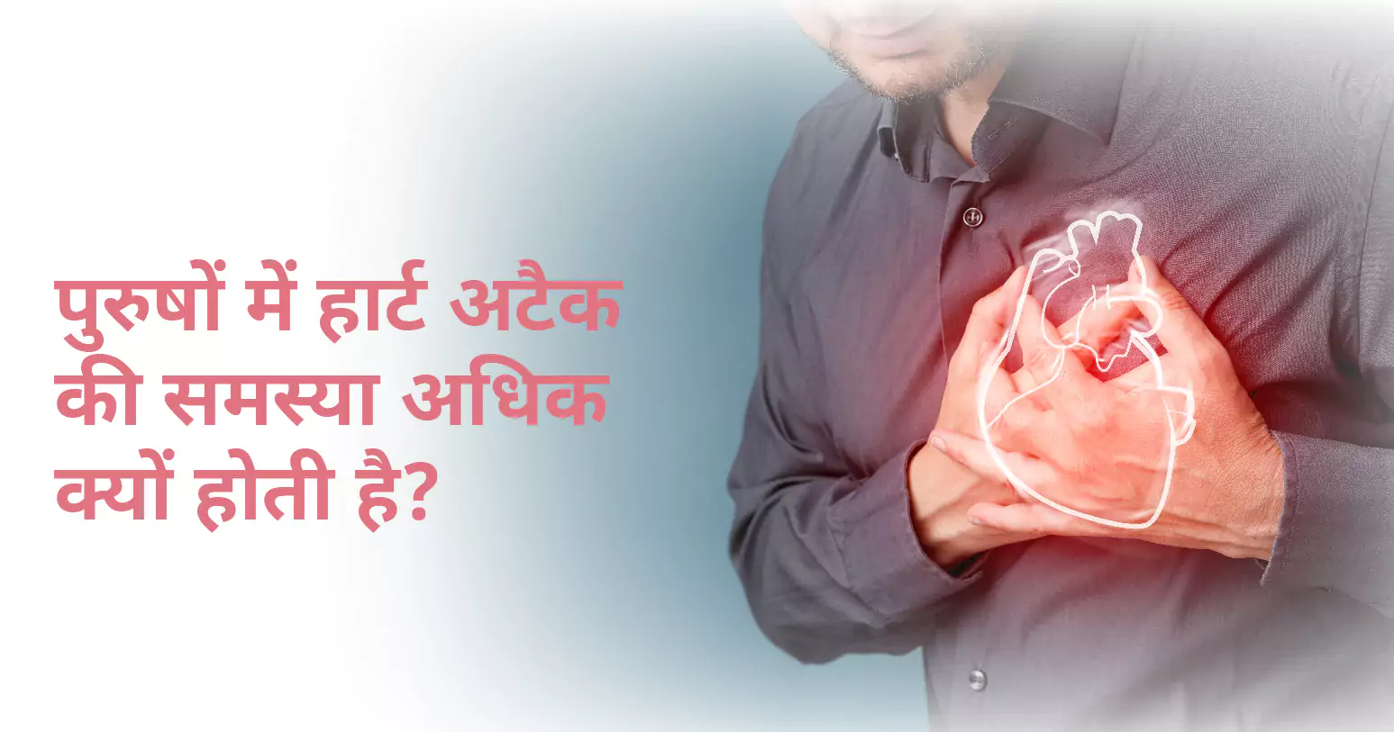
पुरुषों को हार्ट अटैक तब आता है जब हार्ट यानी हृदय के एक हिस्से में खून का प्रवाह बाधित होता है। इसके सामान्य लक्षणों में सीने में दर्द या बेचैनी, सांस लेने में तकलीफ, मतली और चक्कर आना शामिल हैं। पुरुषों को बांहों, गर्दन, जबड़े या पीठ में भी दर्द का अनुभव हो सकता है।
पुरुषों में हार्ट अटैक के जोखिम कारकों में धूम्रपान, उच्च रक्तचाप, उच्च कोलेस्ट्रॉल, मोटापा, व्यायाम की कमी और हृदय रोग का पारिवारिक इतिहास शामिल हैं। हार्ट फेल होने के खतरे को कम करने के लिए तत्काल मेडिकल सहायता महत्वपूर्ण है। इस ब्लॉग में हम पुरुषों में हार्ट अटैक के कारणों, लक्षणों और बचाव के बारे में जानेंगे।
यह भी पढ़े: बीपी (ब्लड प्रेशर) कम करने के 5 उपाय
पुरुषों में हार्ट अटैक विभिन्न कारणों से होता है:
समय के साथ धमनियों में फैट जमा हो जाता है, जिससे हार्ट में रक्त का प्रवाह कम हो जाता है जो हार्ट अटैक का कारण बनता है।
धमनियों में जमा फैट फटने पर थक्का बना जाता है जो हार्ट तक खून को जाने से रोकता है, जिससे हार्ट अटैक का खतरा होता है।
लंबे समय तक हाई ब्लड प्रेशर की समस्या होने से धमनियों को नुकसान पहुंचाता है, जिससे उनमें खून का थक्का बनने और धमियों के सिकुड़ने की संभावना बढ़ जाती है।
खून में अतिरिक्त कोलेस्ट्रॉल धमनियों में खून का थक्का बनने में योगदान कर सकता है, जिससे खून का प्रवाह बाधित होता है।
यह भी पढ़े: कोलेस्ट्रॉल कम करने का रामबाण इलाज
सिगरेट में मौजूद रसायन हृदय और रक्त वाहिकाओं (Blood Vessels) को नुकसान पहुंचाते हैं, जिससे थक्का बनने और हार्ट अटैक आने का खतरा बढ़ जाता है।
वजन अधिक होना यानी मोटापा के कारण हार्ट पर अतिरिक्त दबाव पड़ता है और उच्च रक्तचाप और मधुमेह जैसी स्थितियां पैदा हो सकती हैं।
हाई ब्लड शुगर, रक्त वाहिकाओं को नुकसान पहुंचा सकता है और हृदय रोग का खतरा बढ़ सकता है।
दीर्घकालिक तनाव से रक्तचाप और हृदय संबंधी अन्य समस्याएं बढ़ सकती हैं।
सैचुरेटेड फैट, नमक और चीनी से भरपूर आहार मोटापा, उच्च कोलेस्ट्रॉल और उच्च रक्तचाप में योगदान कर सकता है।
इन सबके अलावा, आनुवांशिकी हृदय रोग विकसित होने की संभावना को प्रभावित कर सकती है, जिससे कुछ पुरुषों को हार्ट अटैक का खतरा अधिक होता है।
यह भी पढ़े: अंकुरित प्याज सेहत के लिए वरदान या जहर?
पुरुषों में हार्ट अटैक के लक्षण अलग-अलग हो सकते हैं, लेकिन इसके सामान्य लक्षणों में निम्न शामिल हैं:
यह सबसे आम लक्षण है। यह छाती के मध्य या बाईं ओर दबाव महसूस हो सकता है। यह कुछ मिनटों से अधिक समय तक रह सकता है या आता-जाता रह सकता है।
यह सीने में दर्द के साथ या उसके बिना भी हो सकता है। ऐसा महसूस हो सकता है कि आप सांस नहीं ले पा रहे हैं या सांस लेने के लिए संघर्ष कर रहे हैं।
पुरुषों को बाहों (आमतौर पर बाएं हाथ), पीठ, गर्दन, जबड़े या पेट में दर्द या असुविधा का अनुभव हो सकता है। यह दर्द अचानक या धीरे-धीरे हो सकता है।
यह भी पढ़े: जोड़ों में दर्द का कारण, लक्षण और उपचार
कुछ पुरुषों को हार्ट अटैक आने पर पेट खराब हो सकता है या उल्टी हो सकती है।
बेहोशी या सिर घूमना हार्ट अटैक आने का संकेत हो सकता है, खासकर अगर यह सीने में दर्द या सांस की तकलीफ के साथ हो।
अत्यधिक थकान, खासकर अगर यह अचानक या गंभीर हो, तो एक संकेत हो सकता है हार्ट अटैक अटैक का।
इन लक्षणों को पहचानना और तुरंत मेडिकल सहायता लेना महत्वपूर्ण है। शीघ्र उपचार से जान बचाई जा सकती है और हार्ट को फेल होने से रोका जा सकता है। अगर आप इन लक्षणों का अनुभव करते हैं तो ह्रदय रोग विशेषज्ञ से परामर्श करें।
यह भी पढ़े: लो बीपी (लो ब्लड प्रेशर) : लक्षण, कारण और उपचार
पुरुषों में हार्ट अटैक को रोकने में स्वस्थ जीवनशैली अपनाना और जोखिम कारकों का प्रबंधन करना शामिल है:
फलों, सब्जियों, साबुत अनाज, लीन प्रोटीन और स्वस्थ वसा से भरपूर संतुलित आहार खाएं। सैचुरेटेड फैट, नमक और चीनी का सेवन सीमित करें।
हफ्ते में कम से कम चार दिन हल्का-फुल्का व्यायाम करें जैसे कि तेज चलना और दौड़ना आदि।
हार्ट पर दबाव कम करने के लिए स्वस्थ वजन बनाए रखें। मोटापा से बचें।
हार्ट हेल्थ में सुधार और हृदय रोग के जोखिम को कम करने के लिए धूम्रपान बंद करें।
अगर आवश्यक हो तो आहार, व्यायाम और दवा के माध्यम से ब्लड प्रेशर की निगरानी और प्रबंधन करें।
स्वस्थ आहार, नियमित व्यायाम और यदि निर्धारित हो तो दवाओं से कोलेस्ट्रॉल के स्तर को कंट्रोल में रखें।
शराब कम मात्रा में पियें या फिर इसका सेवन बंद कर दें।
मेडिटेशन और योग एवं व्यायाम आदि से तनाव कम करें।
जोखिम कारकों का शीघ्र पता लगाने और उन्हें प्रबंधित करने के लिए नियमित स्वास्थ्य जांच के लिए अपने डॉक्टर से मिलें।
जीवनशैली में इन बदलावों को लागू करने से पुरुष ही नहीं बल्कि महिलाऐं भी हार्ट अटैक से बच सकती हैं।
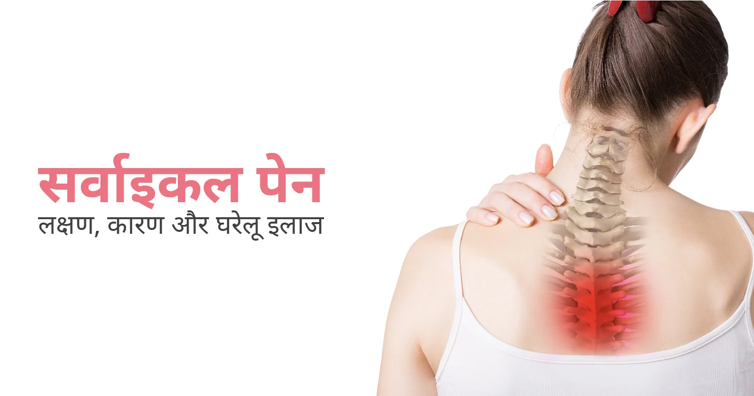
गर्दन क्षेत्र में असुविधा या दर्द को सर्वाइकल पेन कहते हैं। यह अक्सर मांसपेशियों में खिंचाव, चोटों या गठिया जैसी अपक्षयी स्थितियों (degenerative conditions) के कारण होता है। इससे अकड़न, सिरदर्द (headache in hindi) और गर्दन की गति सिमित होना जैसे लक्षण अनुभव होते हैं, जो दैनिक गतिविधियों और जीवन की क्वालिटी को प्रभावित करते हैं।
Table of Contents
सर्वाइकल पेन के लक्षण अंतर्निहित कारण के आधार पर भिन्न हो सकते हैं, लेकिन इसके सामान्य लक्षणों में निम्न शामिल हैं:
ये लक्षण दैनिक गतिविधियों और जीवन की गुणवत्ता को महत्वपूर्ण रूप से प्रभावित करते हैं। कारण निर्धारित करने और उचित उपचार प्राप्त करने के लिए लगातार या गंभीर सर्वाइकल पेन के लिए मेडिकल सहायता महत्वपूर्ण है।
ये भी पढ़े: सिर में भारीपन के कारण और घरेलु इलाज
सर्वाइकल पेन विभिन्न कारणों से होता है, जिसमें मामूली समस्याओं से लेकर अधिक गंभीर स्थितियां शामिल हैं। इन कारणों में निम्न शामिल हैं:
सर्वाइकल पेन दैनिक जीवन को महत्वपूर्ण रूप से प्रभावित कर सकता है। इसलिए इस स्थिति को प्रभावी ढंग से प्रबंधित और इलाज करने के लिए अंतर्निहित कारण की पहचान करना आवश्यक है।
ये भी पढ़े: नसों में दर्द का कारण और उपचार | Nerve Pain in Hindi
घरेलू उपचार सर्वाइकल पेन को प्रबंधित और कम करने में प्रभावी होते हैं, खासकर जब यह मांसपेशियों में खिंचाव या पुअर पोस्चर जैसी छोटी समस्याओं के कारण होता है। नीचे दिए गए घरेलू उपचार आपकी मदद कर सकते हैं:
इन घरेलू उपचारों की मदद से सर्वाइकल पेन को कम करने और दोबारा होने से रोका जा सकता है। हालाँकि, अगर दर्द बना रहता है या बिगड़ जाता है, तो आगे के मूल्यांकन और उपचार के लिए एक्सपर्ट डॉक्टर से परामर्श करना आवश्यक है।
ये भी पढ़े: जाने सिरदर्द के प्रकार, कारण और इलाज अनुभवी डॉक्टर द्वारा
अगर घरेलू उपचार से सर्वाइकल दर्द में आराम नहीं मिलता है, तो आपको एक विशेषज्ञ से परामर्श लेना चाहिए। वे सही निदान और उपयुक्त उपचार की सिफारिश करेंगे।
तनाव गर्दन की मांसपेशियों में तनाव पैदा करके सर्वाइकल पेन में योगदान कर सकता है, जिससे असुविधा हो सकती है और मांसपेशियों में खिंचाव जैसी स्थिति बढ़ सकती है।
हाँ, गलत तरीके से नींद सोने से गर्दन की मांसपेशियों में तनाव पैदा होता है जो सर्वाइकल पेन का कारण बन सकता है।

Are you prepared to face the scorching heat wave this season? With temperatures rising to extreme levels, saving oneself from a heatwave has become of utmost importance.
Typically, India is known for its diverse climate and often experiences extreme weather conditions, including fiery heat waves. According to the India Meteorological Department (IMD) this summer season, particularly in the northern regions like Delhi NCR, Punjab, Rajasthan and other North Indian states, can be brutal, with temperatures soaring well above 45 degrees Celsius. Therefore, staying safe during these intense conditions is critical to avoiding heat-related illnesses and ensuring general well-being.
This article comprises some essential tips and strategies to help you stay safe during a heat wave. But first, let’s understand why heat waves are dangerous.
Table of Contents
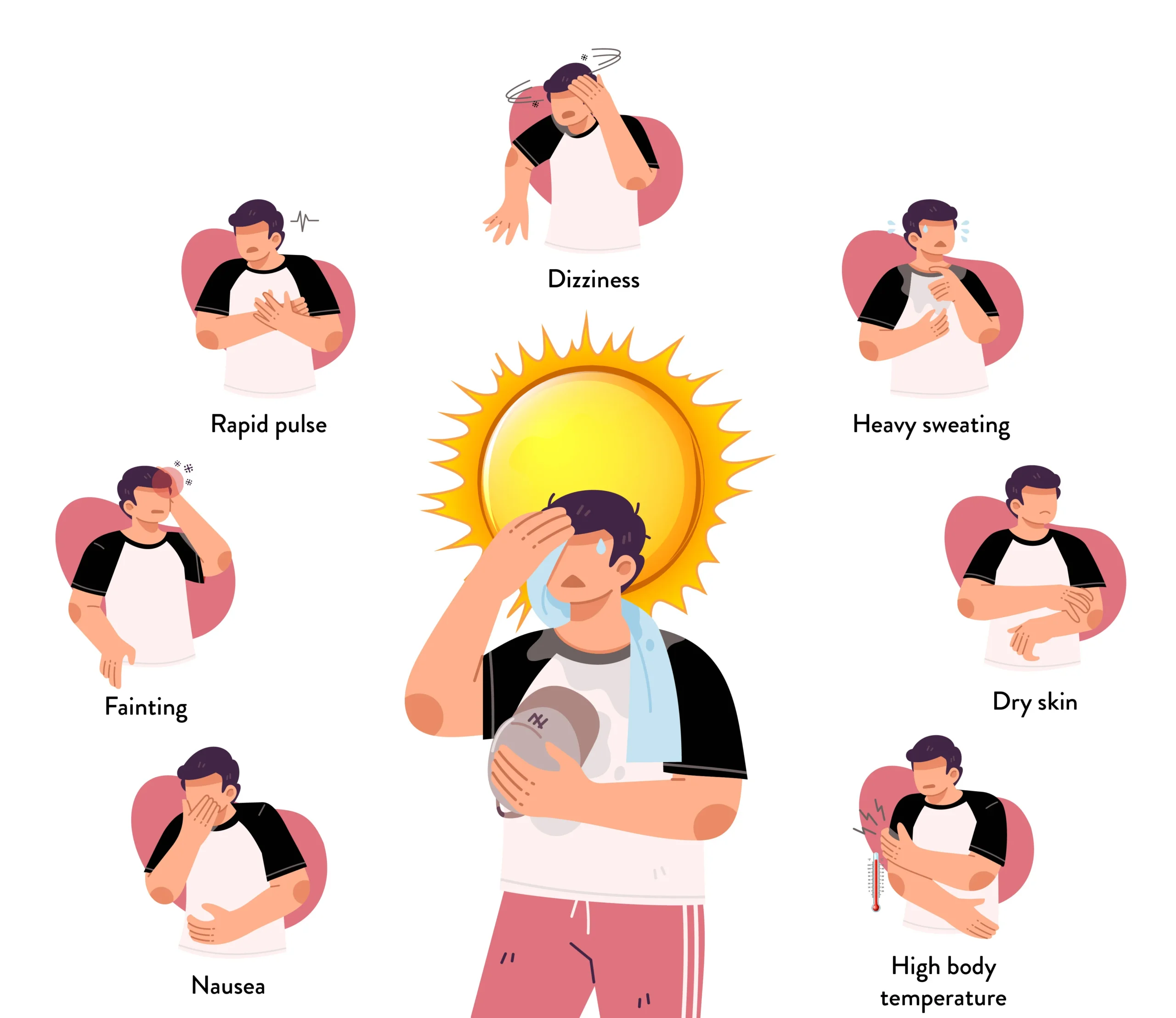
What is Heat wave? It is a prolonged period of excessively hot weather, often accompanied by high humidity. These conditions can be life-threatening, especially for vulnerable individuals. The dangers of heat waves include heat exhaustion, increased mortality, heatstroke, dehydration, and respiratory issues.
Some of the common symptoms of these conditions are:
During heat wave temperatures, preparation becomes the key to staying safe. Here are some steps to take before the temperatures rise:
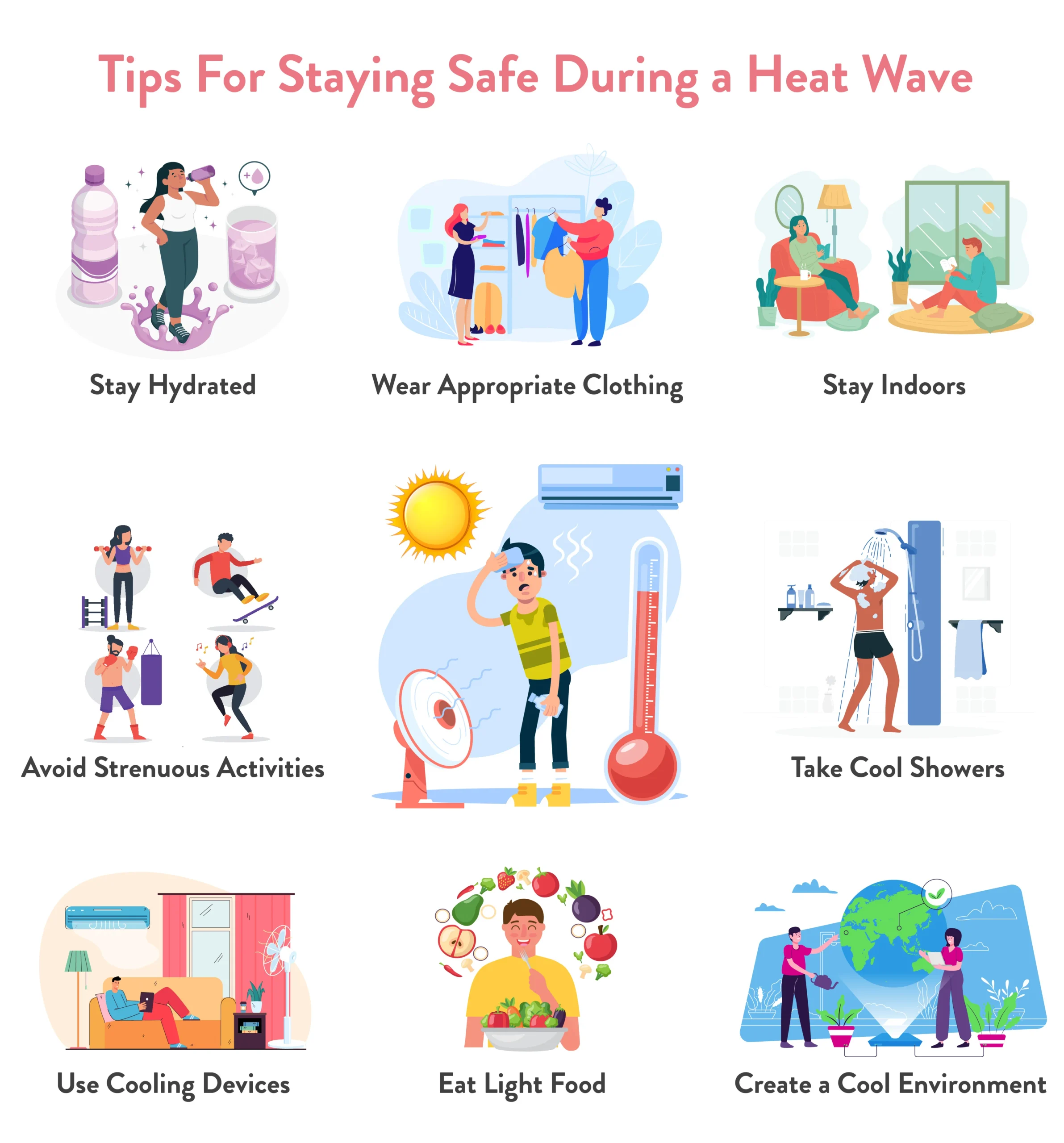
To stay safe during these heat waves these few guidelines can help you:
Some places are red alert for heat waves. To give you an idea of the temperatures you might face, here is a table showing average heat wave temperatures in some regions of India:
| Region | Average Temperature (°C) |
| Heat Wave Temperature in Delhi NCR | 40-45 |
| Heat Wave Temperature in Punjab | 38-44 |
| Heat Wave Temperature in Haryana | 40-46 |
| Heat Wave Temperature in Uttar Pradesh | 38-45 |
| Heat Wave Temperature in Rajasthan | 42-48 |
Certain age groups are more vulnerable to the effects of extreme heat and such high temperatures. While keeping in mind that some areas are on red alert because of heat waves, it’s important to consider these groups:
Planning ahead can make all the difference. Below are a few tips that can help you in case of a heat-related emergency:
Staying cool is essential to prevent heat-related illnesses. Here are some dos and don’ts to follow along with effective cooling techniques to prevent yourself from heat:
Heat waves can be harmful, but with careful planning and safeguards, you can stay safe and healthy. Keep yourself informed about weather conditions, stay hydrated, avoid intense activity during peak heat hours, and protect vulnerable individuals like elders and infants. If you suspect someone is suffering from heatstroke, relocate them to a cooler location, administer cool water to their skin, and contact emergency medical services. All these suggestions mentioned in the article can help you navigate the severe heat wave of Indian summers while protecting yourself and your loved ones from heat-related ailments. Remember that staying cool is more than just about comfort; after facing the heat wave red alert it’s about survival. Stay safe!
Use fans, stay in well-ventilated spaces, keep a wet cloth with you, and a small spray bottle for face misting in order to balance the body temperature.
You should aim for at least 8-10 glasses a day and more if you’re sweating heavily.
Yes, make sure they have plenty of drink and shade, and avoid walking them at high temperatures.
The common signs of heatstroke include high body temperature, confusion, rapid pulse, and unconsciousness. Seek immediate medical help and cool the person with ice packs or a cold bath.
Sources –
https://ncdc.mohfw.gov.in/wp-content/uploads/2024/03/Emergency-Cooling-for-Severe-Heat-Related-Illnesses_March2024_NPCCHH.pdf
https://m.economictimes.com/news/india/delhi-weather-forecast-imd-issues-heatwave-red-alert-in-north-india/amp_videoshow/111086046.cms
https://www.businesstoday.in/latest/trends/story/imd-predicts-heatwave-in-northwest-india-from-may-16-heavy-rainfall-in-these-regions-429378-2024-05-13
https://economictimes.indiatimes.com/news/india/delhi-heat-index-crosses-50-as-imd-issues-red-alert-when-heatwave-in-north-india-will-ease/articleshow/111072409.cms?from=mdr
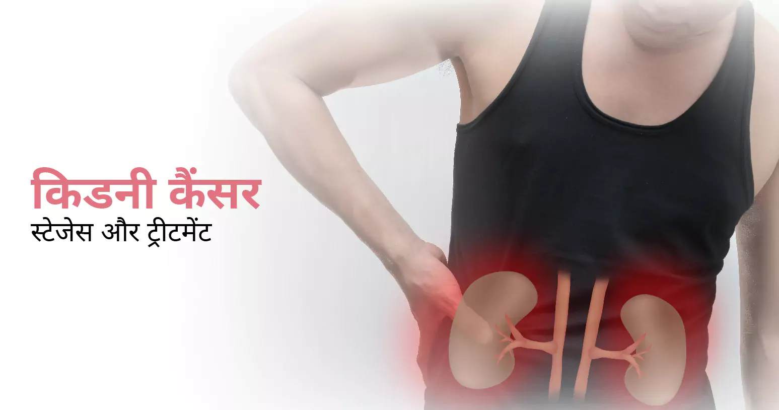
किडनी कैंसर एक प्रकार का कैंसर है, जो किडनी में होता है। इसके मुख्य लक्षणों में पेशाब में खून आना, पीठ दर्द और वजन कम होना शामिल हैं। इस ब्लॉग में हम किडनी कैंसर के स्टेज और ट्रीटमेंट के बारे में विस्तार से जानेंगे।
Table of Contents
किडनी कैंसर आमतौर पर कई स्टेज में बढ़ता है। इसके प्रकार की पुष्टि इस बात से की जाती है कि ट्यूमर शरीर के अन्य हिस्सों में कहाँ तक तक फैल चुका है। किडनी कैंसर को चार स्टेज में बांटा गया है जिसमें शामिल हैं:
आमतौर पर किडनी कैंसर में ट्यूमर व्यास में 7 सेंटीमीटर (लगभग 2.75 इंच) से छोटे होते हैं और ये किडनी तक ही सीमित होते हैं।
स्टेज 1 की तुलना में, ट्यूमर बड़े होते हैं, लेकिन फिर भी ये किडनी तक ही सीमित होते हैं। स्टेज 2 में, ट्यूमर का व्यास आमतौर पर 7 से 10 सेंटीमीटर (लगभग 2.75 से 3.94 इंच) के बीच होता है।
इस स्टेज में, ट्यूमर किडनी के आस-पास के टिशू या संरचनाओं, जैसे कि ब्लड वेसेल्स या एड्रेनल ग्लैंड में फैल सकते हैं। ट्यूमर का आकार अलग-अलग हो सकता है, लेकिन आमतौर पर ये 10 सेंटीमीटर से बड़े होते हैं।
इस स्टेज में ट्यूमर, शरीर के अन्य हिस्सों में फैल जाते हैं। प्राथमिक ट्यूमर का आकार अलग हो सकता है और एक से अधिक ट्यूमर मौजूद हो सकते हैं। किडनी कैंसर के चौथे स्टेज में, डॉक्टर कैंसर के आकार के बजाय इस बात पर ज़्यादा ध्यान देते हैं कि ट्यूमर शरीर के किस हिस्सों में फैल चूका है।
यह भी पढ़े: कमर दर्द का कारण ,उपाय और घरेलू इलाज
यदि आप नीचे दिए गए लक्षणों को अनुभव करते हैं तो तुरंत डॉक्टर से मिलें:
अगर ये लक्षण बने रहते हैं या गंभीर हो जाते हैं तो तुरंत मेडिकल सहायता लें। शीघ्र निदान और उपचार से इन लक्षणों में सुधार होता है।
यह भी पढ़े: किडनी में सूजन का कारण और इलाज | Kidney Inflammation in Hindi
किडनी कैंसर का ट्रीटमेंट इसकी गंभीरता, ट्यूमर के आकार और मरीज़ के समग्र स्वास्थ्य पर निर्भर करता है। इसके मुख्य उपचार में शामिल हैं:
यह किडनी कैंसर का सबसे सामान्य इलाज है, जिसमें ट्यूमर या पूरी किडनी को निकालना शामिल है। इस सर्जरी के दो मुख्य प्रकार हैं:
केवल ट्यूमर और स्वस्थ ऊतक का एक छोटा सा हिस्सा हटाना।
आस-पास के ऊतकों सहित पूरी किडनी को हटाना। कभी-कभी, एड्रेनल ग्लैंड और लिम्फ नोड्स को भी बाहर निकाल दिया जाता है।
ट्यूमर को हटाए बिना उसे नष्ट कर देता है। इन प्रक्रिया में रेडियोफ्रीक्वेंसी एब्लेशन और क्रायोएब्लेशन (ट्यूमर को फ्रीज करना) शामिल हैं।
ट्यूमर में खून का प्रवाह बंद कर देता है, जिससे यह सिकुड़ जाता है। यह ट्यूमर को पोषण देने वाली ब्लड वेसेल्स में छोटे कणों को इंजेक्ट करके किया जाता है।
विशिष्ट जीन या प्रोटीन को टारगेट करने के लिए दवाओं का उपयोग किया जाता है जो कैंसर कोशिकाओं को बढ़ने में मदद करते हैं। ये दवाएं कैंसर कोशिकाओं की वृद्धि को रोक सकती हैं।
इससे कैंसर से लड़ने के लिए शरीर की प्रतिरक्षा प्रणाली को बढ़ावा मिलती है। चेकपॉइंट ब्लॉकर जैसी दवाएं प्रतिरक्षा प्रणाली को कैंसर कोशिकाओं को पहचानने और उन्हें में मदद करती हैं। इम्यूनोथेरेपी थेरेपी एडवांस किडनी कैंसर के लिए उपयोगी है।
इस थेरेपी में कैंसर कोशिकाओं को मारने के लिए उच्च-ऊर्जा किरणों का उपयोग होता है। आमतौर पर इसका इस्तेमाल किडनी कैंसर के लिए नहीं किया जाता है, लेकिन जब कैंसर शरीर के अन्य हिस्सों में फैल जाता है तो लक्षणों से राहत दिलाने में सहायक हो सकता है।
हर मरीज़ की उपचार योजना यूनिक होती है। इसलिए किडनी कैंसर का उपचार करने से पहले डॉक्टर अनेक बातों पर विचार करते हैं जैसे कि मरीज़ का समग्र स्वास्थ्य, किडनी कैंसर का स्टेज और लक्षण आदि।
किडनी कैंसर के सामान्य लक्षण में पेशाब में खून, लगातार पीठ में दर्द, बिना कारण वजन कम होना, थकान और पेट में गांठ आदि शामिल हैं।
धूम्रपान, मोटापा, उच्च रक्तचाप, किडनी कैंसर का पारिवारिक इतिहास और कुछ आनुवंशिक स्थितियाँ किडनी कैंसर के खतरे को बढ़ाती हैं।
इमेजिंग परीक्षण (सीटी स्कैन, एमआरआई, अल्ट्रासाउंड), खून और पेशाब की जांच एवं कभी-कभी बायोप्सी के माध्यम से किडनी कैंसर का निदान किया जाता है।
हालांकि सभी मामलों को रोका नहीं जा सकता है, धूम्रपान जैसे जोखिम कारकों को कम करना, स्वस्थ वजन बनाए रखना, रक्तचाप का प्रबंधन करना और नियमित जांच से जोखिम को कम करने में मदद मिल सकती है।
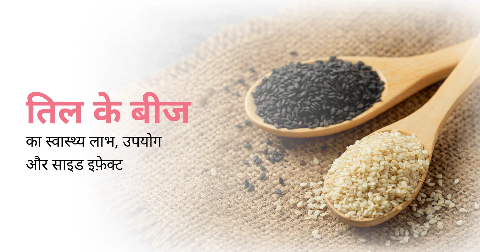
क्या आपको पता है तिल के बीज के फायदों के बारे में? यह आपके स्वास्थ्य और वेलनेस के लिए बहुत फायदेमंद है। इसके अनेक फायदों के कारण ही डॉक्टर भी कई बार मरीज़ को इसे अपनी डाइट में शामिल करने का सुझाव देते हैं। तो आइए, तिल के बीज के फायदों के बारे में जानते हैं।
यह भी पढ़ें: चिया सीड्स के फायदे, नुकसान, और पोषक तत्त्व
अपने छोटे आकार के बावजूद, तिल के बीज प्रोटीन, फाइबर, स्वस्थ वसा, विटामिन और खनिज जैसे आवश्यक पोषक तत्वों से भरपूर होते हैं। इनमें कैल्शियम, आयरन, मैग्नीशियम, फॉस्फोरस, जिंक, विटामिन बी6 और बहुत कुछ होता है, जो उन्हें पोषण का पावरहाउस बनाता है।
तिल के बीज में मोनोअनसैचुरेटेड और पॉलीअनसेचुरेटेड वसा की अधिक मात्रा, खराब कोलेस्ट्रॉल के स्तर को कम करने में मदद कर सकती है, जिससे हृदय रोग और स्ट्रोक का खतरा कम हो जाता है। इसके अलावा, उनमें सेसमोल और सेसामिनोल, एंटीऑक्सिडेंट होते हैं जो हृदय के स्वास्थ्य के लिए आवश्यक होते हैं।
यह भी पढ़ें: कोलेस्ट्रॉल कम करने का रामबाण इलाज
तिल के बीज, कैल्शियम का एक बेहतर स्रोत हैं, जो हड्डियों को मजबूत बनाने और ऑस्टियोपोरोसिस जैसी स्थितियों को रोकने के लिए महत्वपूर्ण हैं। अपने आहार में तिल को शामिल करने से हड्डियों के घनत्व और समग्र स्केलेटल हेल्थ में योगदान मिलता है।
तिल के बीज एंटीऑक्सीडेंट से भरपूर होते हैं, जिनमें सेसमिन, सेसमोलिन और विटामिन ई शामिल हैं, जो शरीर में हानिकारक मुक्त कणों को बेअसर करने में मदद करते हैं। यह एंटीऑक्सीडेंट गतिविधि सूजन, ऑक्सीडेटिव तनाव और कैंसर जैसी पुरानी बीमारियों के खतरे को कम कर सकती है।
तिल के बीज में फाइबर सामग्री नियमित मल त्याग को बढ़ावा देकर, कब्ज को रोककर पाचन स्वास्थ्य का बेहतर बनाता है। फाइबर लाभकारी आंत बैक्टीरिया को भी पोषण देता है, स्वस्थ आंत, माइक्रोबायोम और समग्र पाचन क्रिया में योगदान देता है।
यह भी पढ़ें: पेट साफ करने के घरेलू उपाय
तिल के बीज में मौजूद मैग्नीशियम, इंसुलिन डिस्चार्ज और ग्लूकोज चयापचय में भूमिका निभाती है, जिससे ब्लड शुगर के स्तर को नियंत्रित करने में मदद मिलती है। अपने आहार में तिल को शामिल करने से ब्लड शुगर में वृद्धि और गिरावट को रोकने में मदद मिल सकती है, जिससे टाइप 2 मधुमेह का खतरा कम हो सकता है।
तिल के बीज में जिंक भरपूर मात्रा में होता है, जो कोलेजन उत्पादन, त्वचा की मरम्मत और स्वस्थ बालों को बनाए रखने के लिए आवश्यक खनिज है। अपने आहार में तिल को शामिल करने या शीर्ष पर तिल के तेल का उपयोग करने से स्किन इलास्टिसिटी, हाइड्रेशन और बालों की मजबूती को बढ़ावा मिल सकता है।
तिल के बीज में लिगनेन, पौधे के यौगिक होते हैं जिनका शरीर में एस्ट्रोजन जैसा प्रभाव होता है। ये यौगिक हार्मोन के स्तर को रेगुलेट करने में मदद कर सकते हैं, विशेष रूप से मेनोपॉज़ के बाद की महिलाओं में, संभावित रूप से हॉट फ्लैश और मूड स्विंग जैसे लक्षणों को कम करते हैं।
तिल के बीज में पाया जाने वाला जिंक और विटामिन बी6 स्वस्थ प्रतिरक्षा प्रणाली के लिए महत्वपूर्ण हैं। जिंक प्रतिरक्षा कोशिकाओं को ठीक से काम करने में मदद करता है, जबकि विटामिन बी6 प्रतिरक्षा प्रतिक्रियाओं में शामिल जैव रासायनिक प्रतिक्रियाओं का समर्थन करता है, जिससे संक्रमण और बीमारियों से बचाव में मदद मिलती है।
तिल के बीज अविश्वसनीय रूप से बहुमुखी हैं और इन्हें व्यंजनों की एक विस्तृत श्रृंखला में शामिल किया जा सकता है। चाहे सलाद पर छिड़का जाए, स्टर-फ्राई में मिलाया जाए, मछली या चिकन के लिए लेप के रूप में इस्तेमाल किया जाए, या सॉस और ड्रेसिंग के लिए ताहिनी पेस्ट में पीसा जाए, तिल भोजन के स्वाद और पोषण मूल्य को बढ़ा सकते हैं।
अक्सर खाना पकाने और बेकिंग में उपयोग किए जाने वाले तिल के बीज के कई संभावित दुष्प्रभाव होते हैं, हालांकि कम मात्रा में सेवन करने पर वे आमतौर पर ज्यादातर लोगों के लिए सुरक्षित होते हैं। इसके संभावित साइड इफ्केट में शामिल हैं:
कुछ लोगों को तिल के बीज से एलर्जी प्रतिक्रियाओं का अनुभव हो सकता है, जिसमें खुजली और पित्ती जैसे हल्के लक्षणों से लेकर सांस लेने में कठिनाई या एनाफिलेक्सिस जैसी गंभीर प्रतिक्रियाएं शामिल हैं।
तिल के बीज में फाइबर की मात्रा अधिक होती है, जो पाचन संबंधी असुविधा जैसे सूजन, गैस या दस्त का कारण बन सकता है, खासकर जब बड़ी मात्रा में या संवेदनशील पेट वाले लोग इसका सेवन करते हैं।
तिल के बीज में ऑक्सालेट, यौगिक होते हैं जो अतिसंवेदनशील व्यक्तियों में गुर्दे की पथरी के निर्माण में योगदान कर सकते हैं। जिन लोगों को गुर्दे की पथरी का इतिहास है, उन्हें इसके सेवन को सीमित करने की आवश्यकता हो सकती है।
कई बीजों की तरह, तिल के बीज में फाइटिक एसिड होता है, जो कैल्शियम, जिंक और आयरन जैसे कुछ खनिजों के अवशोषण में हस्तक्षेप कर सकता है। हालाँकि, संतुलित आहार में यह प्रभाव आमतौर पर न्यूनतम होता है।
तिल के बीज में गोइट्रोजेन्स होते हैं, ऐसे यौगिक जो बड़ी मात्रा में सेवन करने पर थायरॉइड फ़ंक्शन में हस्तक्षेप कर सकते हैं, संभावित रूप से हाइपोथायरायडिज्म जैसे थायरॉयड विकारों को बढ़ा सकते हैं।
तिल के बीज हृदय स्वास्थ्य और एंटीऑक्सीडेंट गुणों सहित विभिन्न स्वास्थ्य लाभ प्रदान करते हैं, उनका सीमित मात्रा में सेवन करना और संभावित साइड इफेक्ट से सावधान रहना आवश्यक है, विशेष रूप से विशिष्ट स्वास्थ्य चिंताओं या एलर्जी वाले व्यक्तियों के लिए।
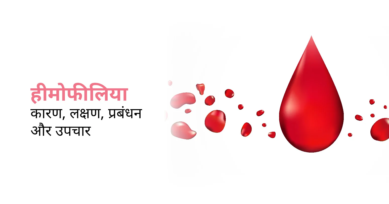
हीमोफीलिया एक आनुवंशिक विकार है। इससे पीड़ित मरीज के खून में थक्के बनने कम हो जाते हैं जिससे मामूली चोट लगने पर भी लंबे समय तक रक्तस्राव (ब्लीडिंग) होता रहता है। यह मुख्य रूप से वंशानुगत (Hereditary) होता है, जो अधिकतर पुरुषों को प्रभावित करता है। इसके लक्षणों में मामूली चोट लगना, जोड़ों में दर्द और लगातार रक्तस्राव होना जिससे जान का खतरा होता है। हीमोफीलिया के उपचार में क्लॉटिंग फैक्टर रिप्लेसमेंट थेरेपी और रक्तस्राव को मैनेज करना शामिल है।
Table of Contents
हीमोफीलिया के लक्षण स्थिति की गंभीरता के आधार पर अलग-अलग होते हैं। इसके सामान्य लक्षणों में निम्न शामिल हैं:
थक्का कम बनने के कारण – चोट लगने, कटने, फटने या सर्जरी के दौरान लंबे समय तक रक्तस्राव हो सकता है।
जोड़ों, खासकर घुटनों, टखनों और कोहनियों में दर्द, सूजन और गतिशीलता में कमी हो सकती है।
यह भी पढ़ें: जोड़ों में दर्द का कारण, लक्षण और उपचार |
आंतरिक रक्तस्राव पेशाब या मल में रक्त के रूप में प्रकट हो सकता है।
खराब थक्के बनने के कारण बार-बार या लंबे समय तक नाक से खून आ सकता है।
बार-बार रक्तस्राव के कारण होने वाले क्रोनिक एनीमिया से थकान और कमजोरी हो सकती है।
हीमोफीलिया से पीड़ित महिलाओं को लंबे समय तक मासिक धर्म हो सकता है। हीमोफीलिया के तुरंत निदान और प्रभावी उपचार के लिए इन लक्षणों की प्रारंभिक पहचान महत्वपूर्ण है। अगर आप खुद में इन लक्षणों को अनुभव करते हैं जल्द से जल्द विशेषज्ञ से परामर्श करें।
हीमोफीलिया मुख्य रूप से आनुवंशिक म्यूटेशन (mutation) के कारण होता है जो रक्त में थक्के जमने वाले कारकों को प्रभावित करता है।
हीमोफीलिया का सबसे आम कारण किसी के माता-पिता से दोषपूर्ण जीन विरासत में मिलना है। पुरुषों को अपनी मां से एक एक्स गुणसूत्र विरासत में मिलता है, और यदि यह एक्स गुणसूत्र हीमोफिलिया के लिए उत्परिवर्तित जीन को वहन करता है, तो उनमें विकार होगा। महिलाओं में हीमोफीलिया विकसित होने के लिए उत्परिवर्तित जीन की दो प्रतियां (प्रत्येक माता-पिता से एक) प्राप्त करने की आवश्यकता होती है, जिससे महिलाओं में यह बहुत दुर्लभ हो जाता है।
कुछ मामलों में, भ्रूण के विकास के दौरान होने वाले थक्के कारक, जीन में स्पॉन्टेनियस म्यूटेशन के कारण हीमोफिलिया का कारण बन सकते हैं। इससे उन लोगों में हीमोफीलिया हो सकता है जिनके परिवार में इस विकार का कोई इतिहास नहीं है।
आनुवांशिक म्यूटेशन, स्पॉन्टेनियस क्लॉटिंग कारकों का उत्पादन करने की शरीर की क्षमता को बाधित करते हैं, जिससे हीमोफिलिया के विशिष्ट लक्षण दिखाई देते हैं, जैसे कि लंबे समय तक रक्तस्राव और मामूली चोटों से भी खून आना आदि।
हीमोफीलिया के उपचार का उद्देश्य रक्तस्राव (ब्लीडिंग) को कंट्रोल करना, जटिलताओं को रोकना और जीवन की क्वालिटी को बेहतर बनाना है। उपचार विकल्प में निम्न शामिल हैं:
हीमोफिलिया उपचार में रक्त में थक्का बनाने वाले कारकों को नसों के ज़रिए शरीर में ट्रांसफर करना शामिल है। क्लॉटिंग कारक, मनुष्य के प्लाज्मा से प्राप्त होते हैं या कृत्रिम रूप से तैयार किए जाते हैं।
ट्रैनेक्सैमिक एसिड या एमिनोकैप्रोइक एसिड जैसी दवाएं रक्त के थक्कों को टूटने से रोकने में मदद कर सकती हैं, खासकर म्यूकोसल रक्तस्राव या डेंटल प्रक्रियाओं के मामलों में।
जीन थेरेपी में वायरल वैक्टर का उपयोग करके, शरीर में दोषपूर्ण थक्के कारक जीन की कार्यात्मक प्रतियां (functional copies) शामिल करना है। इसका उद्देश्य शरीर को स्वतंत्र रूप से अपने थक्के बनाने वाले कारकों का उत्पादन करने में सक्षम बनाना है।
जोड़ों या मांसपेशियों में रक्तस्राव से जुड़े दर्द को कम करने के लिए एनाल्जेसिक और सूजन-रोधी दवाएं निर्धारित की जा सकती हैं।
हीमोफीलिया की गंभीरता, व्यक्तिगत जरूरतों और जीवनशैली कारकों को ध्यान में रखते हुए, अनुकूलित उपचार योजनाएं, हीमोफीलिया से पीड़ित मरीज के समग्र स्वास्थ्य में सुधार के लिए महत्वपूर्ण हैं।
हीमोफीलिया मुख्य रूप से एक आनुवंशिक विकार है। इसलिए रोकथाम में ऐसे लोगों की पहचान करने के लिए आनुवंशिक परामर्श और परीक्षण शामिल हैं, जो इस स्थिति को अपने बच्चों में ट्रांसफर करने के जोखिम में हैं। इससे बचने के निम्न उपाय हैं:
हीमोफीलिया के पारिवारिक इतिहास वाले या दोषपूर्ण जीन के वाहक (carrier) के रूप में पहचाने जाने वाले व्यक्ति, आनुवंशिक परामर्श से लाभ उठा सकते हैं। इससे उन्हें वंशानुक्रम पैटर्न को समझने, अपने बच्चों में इस स्थिति के ट्रांसफर होने के जोखिम का आकलन करने और प्रजनन विकल्पों का पता लगाने में मदद मिलती है।
हीमोफिलिया फैलने के जोखिम वाले दंपति पीजीडी के साथ इन विट्रो फर्टिलाइजेशन (आईवीएफ) करा सकते हैं। यह तकनीक प्रत्यारोपण से पहले हीमोफिलिया जीन म्यूटेशन के लिए भ्रूण की जांच करने की अनुमति देती है, जिससे गर्भावस्था के लिए अप्रभावित भ्रूण का चयन किया जा सकता है।
हीमोफीलिया के पारिवारिक इतिहास वाली गर्भवती महिलाओं के लिए, भ्रूण की स्थिति का निदान करने के लिए कोरियोनिक विलस सैंपलिंग (सीवीएस) या एमनियोसेंटेसिस जैसे प्रसवपूर्व परीक्षण किए जा सकते हैं।
बचाव के इन उपायों का इस्तेमाल करके, आप फैमिली प्लानिंग के बारे में सूचित निर्णय ले सकते हैं, जिससे आने वाली पीढ़ियों में हीमोफिलिया होने की संभावना कम या ख़त्म हो जाती है।
यह भी पढ़ें: प्रेगनेंसी के शुरूआती लक्षण क्या होते हैं|
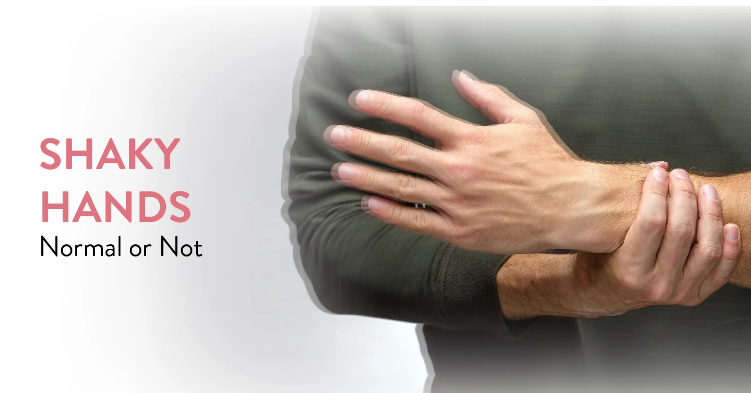
Shaky hands also referred to as hand tremors are among the most common conditions affecting millions of people across the world. This condition occurs at any age due to several underlying causes although is usually linked to growing age. Shaky hands are not life-threatening but can substantially influence regular activities, making tasks like eating, holding any object, or writing difficult.
In this blog, we will dig deep into understanding shaky hands, their types, symptoms, causes, diagnosis, and treatment options to manage this condition efficiently.
Table of Contents
Shaky hands are determined by automatic, rhythmic muscle contractions causing shaky movements in one or both hands. This condition differs in severity, prevalence, and influence on day-to-day life. Hand tremors can be temporary or consistent, and their existence signifies various underlying health problems or physiological conditions.
Everyone experiences physiological tremors at some point in life, but are nearly imperceptible tremors; however, some individuals experience severe shaking making their daily functions challenging such as eating, writing, or holding items.
There are several types of hand tremors, each with distinct characteristics and causes. The most common types include:
| Type of Tremor | Initial Symptoms | Progressive Symptoms |
| Essential Tremor | The most common type, which is congenital and affects both hands. | The condition tends to deteriorate with movement and gets better with proper rest. |
| Parkinsonian Tremor | Associated with Parkinson’s disease, this variant is triggered at rest and starts in one hand. | As the condition goes downhill, it spreads to the other hand and other parts of the body. |
| Cerebellar Tremor | Occurs due to cerebellum damage, this tremor arises during voluntary movements like reaching for an object. | This variant can be serious as it impacts coordination and balance. |
| Dystonic Tremor | Related to dystonia, this type is a movement disorder causing involuntary muscle contractions. | It may impact any part of the body and may turn severe in particular postures or movements. |
| Physiologic Tremor | Usually experienced by every individual, these tend to be nearly unnoticeable | This worsens over time due to caffeine, anxiety, fatigue, or some medications. |
The main symptom of shaky hands is the involuntary tremor or shaking of one or both hands. Other symptoms involved with shaky hands are:
Decreased control over bodily movements is identified to be the main contributor to hand tremors and is related to different issues such as weariness, stress, drug side effects, medical illnesses, or mental health disorders. Additionally, other causes of shaky hands are:
There are several risk factors that increase the probability of shaky hands, such as:
To diagnose shaky hands, the healthcare professional involves comprehensive medical assessment, including:
The underlying causes and the severity of the tremors identify the course of treatment for shaky hands. Some of the commonly involved options are:
Shaky hands can be more than just a little nuisance as they can interfere with your daily life and wear away your level of confidence. Comprehending the causes and treatments helps to manage the condition efficiently. Each type of shaky hands has several causes, whether it’s due to genetics, neurological problems, or lifestyle factors. Fortunately, modern medicine nowadays is offering us several solutions, from medications to innovative therapies. You can find ways to stabilize your hands and regain control over daily tasks by learning more and seeking expert guidance.
At the CK Birla Hospital, we ensure patients get holistic medical support which includes treatment in a compassionate environment. This patient-centric approach not only helps patients heal better but also ensures they are aware of the preventive measures as well. In case you need to consult a cardiologist, reach out to us, or book a direct appointment at the CK Birla Hospital.
Usually, shaky hands don’t signify serious condition, however, they are a symptom of conditions like multiple sclerosis, dystonia, or Parkinson’s disease. Also, they arise due to a stroke or traumatic brain injury. Hence, it is always a good idea to seek professional advice if you are regularly experiencing them.
If shaky hands are disrupting your day-to-day tasks such as drinking, writing, eating, phone dialing, etc. then it is a sign that you must visit Shaky Hands Doctor to test for neurological or metabolic abnormalities.
For essential tremors, no cure is available to date, however, with the right treatment options like medication, lifestyle changes, stress management, etc. you can suppress the tremors.
Natural remedies that can be involved for hand tremors are relaxation methods, avoiding triggers, entailing hand exercises, using assistive devices, and stress management to reduce tremors.
Be calm and keep note of additional signs if you suddenly experience hand tremors. Make sure you get sufficient sleep, limit caffeine intake, and manage your stress. Seek clinical intervention immediately to identify the reason and start therapy if they are accompanied by other symptoms.