Filter :
Diet – a term carrying a lot of misconceptions. To some diets means starving themselves while to some diets is only for weight loss. However, the true meaning of diet is much more diverse and helps people make more informed choices for overall well-being. To help maintain good health a diet chart is a guide that lists foods along with their nutritional value. A diet chart is a guide that lists foods and their nutritional value in order to support good health. It is an organized plan that advises on the type, quantities, and timing of food & beverages to be consumed to meet the body’s nutritional requirements.
There is no ‘one size fits all’ approach when it comes to diet. A diet plan is customised based on its intended use/goal. Although there are generic diet charts, a customised chart is essential for effectively reaching the goal. Everybody is different and so are the nutritional requirements, thus a tailored diet chart seeks to cater to the body’s requirements specifically.
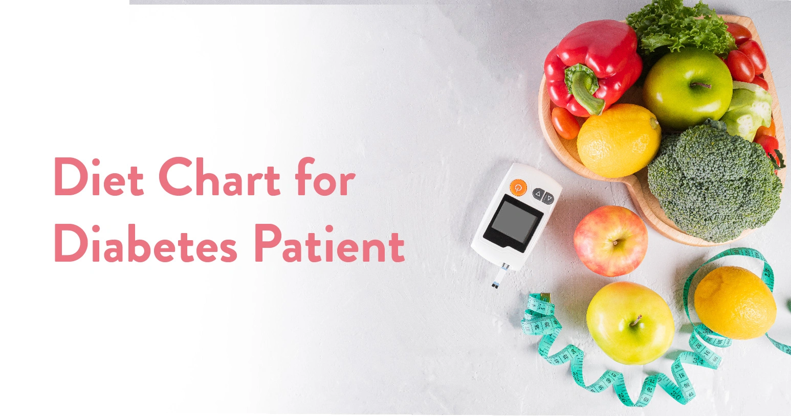
Just as diet plans vary based on the objectives/goals they also vary based on the underlying medical condition. Patients with diabetes have different diet plans. The body’s inability to produce enough insulin or use it efficiently is the cause of this chronic illness. Diet chart for diabetic patients focuses on promoting eating a variety of foods providing adequate nutrients while limiting carbohydrate intake which has an impact on blood sugar levels. These diet charts emphasize guiding people with diabetes in controlling their blood sugar levels through healthy eating.
Table of Contents
Diabetes is a chronic condition that requires maintaining the blood sugar level within a healthy range. When we consume food, it is broken down into glucose (sugar) in the digestive tract, further, the pancreas produces insulin to help regulate this glucose (sugar) into the body’s cells and used as energy. However diabetic patients have a harder time controlling their blood sugar levels after eating because their insulin production is either not adequate or ineffectively used. For this reason food intake is crucial for diabetic patients as the foods they eat have a direct impact on blood glucose levels. There are certain foods that a person with diabetes should avoid including,
| Category | Food Products |
| Refined & Processed Carbohydrates | White bread, white rice, pasts, instant noodles |
| Sugary Food & Drinks | Fruit juices, candies, cookies, cakes, sweetened teas |
| Unhealthy Fats | Fried foods, high fat meats & trans fats |
| High Sugar Fruits | Fruits like mangoes, grapes, pineapple, etc, should be consumed in moderation |
It’s critical for diabetic patients to make informed dietary choices and monitor their intake in order to maintain their blood sugar levels. The foods that are part of the diabetic diet help keep blood sugar levels steady, support general health and lower the risk of complications from diabetes including kidney issues, nerve damage, heart disease, and other related disorders. In addition to assisting with blood sugar regulation, making healthy food choices promotes long-term health by averting the negative consequences of diabetes.
Also Read: Ivy Gourd: Benefits & Side Effects
Diet plays a key role in managing blood sugar levels, supports weight management and reduces the complications associated with diabetes. An ideal diet plan for controlling diabetes includes all the macronutrient and micronutrient components and covers all food groups to ensure the body receives enough nutrition with controlled carbohydrates. There are other factors that help with diet management including:
Various factors affect diet charts for diabetic patients in India including,
| Factors | Description |
| Cultural Preferences | Regional food habits like rice, wheat, or millet-based diets |
| Age & Gender | Nutritional needs vary for children, adults & elderly, as well as between genders |
| Health Condition | Pre-existing issues like hypertension, heart disease, etc, influence food choices |
| Weight Management | Calorie deficit for reducing fat, common among Indians |
| Lifestyle | Diet depends on activity levels, like sedentary office jobs or physical labour |
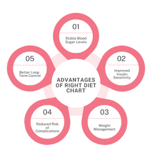
For diabetic patients, a carefully thought-out diet plan is essential to controlling blood sugar levels and maintaining overall health. Patients with diabetes should follow a diet that emphasises well-balanced meals that include the appropriate amounts of fats, proteins, carbs, and other nutrients. Maintaining stable energy levels and improving insulin sensitivity can be achieved by adhering to a customised diet. Under the direction of a nutritionist or specialist a customised diet plan is created with the patient’s overall health in mind. It aids in the efficient management of diabetes and fosters long-term health.
A combination of strategies including adhering to a diabetic-specific diet plan, exercising, drinking plenty of water etc. can help control blood sugar levels.
Diabetic patients can eat rice as a part of their diet however it should be consumed in moderation and small portions.
Dates are low on glycemic index (GI) and release sugar slowly into the blood. Dates can be consumed by diabetic patients in moderation.
Fruits with low to moderate glycemic index (GI), which release sugar slowly into the blood are recommended for diabetic patients apples, bears, berries, etc.
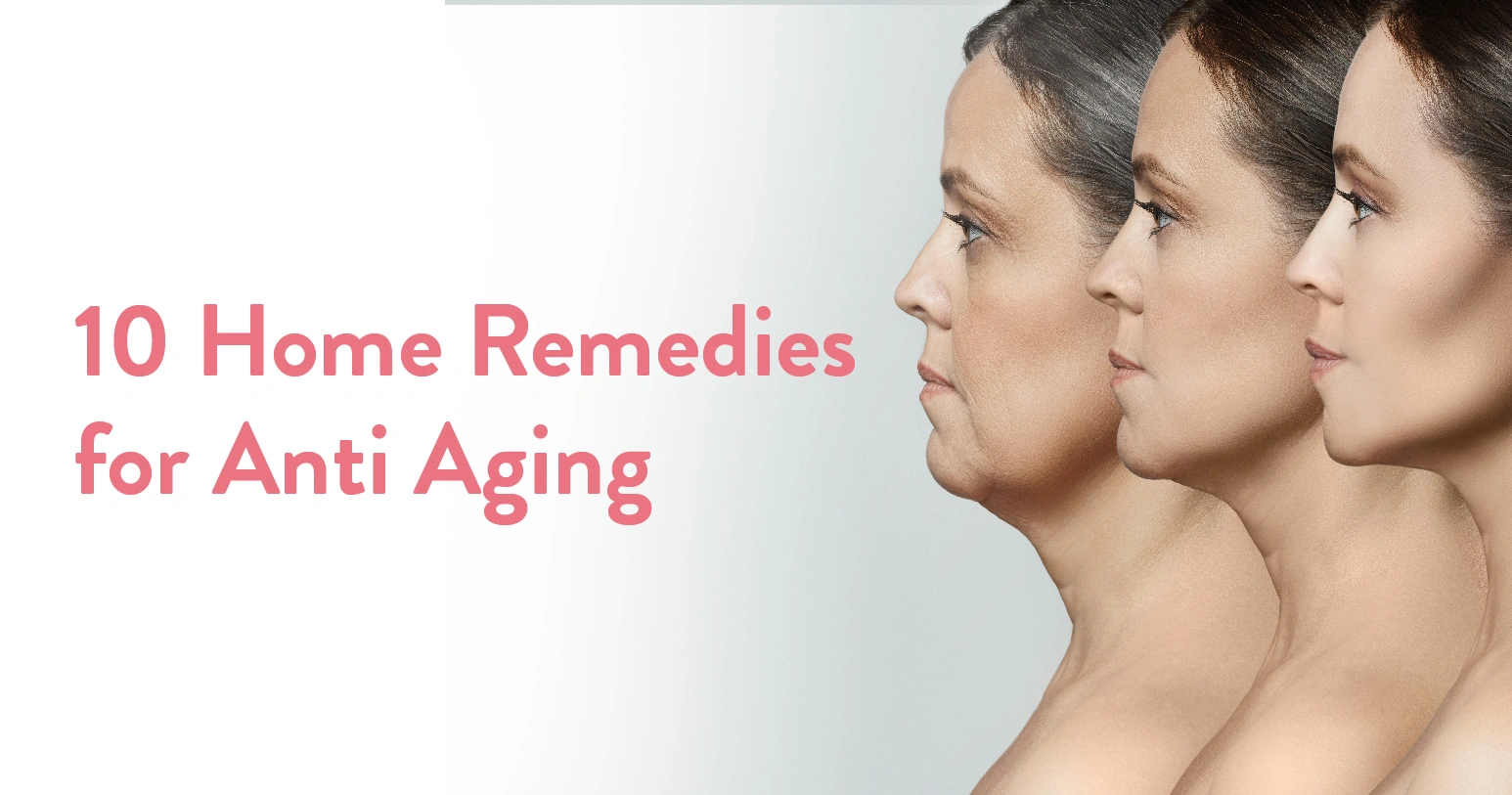
Aging is a natural part of life, but it doesn’t mean you can’t look and feel youthful for longer. Wrinkles, fine lines, and sagging skin are some of the visible signs of aging that many of us wish to delay. While dermatological treatments can be effective, many millennials are opting for home remedies to keep their skin healthy and glowing as they enter their 40s.
This blog explores the causes of wrinkles, types of wrinkles, and ten effective home remedies for combating signs of aging—all at the comfort and familiarity of your home.
Table of Contents
Wrinkles form due to a combination of intrinsic and extrinsic factors, i.e., some causes are internal to our body while others are external and hence more manageable:
Natural aging starts when the body reduces collagen and elastin production. These are the primary causes due to which our skin loses elasticity.
Ultraviolet (UV) rays damage skin cells, accelerating the aging process. So if one is exposed to sun for longer durations their skin will start showing signs of aging faster than others.
A healthy lifestyle shows up on your skin. Which is why smoking, poor diet, and lack of hydration can contribute immensely to premature wrinkles.
Repeated expressions like frowning or squinting can cause dynamic wrinkles. These are often unavoidable because people cannot consciously change their natural movements.
Sleeping on your side or stomach may lead to sleep wrinkles over time.
Surprised? Wrinkles are not just one kind. They can also be categorized into two main types. (yes, there are more than one!)
These occur due to repetitive muscle movements, such as smiling or frowning. Common examples of wrinkles caused by these are crow’s feet and laughter lines, which deepen with loss of skin elasticity and repetitive action.
These are visible even when your face is at rest and result from depletion of elasticity and collagen production internally.
Now that we know the causes and types of wrinkles, let us explore some natural remedies you can try at home.
Packed with vitamins and antioxidants, aloe vera hydrates the skin, reducing fine lines. Applying the raw aloe vera gel can help immensely with wrinkles.
Moisturizes and repairs skin, making it appear smoother and plumper. The vitamin E in coconut oil makes it excellent for dry and damaged skin.
Mash a ripe banana and apply it to your face. The vitamins and natural oils in it will help rejuvenate your skin and try to moisturize your face to its best outcome.
A natural humectant, honey locks in moisture and boosts collagen production. Although you may think it is a sticky bothersome method, honey will work wonders on tired skin textures that are breeding grounds for wrinkles.
Tighten sagging skin by applying a thin layer of egg white and rinsing after it dries.
Rich in vitamin A, it promotes skin renewal and reduces fine lines. Rosehip oil is considered the epitome of luxury skin care as many high-end skin products include this to prevent showing the signs of aging.
Lactic acid in yogurt helps remove the dead skin cells, revealing a smooth and glowing texture that is well hydrated too.
Use cooled green tea bags on your eyes to reduce puffiness and wrinkles. This is especially important if you have dark under-eye patches.
These actually do have lasting benefits as it can hydrate and soothe the skin while reducing under-eye wrinkles.
The enzymes in papaya brighten the skin, while honey keeps it moisturized. Regular use can prevent age spots and wrinkles successfully.
These 10 natural ingredients have been extensively used in various skincare products and even leading dermatologists approve of them. While these are good to use there are certain ways in which we can inculcate a good skin care habit that will ensure that tired, dull and aging skin starts looking healthier naturally. So the next question is…
Reversing aging skin requires consistent care alongside the support of natural remedies. These are a few mindful practices that over time will ensure the best outcomes:
Drinking plenty of water and fluids such as fruit juices, green smoothies etc. improves skin elasticity. Avoid alcohol as it dehydrates the body.
Include berries, nuts, and green vegetables in your diet. Try to consume seasonal fruits and vegetables to optimise the outcomes.
Most of our skin problems crop up due to the accumulation of dead skin on the surface. The best way to care for this is by removing dead skin cells using natural scrubs like sugar and honey.
Sun exposure is not completely preventable. In some cases exposure to sun may be helpful too. However care must be taken to prevent UV damage. It is advisable to use a broad-spectrum SPF to protect your skin from the ill effects.
Blood circulation is one of the best ways to ensure your skin stays healthy and has access to the benefits of all the nutrients consumed by you. Get regular facial massages to ensure proper lymphatic drainage for a tightened skin and healthy glow.
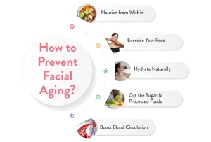
Apart from these, aging can show up on your face first. So it is also important to know how to stop your face from looking older than it should be. To slow down facial aging follow this checklist:
Blood circulation is one of the best ways to ensure your skin stays healthy and has access to the benefits of all the nutrients consumed by you. Get regular facial massages to ensure a tightened skin and healthy glow.
If you are on the other side of 40 it is imperative to follow some tried and tested methods to prevent the formation or deepening of wrinkles on your face and skin.
Cleanse, moisturize, and use anti-aging serums. Make sure you get a dermatologist to sign off on the use of active ingredient serums like vitamin C and retinol before you start using them.
Protect the delicate skin around your eyes from UV rays by wearing sunnies with good coverage. (There is a reason why movie stars wear them all the time!)
Smoking reduces collagen production, accelerating skin aging. Remember this also includes second hand smoke.
Prevent sleep wrinkles caused by friction by using materials like silk which are easy on the skin.
High stress levels can age you both mentally and physically. Take time off to look after yourself.
Home remedies for anti-aging can work wonders when used consistently and in conjunction with a healthy lifestyle. Those of you who are looking for natural solutions can rely on these remedies to keep their skin youthful and radiant without visiting a dermatologist. Remember, taking care of your skin early on is the best way to prevent wrinkles and signs of aging.
Share this blog with friends and family to help them embrace these simple, effective remedies for timeless beauty!
Home Remedies for Wrinkles Near Eyes?
Try incorporating these hacks in your daily lifestyle:
How to Remove Wrinkles From Face Quickly at Home?
There are no 100% quick fixes when it comes to improving your skin. It takes practice and patience. For improved outcomes try the following:
Are Home Remedies Effective?
Yes, home remedies can be effective for mild wrinkles and as a preventive measure. However, they work best when combined with a healthy lifestyle and consistent skincare routine. For deeper wrinkles, dermatological treatments may be more effective.

गर्भावस्था के दौरान उल्टी आना एक सामान्य लक्षण है, लेकिन कभी-कभी यह कुछ महिलाओं के लिए परेशानी का कारण बन सकता है। इस अवस्था को चिकित्सकीय रूप से मॉर्निंग सिकनेस (morning sickness in pregnancy) कहा जाता है। हालांकि, अपने नाम के विपरीत यह परेशानी केवल सुबह ही नहीं, बल्कि पूरे दिन बनी रह सकती है।
आमतौर पर गर्भावस्था के लक्षण (symptoms of pregnancy in Hindi), गर्भावस्था के छठे सप्ताह में शुरू होते हैं और चौदहवें सप्ताह तक उपस्थित रह सकते हैं। हालाँकि, कुछ महिलाओं में ये लक्षण पूरे नौ महीने तक रह सकते हैं जिसके कारण उन्हें कमज़ोरी, थकान और डिहाइड्रेशन जैसी अनेक समस्याओं का सामना करना पड़ता है।
अगर आप भी गर्भावस्था के दौरान होने वाली उल्टी से परेशान हैं तो यह ब्लॉग आपके लिए स्पेशल है। इसमें हम उल्टी को रोकने, उसे मैनेज करने या उसके प्रभाव को कम करने वाले उपायों के बारे में जानेंगे।
गर्भावस्था के दौरान महिला के शरीर में हार्मोनल परिवर्तन होते हैं जो शरीर की फंक्शनिंग को प्रभावित करते हैं जिससे उल्टी की समस्या पैदा होती है। गर्भावस्था के दौरान उल्टी होने के सामान्य कारणों में निम्न शामिल हो सकते हैं:
इन सबके अलावा, अगर गर्भ में जुड़वाँ बच्चे हैं, तो उल्टी का खतरा बढ़ सकता है। हालाँकि, कुछ घरेलू उपाय हैं, जिनकी मदद से उल्टी की संभावना को कम किया जा सकता है।
ये भी पढ़े: महिलाओं में थायराइड के कारण, लक्षण और इलाज
गर्भावस्था के शुरुआती दिनों में उल्टी होना सामान्य है, लेकिन समय के साथ यह धीरे-धीरे कम हो जाता है। अगर आप उल्टी से परेशान गयी हैं तो अपने गायनेकोलॉजिस्ट से परामर्श करने के बाद कुछ ख़ास घरेलू नुस्खों का इस्तेमाल कर सकती हैं। ये आपको उल्टी से राहत दिलाने में मदद कर सकते हैं। गर्भावस्था में उल्टी के उपायों में निम्न शामिल हैं:
अदरक का छोटा सा टुकड़ा लें, उसमें काला नमक लगाएं और अपने दांतों के नीचे दबा कर 5-10 मिनट तक रखें। अदरक में कुछ ऐसे एंटीऑक्सीडेंट मौजूद होते हैं जो मतली की भावना को कम करने में मदद करते हैं। साथ ही, काला नमक पेट और फ़ूड पाइप में मौजूद एसिड को संतुलित करता है जिससे गर्भावस्था के दौरान होने वाली उल्टी को रोकने में मदद मिलती है।
पुदीने की पत्त्तियों में मौजूद रिलैक्सिंग गुण गर्भावस्था के दौरान होने वाले मिलती और उल्टी से राहत प्रदान करते हैं। पुदीना का सेवन पाचन एंजाइम में भी मदद करता है। आप पुदीने की पत्तियों को धोकर ऐसे ही चबा सकती हैं या फिर उसकी चटनी बनाकर खा सकती हैं। आप चाहें तो पानी में अदरक और पुदीने की पत्ती डालकर उसे गर्म करके, फिर उस पानी को छानकर पी भी सकती हैं।
निम्बू में मौजूद अनेक औषधीय गुण कई तरह से एक प्रेगनेंट महिला को लाभ पहुंचा सकते हैं। उन्हीं में से एक है मितली और उल्टी से छुटकारा पाना। निम्बू काटकर उसपर काला नमक लगाएं और खाएं या फिर लेमोनेड बनाकर पी सकती हैं। निम्बू में मौजूद विटामिन सी और साइट्रिक एसिड उल्टी कम करने के साथ-साथ अपनों एनर्जी भी देता है और आपकी इम्युनिटी को बूस्ट करता है।
इलायची में कुछ ऐसे औषधीय गुण होते हैं जो पाचन एंजाइम को बेहतर बनाने के साथ-साथ पेट की समस्या जैसे कि मितली और उल्टी को रोकने का काम भी करते हैं। अगर आप गर्भावस्था के दौरान अनुभव होने वाले मितली और उल्टी से छुटकारा पाना चाहती हैं तो इलायची आपके लिए एक बेहतर विकल्प है। इलायची को चबा सकती हैं, पानी में गर्म करके पी सकती हैं या ब्लैक या लेमन टी में इस्तेमाल कर सकती हैं।
भारतीय मसले औषधीय गुणों से भरपूर होते हैं जो कई बीमारियों और स्वास्थ्य संबंधित समस्याओं में प्रभावशाली साबित होते हैं। गर्भावस्था में मितली और उल्टी जैसी समस्याओं को दूर करने का भी गुण होता है। आपको बस सौंफ का पाउडर, दालचीनी का पाउडर और जीरे का अर्क लेना है और उन्हें मिलाकर चाय बनाना और पीना है। इससे मिलती और उल्टी की समस्या मिनटों में ख़त्म हो सकती है।
गर्भावस्था के दौरान, कुछ समय के लिए उल्टी होना सामान्य है और इससे घबराने की ज़रूरत नहीं है। हालाँकि, अगर ये लगातार बने रहते हैं, ये समय के साथ गंभीर होते हैं या इनके कारण आपको कोई और समस्या होती है तो बिना देरी किए गायनेकोलॉजिस्ट परामर्श करना चाहिए।
घरेलु नुस्खों के भरोसे लंबे समय तक बैठना सही नहीं है। साथ ही, आपको इस बात का भी ध्यान रखना है कि गर्भावस्था के दौरान ऊपर दिए गए घरेलू नुस्खों का इस्तेमाल गायनेकोलॉजिस्ट से सलाह लेने के बाद ही करनी है।
आपके सम्पूर्ण स्वास्थ्य, गर्भावस्था की अवधि और उल्टी की गंभीरता आदि को ध्यान में रखते हुए डॉक्टर कुछ दवाओं के सेवन का सुझाव भी दे सकते हैं। डॉक्टर की दिशा-निर्देशों का पालन करें और खुद का ध्यान रखें और खुद में जिस भी लक्षण को अनुभव करती हैं अपने गायनेकोलॉजिस्ट से उस बारे में खुलकर बात करें।
ये भी पढ़े: एसिडिटी से तुरंत राहत पाने के घरेलू नुस्खे
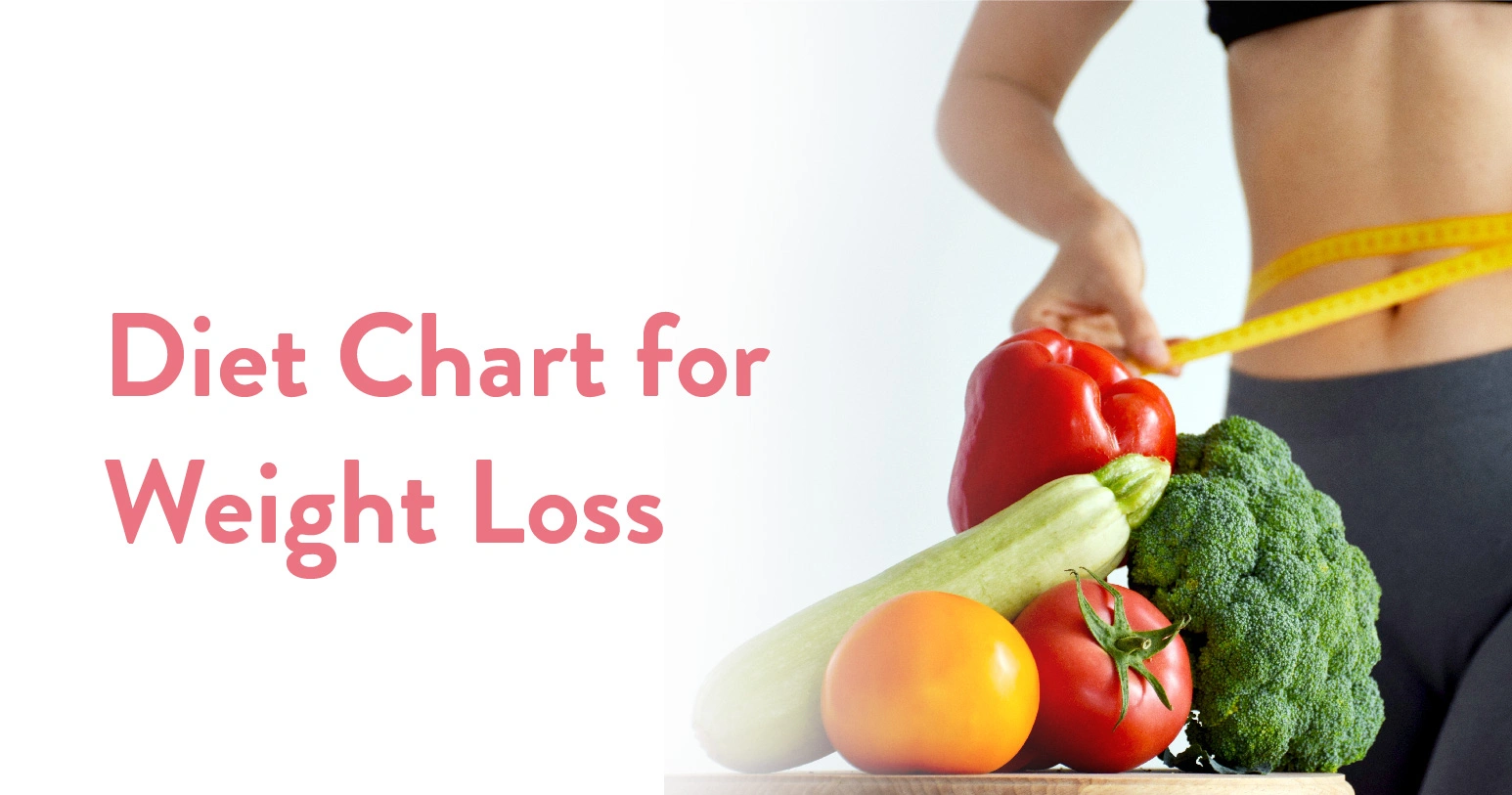
The holidays have arrived, the new year is drawing near and yes along with it the ‘new resolutions’ too! Being healthy, losing weight, and being in shape are already on the list of resolutions for many, to accomplish this goal and demonstrate it, one must adhere to a diet chart. To help maintain good health a diet chart is a guide that lists foods along with their nutritional value. A diet chart is an organized plan that guides a person on the type, quantity, and timing of food and beverages to be consumed in order to meet their health and nutritional needs.
A diet chart for weight loss is a chart tailored to an individual’s specific goals, body type, health conditions, and lifestyle. The goal of this type of diet plan is to help people lose weight by promoting calorie deficit – consuming fewer calories than the body burns – while maintaining a healthy level of nutrition.
Table of Contents
A healthy diet chart is a structured plan with a balanced combination of food required from all the major food groups ensuring the body gets the right nutrients. Proteins, carbs, healthy fats, vitamins, minerals, and other vital nutrients are all balanced in a healthy diet chart. The recommended amount of these nutrients depends on several factors including:
A balanced diet plan for weight loss promotes calorie deficit while providing the body with all essential nutrients in the right proportions. The key components of a healthy diet chart include:
Also Read: Ivy Gourd: Benefits & Side Effects
It all starts with eating the right food. But this itself sounds like a challenge to many. According to NFHS 2019-21, every one in four people in India is obese. Diet charts are tailored based on individual goals, for weight loss and weight gain the science revolves around calorie consumption and expenditure. Simply put, eating more calories than you burn or spend results in weight gain and eating fewer calories than you burn or spend results in weight loss. Making sure the diet plan is balanced is essential for healthy weight loss, i.e. all of the main food groups—fruits, vegetable grains, dairy, and proteins—are consumed in sufficient amounts.
| Category | Do’s in a Diet Chart for Weight Loss | Don’ts in a Diet Chart for Weight Loss |
| Food Choices | Eat whole, unprocessed foods | Avoid processed and junk foods |
| Portion Control | Control portion sizes | Don’t overeat, even healthy foods |
| Fiber & Nutrition | Increase fiber intake with fruits/veggies | Avoid high-calorie, low-nutrient foods |
| Macronutrients | Prioritise lean proteins | Limit refined carbs like white bread/pasta |
| Hydration | Drink plenty of water daily | Avoid sugary drinks & excessive alcohol |
| Fats Consumption | Consume healthy/good fats | Avoid unhealthy/bad fats |
| Meal Timing | Eat at regular intervals | Don’t skip meals or follow extreme diets |
It’s a common misconception that ‘if you don’t eat you will lose weight’. However, when you don’t eat you will gradually become weaker and your daily lifestyle will be affected by inadequate nutrition which can lead to health problems. It’s also common to think of following a generic diet but there is no one-size-fits-all diet plan for weight loss because each person’s nutritional needs vary depending on their age, gender level of activity, health, body weight and other factors.
The idea behind weight loss is based on the number of calories we consume and burn. If our caloric intake is less than our expenditure we lose weight. Eating a balanced diet is the top priority when it comes to diet charts, i.e. covering every food group to ensure the body receives enough nutrition while maintaining a calorie deficit. Choosing foods that are high in nutrients and low in calories and unhealthy fats is crucial when following a diet plan with the aim of losing weight. Weight control and general health maintenance can be achieved by a balanced diet that consists of lean proteins, healthy fats, whole grains and fruits and vegetables high in fibre. Avoiding processed foods, refined carbohydrates and sugary drinks helps with unnecessary calorie intake.
| Category | What to Eat | What to Avoid |
| Proteins | Lean meats, fish, eggs | Processed meats |
| Carbohydrates | Whole grain | Refined carbs |
| Fats | Good fats – olive oil, avocados | Bad fats – trans fats, fried foods |
| Vegetables | Leafy greens, broccoli | Excessive starchy vegetables |
| Fruits | Apples, berries, grapefruits | Fruits high in sugar |
The body changes as we age not only in terms of appearance but also in terms of nutritional needs, immunity, digestion, and many other aspects. Similarly, as we age our body’s calorie requirement also decreases, the fat percentage rises and our muscle mass declines. To lose weight, a calorie deficit has to be maintained, i.e. consuming fewer calories than you spend/expend. By understanding the total daily energy expenditure, one can determine the number of calories needed every day. A nutritionist or specialist then creates a customised diet plan based on the body weight and defines the number of calories needed.
An ideal diet plan for weight loss must include all the macronutrient and micronutrient components and cover every food group to ensure the body receives enough nutrition while maintaining a calorie deficit. It must be noted that there is no one-size-fits-all method for losing weight; rather there are several factors that determine an individual’s nutritional needs including age, gender activity level, health, body weight and other related factors. Thus, it is best to get customised guidance on diet plans to achieve your goals.
Chia seeds are considered a superfood because of their fibre and nutrient content. The consumption amount, frequency, and timing can be determined by an expert/nutritionist post analysing the body’s calorie requirement.
Bananas can be good for weight loss when consumed in moderation, as it is high in natural sugars & calories compared to other fruits.
It depends upon the type of rice, i.e. white rice is low on nutrition compared to brown or black rice. It can help with weight loss if consumed in controlled portions, and balanced with other nutritional-rich food.
Losing weight in 7 days requires following a strict weight-loss diet (one that helps maintain the calorie deficit), exercising, and adopting active lifestyle habits.
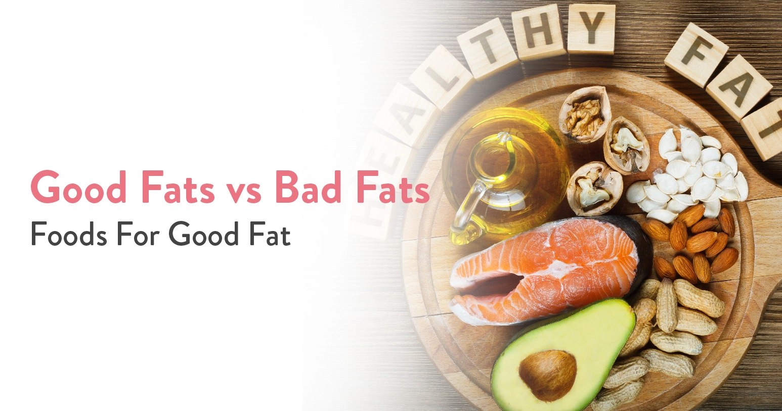
Table of Contents
The big “Fat” wedding season is back! 4.8 Million weddings are expected to take place between November – December 2024 itself. (Business Standard). You must have been invited and you must be preparing to look your best. You are most likely already on a cut or searching for a change in your diet that will help you get in the best possible shape. And like most people you have decided to steer clear from fats till you reach your desired fitness goals. But is that a winning formula? When it comes to dieting, are all fats making your dream of fitness coming true or impacting your health? The answer is no.
Not all we consume is bad. The optimal functioning of the human body depends on a variety of macronutrients and micronutrients. Macronutrients consist of carbohydrates, fats, and proteins and are required in high quantities while micronutrients include vitamins and minerals needed in relatively smaller quantities. Each of these macronutrients and micronutrients plays a unique role in maintaining health and overall well-being. Our body produces the majority of fats we need, but certain fats can only be procured through the food we eat.
Fats are an important component found in food and help provide energy to the body for normal functioning. These fats are stored in the body and used for energy when required. Fats have a variety of roles to play including,
Fats that we get from food we consume are further divided into good fats and bad fats. Good fats are healthy fats that help maintain cholesterol levels, and support the overall bodily functions, while bad fats are fats that negatively affect the health and impact the normal functioning of the body.
Not all of the fats we consume are unhealthy. Bad fats are characterised as fats that have a negative effect on health when consumed excessively or for prolonged periods.
These fats pose a threat to various organs by impacting the blood vessel flow and increase in the body’s production of cholesterol. These fats make the body more prone to heart attack, obesity, and other health issues. Two types of fats are generally bad for health:
These are usually found in animal products like butter, cheese, fatty meats, etc. Saturated fats may not pose a direct threat to health if consumed sparingly, but excess or too much consumption of these fats can lead to an increase in bad cholesterol leading to heart conditions.
Consumption of these fats has a major impact on the body as these contain hydrogenated vegetable oils, and are found in processed food, deep-fried food, etc. Trans fats like saturated fats increase the body’s production of bad cholesterol and also suppress good cholesterol levels.
Also Read: Ivy Gourd: Benefits & Side Effects
These fats as the name implies are good for the body as they help lower blood cholesterol levels. However, consuming too much of these healthy fats can also have negative health effects because they are high in calories. These fats must be consumed within limits to extract the ‘good’ out of them.
The types of good fats include:
Commonly called MUFA, these fats are found in vegetable oils that stay liquid at room temperature like peanut oil, olive oil, and other products like avocados, peanuts, etc.
PUFA or polyunsaturated fats are the ‘essential fats’ that the body cannot produce and are dependent on the food intake. These fats are usually found in fatty fish, soybeans, sunflower oils, etc.
These fats are considered the healthiest for the body and are found in fatty fish, chia seeds, flax seeds, walnuts, etc.
These good fats if consumed adequately help maintain good health and lower the level of bad cholesterol.
Good fats are an important component of a balanced diet which is directly related to overall good health of the body. Good fats have various roles to play including,
You must have come across the phrase – Anything in excess is bad, this holds true for good fats too.
Good fats play an important role in maintaining good health, but when consumed in limited amounts. Fats are high in calories, the more you intake the more the body stores it in the form of body fat which leads to weight gain and other complications. The key lies in the right amount of consumption, i.e. maintaining a balance between the calorie intake and calorie expend.
Both healthy and unhealthy fats are present in the food we eat. While bad fats have a negative impact on the body, good fats are healthy fats that promote heart and brain health and regulate bad cholesterol levels.
| Aspect | Good Fats | Bad Fats |
| Types | Unsaturated (MUFA, PUFA) | Saturated Fats, Trans Fats |
| Source | Fish, Avocados, Seeds, Nuts, etc. | Fatty Meats, Processed Food, etc. |
| Cholesterol Effect | Lowers Bad & Raises Good Cholesterol | Raises/Increases Bad Cholesterol |
| Health Impact | Improves Heart Health, Supports Brain Function. | Increases Risk of Heart Disease, Stroke, and Obesity. |
Good fats or bad fats – It doesn’t sound healthy right? After all, these are all fats. This is the common perception the majority of people have.
Good fats if consumed in moderation don’t get you fat. The best part about good fats is they help with weight control if consumed in the right quantities. Good fats must be consumed within ‘limits’ to extract the ‘good’ out of them.
Good fats are a promoter of good health and help maintain cholesterol levels. Fats are high in calories, more than the other macronutrients i.e. proteins and carbohydrates. Regardless of their nature, good or bad, fats have 9 calories per gram. These calories when consumed excessively get stored in the body as energy reserve in the form of body fat that increases body weight.
If you are concerned about whether your diet has the optimum fat percentage, feel free to reach out to our dietician and get a personalised diet chart, because every anatomy is different.
Good fats are promoters of good health if consumed in moderation. Good fats help lower bad cholesterol levels, improve heart health, and maintain overall well-being.
Saturated fats are found in animal products like fatty meats, butter, cheese, etc. Excess consumption of saturated fats can lead to an increase in bad cholesterol leading to heart conditions.
Fats are an important component found in the food we eat and help provide energy to the body for normal functioning.

Table of Contents
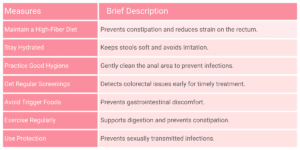
The digestive system is made up of many crucial organs, some playing a major role in digesting the food while some with managing and extracting the waste. Two such vital parts of the digestive tract are the rectum and anus. The rectum forms the last part of the large intestine and on average is sized between 10 to 15 centimeters. The primary function of the rectum is to store and release stool/waste. The food when it reaches the rectum is already in a solid waste state, the rectum absorbs the remaining water and electrolytes and extracts the waste. Rectum plays certain crucial roles including,
The solidified waste is further transferred for extraction through the anus, a part through which the waste exits the body. The anus is the opening at the end of the digestive tract and consists of sphincters that open up when there is a bowel movement.
Rectal discharge also known as anal discharge is a condition in which unusual fluids, mucus, pus, blood, etc come out of the anus. Rectal discharge can be a painful condition with common symptoms like dampness around the anus area, stains on inner wear, pain during bowel movement, abdominal cramps, foul-smelling discharge, and other similar conditions. There are several causes of rectal discharge and it can differ in color consistency and appearance.
Rectal discharge can be occasional or persistent, can vary in appearance and typically signal underlying health problems. The common causes of rectal discharge include,
Rectal discharge is a condition in which unusual fluids, mucus, pus, blood, etc come out of the anus. Rectal discharge is an indication of an infection or underlying medical condition. Below are the symptoms associated with rectal discharge:
Rectal discharge is a symptom of an underlying medical condition and can be caused by varying factors. The way rectal discharge is treated varies according to its cause. A combined approach involving medications, lifestyle changes, and sometimes surgery effectively resolves the issues.
Rectal discharge is caused by underlying medical conditions. Identification and treatment of these issues/medical conditions can help with rectal discharge. Following a comprehensive diagnosis, a specialist can treat these conditions. A combination of medication, surgery and lifestyle modifications can be an effective way to help with rectal discharge.
Yes, rectal discharge can be a symptom of colon cancer. Colon cancer can cause rectal discharge as it irritates the bowel lining, partially blocks the bowel and may cause inflammation.
Rectal discharge is caused by underlying medical conditions. The seriousness of rectal discharge depends on its cause. While some underlying conditions are easily treated others necessitate significant medical intervention.
ठंड का मौसम शुरू हो गया है और अब हर रोज टाइफाइड बुखार के मामले सामने आ रहा हैं। अगर आप टाइफाइड के बारे में विस्तार से जानना चाहते हैं तो यह ब्लॉग आपके लिए है। इस ब्लॉग में टाहम इफाइड के कारण, लक्षण और खानपान के बारे में विस्तार से जानेंगे।
Table of Contents
टाइफाइड (Typhoid) एक बैक्टीरियल इंफ्केशन है जो मुख्य रूप से साल्मोनेला टाइफी नामक बैक्टीरिया के कारण होता है। हालाँकि, यह अन्य कारणों से भी हो सकता है जैसे कि:
इन सबके अलावा, उन जगहों पर जाना जहां बैक्टीरिया मौजूद होते हैं जैसे कि लेबोरेट्री या हॉस्पिटल।
टाइफाइड होने पर आप बुखार के अलावा खुद में अन्य लक्षणों को भी अनुभव कर सकते हैं। इसमें सामान्य तौर पर निम्न शामिल हो सकते हैं:
इन सबके अलावा, आप अन्य लक्षण भी अनुभव कर सकते हैं जैसे कि चक्कर आना और चलने-फिरने में असमर्थ होना आदि। आमतौर पर टाइफाइड के लक्षण बैक्टीरिया के सम्पर्क में आने के 1-3 सप्ताह के बाद शुरू होते हैं।
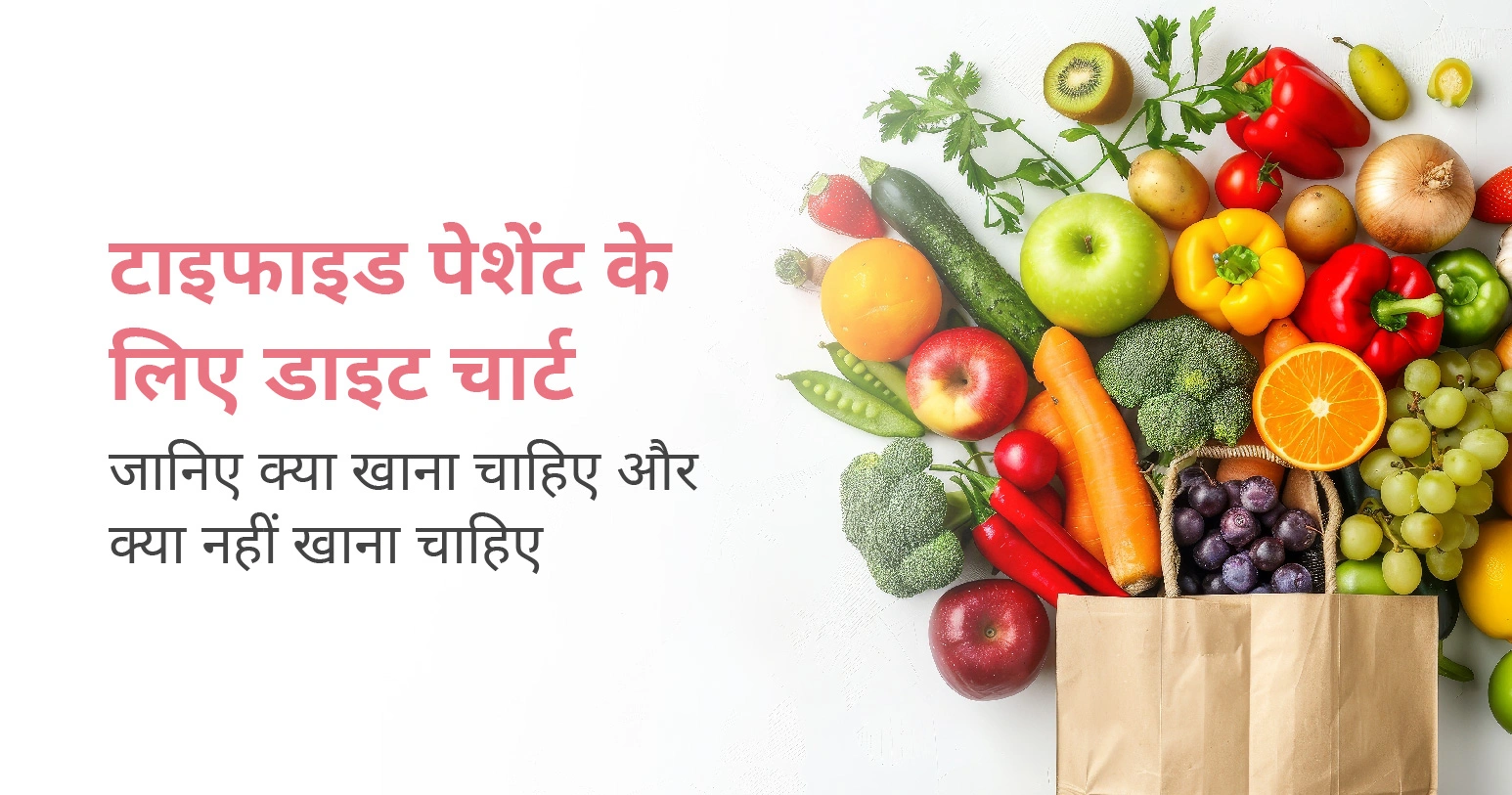
टाइफाइड में डाइट बहुत महत्वपूर्ण होता है। इसलिए इस दौरान आपको अपनी डाइट यानी खानपान का ख़ास ध्यान रखना चाहिए। टाइफाइड होने पर डॉक्टर आमतौर पर निम्न का सेवन करने का सुझाव देते हैं:
ये सब आपको आपके शरीर को आवश्यक पोषक तत्व प्रदान करते हैं जिससे कमज़ोरी दूर होती है और आपको जल्दी ठीक में मदद मिलती है।
टाइफाइड होने पर आपको कुछ चीज़ों से परहेज़ करने की सलाह दी जाती है, क्योंकि इससे आपके लक्षण बेहतर होते हैं और जल्दी रिकवरी में मदद मिलती है। इसमें मुख्य रूप से निम्न शामिल हैं:
साथ ही, मीठी चीज़ें, गाढ़ी मलाई, घी, मक्खन, चिप्स, बिस्कुट आदि का सेवन करने से बचें। खानपान को लेकर अगर आपके मन में कोई प्रश्न हो तो डॉक्टर से बात करें।
टाइफाइड से पीड़ित मरीज़ को कुछ बातों का ख़ास ध्यान रखने की आवश्यकता होती है ताकि लक्षण और खराब न हों और रिकवरी जल्दी हो। अगर आपको टाइफाइड है तो निम्न बातों का ध्यान रखें:
टाइफाइड के उपचार में दवाओं के सेवन के साथ-साथ डॉक्टर लाइफस्टाइल में भी कुछ बदलाव लाने का सुझाव देते हैं। डॉक्टर की दिशानिर्देशों का पालन करें और किसी भी तरह की कोई समस्या होने पर बिना देरी किए अपने डॉक्टर से मिलें। टाइफाइड होने पर प्लेटलेट्स बहुत तेज़ी से कम होते हैं, ऐसे में इन सभी बातों का ध्यान रखना आवश्यक है, ताकि किसी भी संभावित गंभीर समस्या को रोका जा सके।
अगर टाइफाइड के लक्षण अनुभव होने के तुरंत बाद ही उपचार शुरू हो जाए तो यह आमतौर पर 10 दिनों तक रहता है, लेकिन उपचार में देरी होने पर यह 3-4 सप्ताह या उससे अधिक समय तक रह सकता है। इस दौरान उसके लक्षण खराब भी हो सकता हैं और आपको अन्य परेशानियों का सामना करना पड़ सकता है।
टाइफाइड होने पर सीमित मात्रा में चाय का सेवन करने का सुझाव दिया जाता है। आमतौर पर इस दौरान डॉक्टर हर्बल चाय पीने की सलाह देते हैं, क्योंकि इससे शरीर का दर्द कम होता है और यह आपकी बॉडी को हाइड्रेट रखता है। चाय के अलावा, आप काढ़ा भी पी सकते हैं।
हाँ, टाइफाइड होने पर दूध का सेवन कर सकते हैं। दूध में मौजूद प्रोटीन आपको ताकत प्रदान करता है। दूध को पीने से पहले उसे हल्का गर्म कर लें। अगर आपको दूध से एलर्जी है या इससे कब्ज या गैस का खतरा है तो इससे परहेज़ करें।
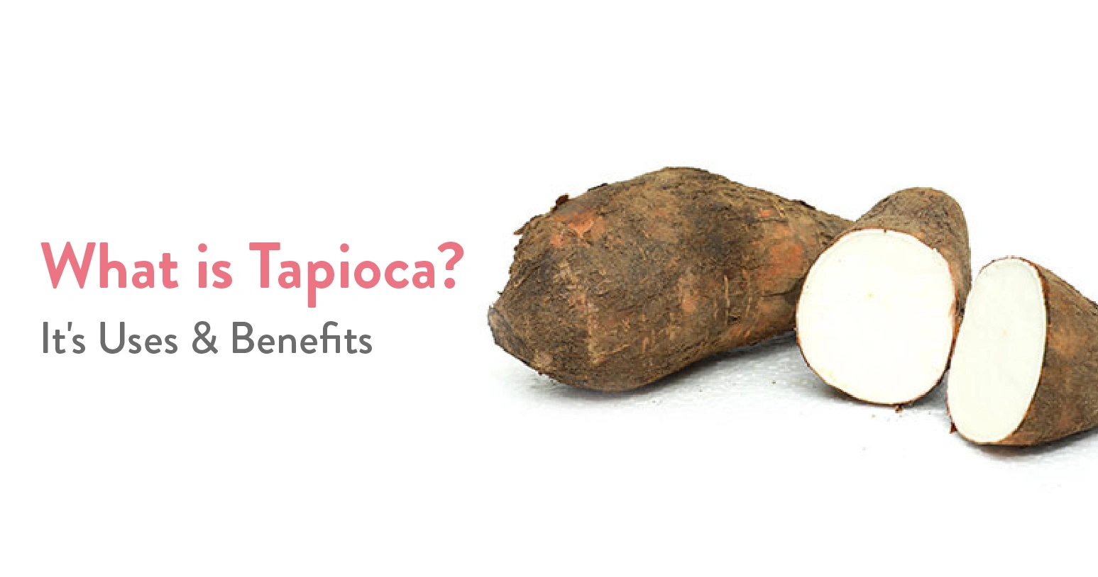
Do you love bubble tea, especially its chewy pearls? So, have you ever wondered what they are and what they are made of? They are tapioca. It is a starchy delicacy extracted from cassava root. While bubble tea has taken the world by storm, tapioca has a rich history of its own. However, besides adding a fun texture to your drink, tapioca has many more uses. In this blog, we’ll discuss what tapioca is, its benefits, and its nutritional value.
Table of Contents
Tapioca is a starch extracted from the cassava root, which contains essential nutrients, fibre, vitamins, and protein. As it has become a popular gluten-free alternative to wheat and other grains, it is being used extensively in cooking and baking worldwide.
| Baking | Since it has been proven to be gluten-free, tapioca is used in baking as a thickening agent. It helps to make light and airy textures in cakes, muffins, and pancakes. |
| Thickening Agent | It is used as a thickener in soups, sauces, and gravies. |
| Puddings | Traditional puddings are made of tapioca. It helps to improve their texture and look. |
| Bubble Tea | In bubble tea, tapioca is an essential ingredient that gives it a chewy texture, which many people enjoy. |
| Savoury Dishes | In some cultures, tapioca is used in savoury dishes like casseroles or fritters, where it helps hold the ingredients together. |
Tapioca is a starchy delicacy extracted from cassava root that has several nutritional benefits, such as:
| Nutrient | Amount per 1 cup (240 grams) | Notes |
| Calories | Approximately 330 calories | High in calories due to carbohydrate content. |
| Carbohydrates | About 80 grams | Primarily composed of starch for energy. |
| Protein | About 1 gram | Low protein content, not a significant source. |
| Fat | Less than 1 gram | Virtually fat-free. |
| Fiber | Very low | Lacks significant fibre content. |
| Vitamins & Minerals | Small amounts of iron, calcium, and B vitamins | Not a major source of vitamins or minerals. |
| Gluten-Free | Yes | It is safe for those with gluten intolerance. |
In the market, tapioca is available in various forms, such as flour, meal, flakes, and pearls. People consider it a healthy alternative to some foods because it is low in sodium and rich in calcium and iron. Let’s discuss the health benefits of tapioca that are:
One of the most significant benefits of tapioca is that it is naturally gluten-free. This makes it an excellent alternative for those with coeliac disease or gluten sensitivity. It can be used in various recipes, from baked goods to soups, without the risk of gluten-related issues.
Tapioca is rich in carbohydrates, which are the body’s primary source of energy. Consuming tapioca can provide a quick energy boost, making it a popular choice for athletes and active individuals.
Tapioca is generally low in common allergens, such as gluten, dairy, and nuts. This makes it suitable for people with dietary restrictions or food allergies.
While tapioca is low in fibre, it is easy to digest. This can be beneficial for individuals with digestive issues or those recovering from gastrointestinal illnesses. Tapioca pudding is often recommended for those with sensitive stomach.
Tapioca can be used in various dishes, from sweet desserts to savoury meals. Its versatility makes it easy to incorporate into your diet, allowing you to enjoy its benefits in many different forms.
Since tapioca is low in protein and fat, it can be included in a balanced diet without contributing to excessive calorie intake. However, it’s essential to pair it with nutrient-dense foods to create a well-rounded meal.
Although tapioca is not a significant source of vitamins and minerals, it does contain small amounts of essential nutrients like iron and calcium. Including tapioca as part of a balanced diet can contribute to your overall mineral intake.
Tapioca is a versatile ingredient extracted from the cassava root. While it’s primarily known for being high in carbohydrates and calories, it also offers several benefits, particularly for those with dietary restrictions, such as gluten intolerance. Its chewy texture makes it a popular choice in various dishes, from bubble tea to puddings and savoury meals. Tapioca can be a fun and tasty addition to your diet, especially when enjoyed in moderation.
By pairing it with other nutrient-dense ingredients and choosing healthier preparation methods, you can make the most of what tapioca has to offer. Whether you’re looking for an energy boost or a gluten-free option, tapioca has something to provide for everyone.
Tapioca is high in carbohydrates and calories, so it may not be the best choice for weight loss. However, if consumed in moderation as part of a balanced diet, it can fit into a weight management plan.
Tapioca has a high glycaemic index, which means it can cause a rapid spike in blood sugar levels. People with diabetes should enjoy it in moderation and pair it with protein or fibre to help stabilise blood sugar.
Some people may experience gas or bloating after eating tapioca, especially if they are sensitive to starches. It’s best to introduce it gradually into your diet to see how your body reacts.
Tapioca is low in potassium and phosphorus, making it a safe option for many kidney patients. However, it’s always best for individuals with kidney issues to consult their healthcare provider before making dietary changes.

दिल्ली एनसीआर में लगातार बढ़ रहा वायु प्रदुषण गंभीर स्वास्थ्य संबंधित प्रश्न खड़े कर रहा है। स्वस्थ जीवन जीने के लिए साफ़ हवा और पानी – दोनों ही अतिआवश्यक हैं। ऐसे में प्रदूषित हवा से खुद के साथ-साथ अपने परिवार वालों को बचाना चुनौतीपूर्ण हो सकता है। वैसे तो वायु प्रदुषण सभी उम्र के लोगों को प्रभावित करता है, लेकिन छोटे बच्चे और बुजुर्ग इसकी चपेट में आसानी से आ सकते हैं, क्योंकि उनकी इम्युनिटी कमज़ोर होती है।
एयर क्वालिटी इंडेक्स (एक्यूआई) को छह भागों में बांटा गया है, जिनमें से प्रत्येक में स्वास्थ्य संबंधी चिंता का एक अलग स्तर है:
ये भी पढ़े: ठण्ड में अस्थमा के मरीज़ो को क्या सावधानियां रखनी चाइये?
इस समय दिल्ली एनसीआर में वायु प्रदुषण (Air pollution in Delhi) अपनी चरम सीमा पर है। अगर एयर क्वालिटी इंडेक्स (Air Quality Index – AQI) की बात करें तो पिछले कुछ दिनों से AQI लगभग 500 के पार ही है। प्रदूषित वायु (Polluted Air) के कारण अनेक स्वास्थ्य संबंधित समस्याएं पैदा होती हैं जिसमें मुख्य रूप से सांस लेने में दिक्कत होना, एलर्जी की समस्या, अस्थमा (Asthma)और कुछ मामलों में दिल की बीमारियां (Heart Disease) आदि शामिल हैं। इस सभी और अन्य बीमारियों से बचने के लिए कुछ बातों का ध्यान रखना ज़रूरी है।
ये भी पढ़े: झड़ते, गिरते, कमजोर बालों के लिये वरदान हैं ये घरेलु उपाय
तेजी से बढ़ रहे वायु प्रदुषण (Air Pollution) से बचने के अनेक तरीके हैं जिनके बारे में हम इस ब्लॉग में चर्चा करेंगे। अगर आप प्रदूषित हवा से खुद को बचाने की सोच रहे हैं तो निम्न 5 कारगर उपायों का पालन करें:
इन सबके अलावा, खुद को प्रदुषण और ठंड में होने वाली सीजनल बीमारियों से बचाने के लिए विटामिन सी से भरपूर चीज़ों को अपनी डाइट में शामिल करें। ये आपकी इम्युनिटी को बढ़ाने के साथ-साथ आपके फेफड़ों को भी मज़बूत बनातेहैं जिससे आपको सांस लेने में किसी तरह की कोई दिक्कत नहीं होती है।
प्रदुषण का बढ़ता स्तर (Rising Pollution Levels) स्वास्थ्य के लिए कई चुनौतियाँ खड़ी कर रहा है। अगर इस स्थिति में आप खुद के साथ-साथ अपने घरवालों की सेहत का ख्याल नहीं रखते हैं तो आपको अनेक बीमारियों का सामना करना पड़ सकता है। अगर ऊपर दिए गए उपायों के बाद भी आप प्रदूषित हवा की चपेट में आ जाते हैं तो डॉक्टर से तुरंत परामर्श करें।