Filter :

Asthma is a relatively common chronic lung disease that can develop even in infancy. This chronic condition has become increasingly common in the past decade. It is believed that the increase in air pollution, environmental smoke etc. might have contributed to the rising incidence of this problem. It is believed that in India, approximately 6% of children have asthma, this statistic may not be accurate as even today, a large number of cases go unreported or undiagnosed. In this article, we will learn about Asthma in children, how to identify its early signs and the best ways to manage this condition in children.
Table of Contents
Asthma is a chronic condition which causes inflammation in the airways and lungs, especially when you inhale any “triggers”. Triggers are any stimuli which can increase the inflammation in the lungs and airways, making it difficult or impossible to breathe. These triggers range from inhaling pollen, infections, second-hand smoke etc.
Asthma can affect people of any age; however, childhood asthma has unique challenges associated with it. This is because the “patient” (child) does not really understand the condition to take precautions and care that adults with the condition would. It can also decrease the quality of life of the child as it can severely limit their lifestyles such as participation in play and sports. It can also interfere with their normal sleep. Hence as parents, it is extremely important to know the early signs of the condition and get the proper care to keep the symptoms under control. Asthma attacks can be extremely dangerous and might require hospitalisation.
Causes of asthma in children is still quite unclear. Research suggests that it could be a combination of genetic and environmental factors such as inherited allergies, parents with asthma, ethnicity, any infection affecting the airways during childhood, second-hand smoke inhalation, air pollution etc. Underlying conditions such as childhood obesity, GERD also have been linked to childhood asthma.
One of the main challenges with childhood asthma is that the child himself/herself will be unable to identify the symptoms or early signs of asthma. Many children will start showing signs of asthma before the age of five. Here are some of the early symptoms of asthma in children that you should watch out for:
These symptoms will worsen when the child is exposed to any triggers. Such instances are called asthma “flare-ups”. These triggers can be any of the following and vary from child to child:
In some cases, you may have to rush your child to the hospital for emergency treatment. If you notice the following signs, take your child to the emergency room immediately:
Asthma is a chronic condition, and treatment protocol is aimed at managing the symptoms and flareups. This includes:
If the child becomes overly dependent on short-acting medications and has to consume them too often, long term medications can be used to manage the symptoms over time. These include inhaled corticosteroids, leukotriene modifiers, long-acting beta-agonist, cromolyn and oral corticosteroids. Consult your child’s paediatrician before giving your child any type of medication, long term or short term.
Most of the aforementioned medications are generally administered via metered-dose inhalers. You can use attachments such as a valved holding chamber with a mask or a nebulizer to make it easier for your child to use the inhaler.
Read: Vaccinating your child – What you need to know?
For parents with asthmatic children, managing symptoms of asthma as well as preventing future flareups can be quite difficult. It is also extremely important to take steps during childhood itself to stop further inflammation of the airways and lungs. The following tips can help you stay ahead and let your child live and play to the fullest:
An asthma action plan will help you react faster and better to any asthma symptoms as and when they manifest. Your child’s paediatrician is the best person to help you come up with this plan. The plan should include step by step instructions about the short-term and long-term medications and when to take them, things to do between flareups, how to identify and manage triggers as well as how to identify when a trip to a hospital is required.
Diligently recording asthma attacks as well as the treatments that your child is undergoing will help you identify what triggers your child’s asthma as well as what form of treatment is working best for him/her. These records will also help your child’s doctor determine whether short-term medication or long-term medication is best suited for your child. The record should include:
Identifying and minimising exposure to all the stimuli which trigger an asthma attack in your child is perhaps one of the most essential aspects of long-term management of asthma. Taking into account the environmental factors in play whenever your child experiences an asthma attack can help identify if they are triggered by dust, pollen, animal fur, smoke, infection etc. In some cases, excessive crying or laughing can also cause an attack. The following steps can help you minimise this risk:
As your child grows older, teaching them what to stay away from, how to react to asthma attacks and precautions to take is extremely important. You can also do an allergy panel to identify what other allergens can be harmful for your child. Routine visits to the paediatrician would help in monitoring your child’s condition and prevent it from becoming worse. You can book a consultation and a comprehensive health check-up for your child under the care of our paediatric experts at the CK Birla Hospital, Gurgaon.
Read: What makes kids picky eaters and what may help them get over it

Left with a bitter taste after a great meal and don’t know why? It’s probably acid reflux. Acid reflux is an uncomfortable backflow of gastric juices up the oesophagus (food pipe). It is usually triggered by what we eat and in severe cases can even be confused for a heart attack! Thankfully, you can simply avoid certain foods to prevent acid reflux and spare yourself this extremely uncomfortable feeling.
Diet is one of the significant contributors to acid reflux. Like it or not, certain types of food, no matter how great they taste, can worsen the condition. These foods can either cause delayed stomach evacuation, decrease the tension of the lower oesophageal muscle or stimulate the production of stomach acids. Here is a list of foods to avoid to prevent acid reflux:
Read: Causes of stomach ache after eating
Chronic acid reflux can also be a symptom of GERD (Gastroesophageal reflux disease). This is a far more serious condition that occurs when the stomach acids and juices start flowing back from the stomach into the oesophagus. If you don’t control acidity right away, it can develop into GERD. You can prevent this by making acid reflux dietary modifications such as:
If your symptoms are not fading away after a few days after these dietary changes as well, you should visit your doctor to figure out what is causing these symptoms. Acidity can be extremely uncomfortable, but it is easily preventable by making healthy dietary and lifestyle choices.
Read: What to eat and avoid if you have an upset stomach
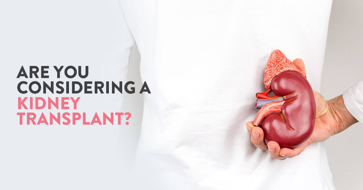
Kidneys are two bean-shaped organs, which are responsible for filtering out toxins and waste fluids from our blood. This waste is then excreted from our body in the form of urine. They are present on either side of the spine, right below the rib cage.
In some situations, the kidneys may lose their filtering ability. When they lose more than 90% of their functional ability, they are considered to be in end-stage kidney disease (renal/kidney failure). In such cases, the patient would have to undergo routine dialysis to artificially filter their blood, or get a kidney transplant. In this article, we will look closer at the causes of kidney failure, caring for a kidney transplant and what all to keep in mind when considering kidney transplants during the COVID-19 pandemic.
Table of Contents
Kidney failure can occur due to a number of reasons. Underlying health conditions which damage the kidneys over a period of time is one of the most common causes of kidney failure. Diabetes, uncontrolled high blood pressure, autoimmune diseases, genetic conditions, urinary tract problems, nephrotic syndrome are some such conditions which can damage your kidneys over time. If these conditions are not kept in check, they continue to damage the kidneys, and the patient is said to have chronic kidney disease (CKD).
In some cases, the kidneys can suddenly stop working. This is called Acute Kidney Injury (AKI) or Acute Kidney Disease (AKD). This can be the result of any trauma, heart attack, drug abuse, lack of blood supply to the kidneys and urinary tract problems. This may or may not be permanent.
Read: Chronic Kidney disease: What is it and how can it be prevented?
Some of the most common symptoms of kidney failure are
Fortunately, patients with kidney failure can still lead completely normal lives with routine dialysis or a kidney transplant.
Dialysis is a process to artificially filter the blood of waste and toxins. There are two types of dialysis: Haemodialysis and Peritoneal dialysis. Based on the type of dialysis you are recommended, you would have to undergo this treatment daily (peritoneal dialysis) or 3-4 times a week (haemodialysis).
Kidney transplant on the other hand, offers a more permanent solution to kidney failure. Kidney transplant is a surgical procedure in which one or two healthy kidneys are placed inside your body when your own kidneys are no longer functioning properly. The damaged kidneys may or may not be removed.
There are many factors to consider before deciding between kidney transplants and dialysis. These include your age, general health, lifestyle, pre-existing conditions and availability of a donor kidney. Another major thing to consider is that kidney damage may not be permanent. Your doctor will take all these factors into consideration while discussing all viable treatment options with you.
In many cases, where the kidney damage is irreversible, patients would require dialysis for the rest of their life. Kidney transplant can be a more permanent solution. Research indicates that kidney transplant is associated with a better quality of life, lower risk of death, fewer dietary restrictions and ultimately lower treatment cost (in the long run). Some people may also choose to opt for a kidney transplant before they require dialysis. This process is called a preemptive kidney transplant.
Kidney transplant is not suitable for everyone. Your doctor might rule out kidney transplant in the following cases:
Apart from the risks associated with any form of surgery such as infection, excessive bleeding, complications from general anaesthesia, etc, there is also a risk of rejection of the donor organ and side-effects of the anti-rejection medication that can occur with kidney transplant. Talk to your doctor in detail about all these risks before opting for a kidney transplant.
After the procedure is completed, you would be required to stay in the hospital under observation for several days up to a week, based on how the procedure went as well as your recovery rate. During this time, your healthcare providers would monitor you closely to ensure that you are recovering fine. In some cases, you may also require dialysis till the time your new kidneys start functioning satisfactorily. You would be advised to avoid all unnecessary physical exertion till your surgical wounds have healed completely.
After you are discharged from the hospital, you would be asked to come for routine checkups. This is just to make sure that your new kidneys are functioning right and your body is not rejecting the donor kidney(s). You would also be required to take several medications which are designed to stop your immune system from attacking your new kidneys. These medicines are called immunosuppressants.
Diet plays an important role in determining the success of your kidney transplant. It is essential to maintain healthy body weight and follow a balanced diet. The major points to keep in mind while choosing what to eat is to avoid excess salt, limit your fat intake and consume more fibre rich food. Ideally, you should have fresh fruits, vegetables, lean meat, low-fat dairy products, whole grains and lots of water. Apart from this, your doctor and/or dietician would give you a more personalised diet plan that would be best suited for you.
Because of the ongoing global pandemic, “Does transplant increase the risk of COVID-19” is one of the most common questions asked today. Due to the anti-rejection medications which are required after the transplant, there is a higher risk of any infection, including COVID-19. Hence, it is that much more essential that you follow the precautions laid out by healthcare advisories regarding COVID-19. This includes:
While these are general precautionary guidelines against COVID-19, these are especially important for people who are at a higher risk, including people who are immunocompromised, have had any type of surgery recently and any type of transplant patient.
Any individual living with a transplant patient, will need to be more cautious, limit their exposure, avoid crowds and strictly follow social distancing whenever they step outside. They will have to be diligent about their sanitisation and should never interact with the transplant patient without taking the necessary precautions. For further information regarding kidney disease and kidney transplant, you can speak to our nephrology team.
Also, read: Types of Kidney & Renal stones, and it’s symptoms

While some couples prefer to leave pregnancy to chance (“let’s just stop trying not to get pregnant and see what happens”), others find themselves meticulously planning every aspect of their pregnancy.
Whichever of these categories you fall in, there are a few things you must do if you are even thinking of becoming pregnant in the near future. These will not only help increase your chances of conceiving, but also make your pregnancy healthier. They significantly reduce the risk of pregnancy complications and aid in maintaining your baby’s health both before and after birth.
Table of Contents
10 things all couples must do before getting pregnant are:
There is no grey area with regard to smoking. If you are not a smoker, do not start. And if you are a smoker, it is always a good time to stop.
Smoking during pregnancy is a strict no-no. It has been linked to a wide range of pregnancy complications including preterm labour, premature birth, low birth weight amongst others. It also increases the baby’s chances of developing chronic conditions such as obesity later on in life.
It is a lesser known fact that smoking also has extremely adverse effects on both male and female fertility. Couples can significantly increase their chances of becoming pregnant by simply kicking this habit.
Finding a good maternity hospital can be very tough. And this decision is one of the most important ones you will make for your pregnancy.
Do extensive research into their qualifications and experience before making your choice. It is essential that you be comfortable with your doctor. You should be able to call them for any question or concern at any time without hesitation.
Try finding your doctor before your pregnancy itself. Schedule a visit with your doctor as soon you decide to start trying to conceive. They will probably assess your general health; review the medications you are currently on, test for infections and advise you to lose or gain weight as required.
Read: Early signs of pregnancy
Certain vaccines that contain live viruses are not advised for pregnant women. These include rubella and the chicken pox vaccine. However, protecting oneself against these diseases can go a long way in preventing them from causing complications in your pregnancy.
If you have never had measles, mumps, rubella or chickenpox, your doctor might advise you to get these vaccinations before you become pregnant.
During pregnancy, women often report that they can feel the effects of caffeine on their unborn child. Women who consume a lot of caffeinated products might find it tougher transitioning to “decaf” during pregnancy. Common symptoms of caffeine withdrawal include headaches, irritability and constipation. Try reducing your caffeine intake before you conceive to avoid experiencing any of these unpleasant symptoms during your pregnancy.
Unfortunately, raw meat such as sushi and certain cheeses can cause mild to severe infections. Pregnant women are advised to steer clear of unprocessed cheese and any type of raw or undercooked meat. If your favourite dish falls into either category, try weaning yourself off gradually or simply go cold turkey before your pregnancy.
Most of us confuse becoming fit with just losing weight. However, being fit and healthy is more than just losing weight. Start exercising regularly to make your body stronger for the pregnancy. It is also advised to start Kegel exercises to avoid complications such as fallen or prolapsed bladder. Remember to continue exercising throughout your pregnancy unless advised otherwise. If you are concerned about what exercises are or are not safe for your baby, consult your obstetrician.
Folic acid is probably one of the most essential and well-known vitamin supplements associated with pregnancy. Women are advised to start folic acid as early as one month before conception. It is generally consumed for the first three months of pregnancy. Folic acid is known to promote the development of your baby’s brain, skull and spinal cord. It helps prevent neural tube defects such as spina bifida in the baby.
Based on your general health, you should also consider taking iodine, iron, fish oil and vitamin D3 supplements.
For the male partner, the following supplements have been shown to have a positive impact on sperm health.
Do not wait for your baby to come before you go for a dental visit. You will not have the time. During pregnancy, your hormones will go haywire and can sometimes cause unpleasant dental issues. It is a good idea to get your teeth cleaned and healthy before you start your pregnancy.
Read: How to prepare your body for Pregnancy: A complete how-to-guide
High stress levels can severely lower your chances of becoming pregnant. This can invariably result in greater stress. Get out of this vicious cycle and start taking care of your psychological health. Try doing some breathing exercises, yoga, meditation etc. to manage stress. Some couples also find short vacations and getaways extremely effective. Also remember to talk openly to your partner as well as your doctor.
Diet plays a key role in improving one’s fertility. Both partners must be conscious about what they are consuming. You and your partner can increase the chances of conception by following a healthy balanced diet. Be sure to include plenty of whole grains, unsaturated fats and vegetable proteins. Your obstetrician will also guide you with your diet based on your nutritional deficiencies.
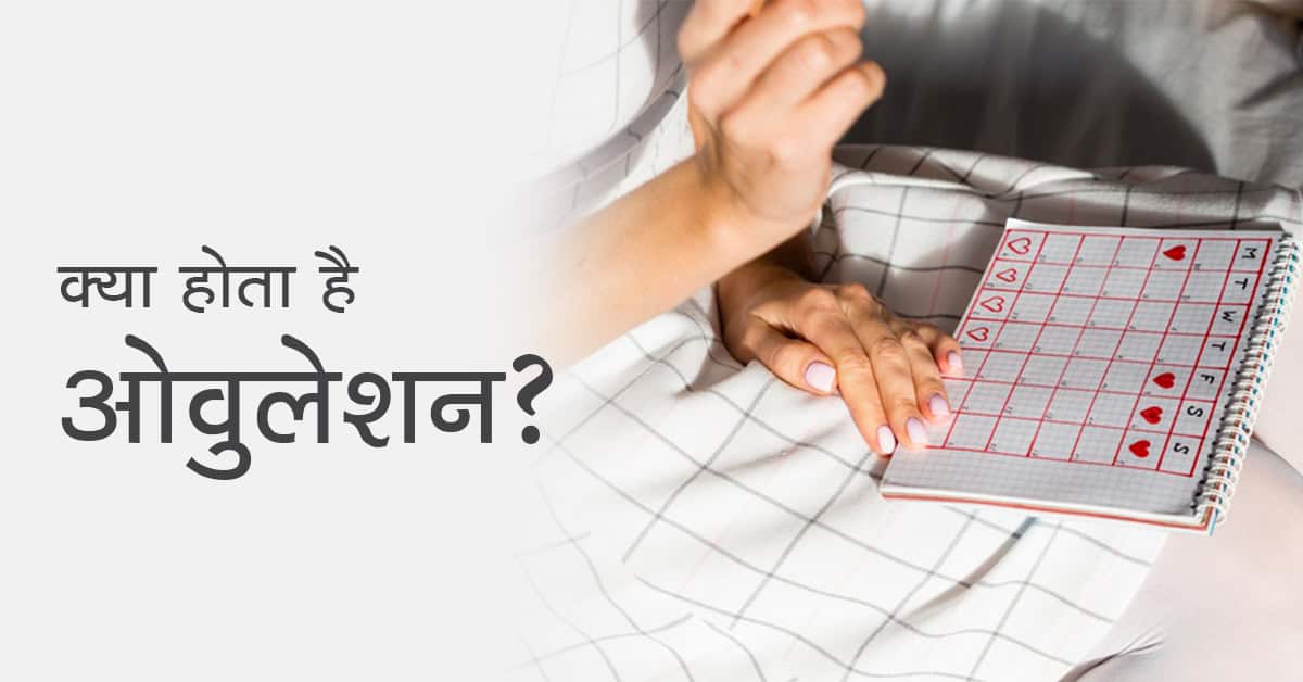
ओवुलेशन के लक्षण यदि आप माँ बनना चाहती हैं या इसके लिए प्रयास कर रही हैं, तो आपके लिए यह जानना अत्यंत आवश्यक है कि आपके मासिक चक्र यानि मेंस्ट्रुअल साइकिल के दौरान वो कौन से दिन होते हैं
Table of Contents
ओवरी से परिपक्व एग रिलीज़ होने की प्रक्रिया को ही ओवुलेशन कहते है, यह प्रक्रिया हर महीने होती है। इस समय के आसपास एक महिला के प्रेगनेंट होने की सम्भावना सबसे अधिक होती है।
आमतौर पर महिलाओं का मासिक चक्र 28 से 35 दिन का होता है, इस साइकिल में कुछ विशेष दिन ही ओवुलेशन पीरियड में आते हैं। सामन्य रूप से महिला के पीरियड्स खत्म होने के 12 से 16 वें दिन का समय ओवुलेशन पीरियड (Ovulation period) कहलाता है।
जब आपके गर्भ धारण करने की संभावना सबसे अधिक होती है, इन संभावित दिनों को ओवुलेशन पीरियड (ovulation in Hindi) कहा जाता है। यदि आप किसी प्रकार के जन्म नियंत्रण तरीके या कॉन्ट्रासेप्टिव दवाइयों का उपयोग नहीं कर रहे हैं तो आमतौर पर आपके गर्भ धारण करने की संभावना 25 से 30 प्रतिशत होती है, हालांकि यह परिस्थितियों के आधार पर व्यापक रूप से भिन्न हो सकता है।
ओवुलेशन के दौरान और उस समय के आसपास (मासिक धर्म चक्र का वह समय जब आप सबसे अधिक फर्टाइल होती हैं और निषेचन (फर्टिलाइजेशन) की संभावना सर्वाधिक होती है) आपके गर्भ धारण करने की संभावना सर्वाधिक होती है।
मासिक चक्र के दौरान जब ओवरी से एग निकलता है तब ओवुलेशन होता है। अब यह एग, स्पर्म से निषेचित (फर्टिलाइज) हो भी सकता है और नहीं भी। यदि एग फर्टिलाइज हो जाता है तो आप गर्भ धारण कर लेती हैं और यदि यह फर्टिलाइज नहीं होता है तो एग टूट जाता है और आपके मासिक धर्म के दौरान यूटेराइन लाइनिंग गिर जाती है और यह निकल जाता है।
अतः गर्भ धारण करने के लिए ओवुलेशन के संकेतों (Ovulation symptoms in Hindi) को पहचानना अत्यंत आवश्यक है।
अब प्रश्न यह उठता है कि – आपको कैसे पता चलेगा की आपका ओवुलेशन पीरियड आने वाला है या चल रहा है? यहां हमने ओवुलेशन के कुछ मुख्य लक्षण (Ovulation symptoms in Hindi) दिए हैं जिनके द्वारा आप पता लगा सकती हैं कि आपका ओवुलेशन पीरियड है या नहीं, साथ ही आप यह भी जान सकती हैं कि आप ओवुलेट कब करेंगी।
ये भी पढ़े: लिकोरिया (Likoria) का कारण, लक्षण और उपचार
डॉक्टर्स के अनुसार – महिलाओं में उनके जन्म के समय उनकी ओवरी में करीबन 2 करोड़ एग्स होते हैं और युवावस्था यानी की उनके मासिक धर्म शुरू होने के समय तक करीब 5००००० एग्स ही बचते हैं।
प्रत्येक माह मेंस्ट्रुअल साइकिल के पहले भाग में एग विकसित होता है और धीरे धीरे परिपक्व होने लगता है। पीरियड्स खत्म होने के कुछ दिन बाद महिलाओं के शरीर में एस्ट्रोजन हार्मोन बनने लगता है जो यूटेरस की लाइनिंग को मोटा करने में सहायता करता है जिससे यह स्पर्म के लिए अनुकूल वातावरण बना देता है।
धीरे धीरे एस्ट्रोजन में वृद्धि होने लगती है और यह एक अन्य हार्मोन ल्यूटिनाइजिंग हार्मोन (LH) में वृद्धि को ट्रिगर करता है। LH की वृद्धि एग के ओवरी से रिलीज होने का कारण होती है और महिला ओवुलेट (LH वृद्धि के 24 से 36 घंटे बाद) करती है।
इस दौरान यदि एग को स्पर्म मिल जाता है और स्पर्म एग को निषेचित करने में सफल हो जाता है तो महिला गर्भ धारण कर लेती है, और यदि एग फर्टिलाईज़ नहीं होता है तो यह यूटेरस की लाइनिंग में मिल जाता है और पीरियड्स के दौरान महिला के शरीर से निकल जाता है।
निषेचन के न होने की स्थिति में यह प्रक्रिया फिर से होती है जो अगले मासिक धर्म/पीरियड्स की शुरुआत होती है।
नोट: गर्भधारण के लिए आवश्यक है की आप फर्टाइल विंडो (एग रिलीज होने के बाद स्पर्म के प्रजनन मार्ग में रहने वाले 6 दिन) में ही सेक्स करें, इसमें ओवुलेशन वाला दिन और इसके बाद के 2 दिन सबसे उचित होते हैं।
जैसा की हमने बताया की यदि आप ओवुलेशन पीरियड में या उसके आस पास सेक्स करते हैं तो आपके गर्भ धारण करने की सम्भावना बढ़ जाती है, अतः आपके लिए ओवुलेशन के लक्षणों (Ovulation ke lakshan) को जानना बहुत महत्वपूर्ण है। यहाँ हमने ओवुलेशन के सात मुख्य लक्षण (Ovulation symptoms in Hindi) दिए हैं, जिनकी सहायता से आप अपने ओवुलेशन पीरियड के बारे में जान सकती हैं :
Also, read in English: Quick guide on understanding of ovulation cycle
उन दिनों के बारे में जानकर जिनमें आप सबसे अधिक फर्टाइल होती हैं, आप अपने गर्भवती होने की संभावना को बढ़ा सकती हैं। सामन्यतः एक महिला का मासिक धर्म चक्र 28 दिन लंबा होता है, लेकिन यह प्रत्येक महिला के लिए अलग हो सकता है।
प्रत्येक मासिक धर्म के दौरान लगभग 6 दिन होते हैं जब आप गर्भवती हो सकती हैं, इसे आपकी फर्टाइल विंडो कहा जाता है। ओवुलेशन कैलकुलेटर (Ovulation Calculator in Hindi) का उपयोग करके आप आसानी से जान पाएंगी कि किस दिन आपके फर्टाइल होने की सबसे अधिक संभावना होगी, इसके लिए आप किसी ऑनलाइन कैलकुलेटर या किसी मोबाईल एप्लीकेशन का प्रयोग कर सकती हैं।
यदि आप अपनी फर्टाइल विंडो/ओवुलेशन के दौरान गर्भधारण कर लेती हैं तो आप निम्नलिखित लक्षणों को महसूस कर सकती हैं:
याद रखें, जब आप गर्भधारण का प्रयास कर रही हों तो धैर्य और दृढ़ता महत्वपूर्ण है, साथ ही इसकी भी कोई गारंटी नहीं है कि आप निश्चित रूप से गर्भ धारण कर ही लेंगी भले ही आप ओवुलेट कर रही हों। लेकिन इन सामान्य ओवुलेशन के लक्षणों (Ovulation symptoms in Hindi) पर नजर रखना अत्यंत आवश्यक होता है और लगातार प्रयास करते रहें।
यदि आपको ओवुलेशन (Ovulation in Hindi) नहीं होता है तो शीघ्र ही अपने नजदीकी हॉस्पिटल जायें और स्त्री रोग विशेषज्ञ से सलाह लें। इसका कारण PCOD या कोई अन्य इनफर्टिलिटी सम्बंधित कारण हो सकता है।
इसे भी पढ़ें: जानिए क्या होता है IVF (IVF kya hai) और कैसे की जाती है IVF प्रक्रिया
1. अंडा फटने के बाद कितने दिन हम गर्भवती प्राप्त करने की कोशिश कर सकते हैं
आपके अंडाशय(ovary) से एक परिपक्व अंडा निकलने के बाद, यह फैलोपियन ट्यूब में जाता है जहां इसे शुक्राणु के साथ निषेचित(fertilized) किया जा सकता है। आप गर्भधारण करने के लिए अंडे के निकलने के 12-24 घंटों के भीतर गर्भधारण करने की कोशिश कर सकती हैं।
2. पीरियड के कितने दिन बाद ओवुलेशन होता है
यदि आपका मासिक धर्म (पीरियड) नियमित है, तो आपकी अगली माहवारी शुरू होने से 12-14 दिन पहले आपका ओव्यूलेशन शुरू हो जाता है।
3. फर्टिलाइजेशन के लक्षण क्या हैं?
ओव्यूलेशन की पहचान सर्विकल डिस्चार्ज में बदलाव, यौन इच्छा में वृद्धि, स्तन कोमलता और शरीर के बेसल तापमान (बी.बी.टी) में वृद्धि से होती है।
4. महिलाओं में अंडा कब बनता है?
आपके मासिक धर्म (पीरियड) के 6-14 दिनों के बीच एक अंडा परिपक्व होना शुरू हो जाता है।

A lot of us today, regardless of age, are involved in sports to stay fit and active. Due to our hectic schedules during the week, many of us fall in the “weekend warrior” category. Basically, we indulge in physical activity during weekends and holidays. In such cases, due to the fact that our bodies are not used to regular physical activity, performing sports without adequate warm-up and training can result in injuries. Foot and ankle regions are one of the most common areas of sports injuries. According to American Academy of Orthopaedic Surgeons, it accounts for 25% of athletic injuries.
In our country, sports injuries are commonly seen in sports such as running, soccer, badminton, cricket and basketball. Most of these injuries are caused by accidents and trauma. However, poor training practices, improper equipment, insufficient warm up and lack of condition can also result in serious injuries.
It is hence extremely important that one should warm up before starting any type of sport, wear shoes that are designed for your foot type and change them when they wear out. Proper conditioning of the muscles by gradually increasing intensity of the sport as well as playing in appropriate conditions (such as running on an even surface) can also significantly reduce the risk of injury.
While sportspersons can suffer from a wide range of orthopaedic problems based on the type of sport they play. There are some injuries that are more common, especially in athletes. Here are the 5 most common foot and ankle injuries that sportspersons experience:
The ankle is made up of complex connections between tiny delicate bones and soft tissues (muscles, tendons and ligaments) that keep the joint in its place. Ankle sprains are considered to be one of the most common injuries seen amongst athletes. Sprains generally occur due to the twisting movement of the foot resulting in damage to the ligaments present in the ankle. Ankle sprains are further categorised into two types: Inversion and Eversion.
Amongst the two, inversion sprains are more common and involved the inward twisting of the ankle. In such cases the ligaments on the outer aspect of the ankle are damaged. On the other hand, eversion sprains are caused by the outward twisting of the ankle, affecting the ligaments on the inner side. These are significantly rare as compared to inversion sprains.
Most sprains are minor and require rest, ice and protection to heal. In some cases, however, lack of treatment can result in lifelong pain and disability. Especially if the ankle isn’t healing correctly. In such cases, it is essential to consult a foot and ankle specialist.
Read: Foot and ankle pain: everything you need to know
Achilles is the largest tendon present in the body. It runs from the back of the leg to the heel. Achilles tendinitis is a condition that affects this tendon. It usually occurs due to exertion and extreme stress and hence is common among sportspeople. It can also be caused due to aging and degeneration. It presents as pain and swelling in the back of the heel that can make it difficult for the affected person to walk and run. Patients suffering from achilles tendinitis are also prone to develop a tear in the achilles tendon. This condition can be effectively managed with rest, ice, analgesia, appropriate footwear and physical therapy.
As the name suggests, stress fractures are fine fractures or cracks that are caused by repetitive force or overuse. They have commonly seen in runners as well as people engaging in sports that involve the legs such as dancing, gymnastics etc. The main reason behind this is the fatigue of bones in the ankle and feet due to excessive repeated loading. The bones in the feet (metatarsals) are most commonly affected bones when it comes to stress fractures. Patients with stress fractures can experience soreness, pain and bruising over the area of stress without any history of trauma. Treatment of stress fractures involves immobilising the foot with the help of plaster/boot as well as staying away from exertion till the fracture heals.
Plantar fasciitis occurs when excessive stress is placed on the plantar fasciitis (band of tissues supporting the arch of the foot). It causes inflammation in the tissues resulting in pain at the bottom of the heel. Many patients complain of severe pain in the heel after the first steps taken in the morning or after long periods of inactivity. In many cases, a heel spur can be seen on the X-ray. Ice, heel pads in shoes and physical therapy involving calf and plantar fascia stretches are used to manage this condition.
Also, read: Severe pain in heels during the morning: do I have Plantar Fasciitis?
Another common sports-related injury that occurs amongst sportspeople is the “turf toe”. Turf toe is a term used to describe a sprain in the ligaments of the big toe. This medical condition can occur when the heel is lifted off the ground and repetitive stress is applied to the big toe due to its hyperextended position. It was so named as it was commonly seen amongst American footballers who used to play on artificial turfs. However, it is also commonly observed in players of other sports such as basketball, gymnastics, wrestling and dance. It results in pain, swelling and stiffness in the base of the big toe that can manifest either after a sudden trauma or with repetitive loads. Doctors generally treat this condition with rest, anti-inflammatory medication, ice and taping.
It is easy for most of us to overlook problems with our feet. In reality, most conditions involving the feet manifest gradually over a period of time. If they are not caught early on and give the proper treatment, it can worsen to severely impact the quality of life. So, pay attention to your feet and make sure you are wearing the footwear designed for the sport to minimise the risk of injury.
If you notice any of such condition, do not ignore, consult the best orthopaedics doctor in Gurgaon today or book your visit to the CK Birla Hospital for your foot or ankle related problems.
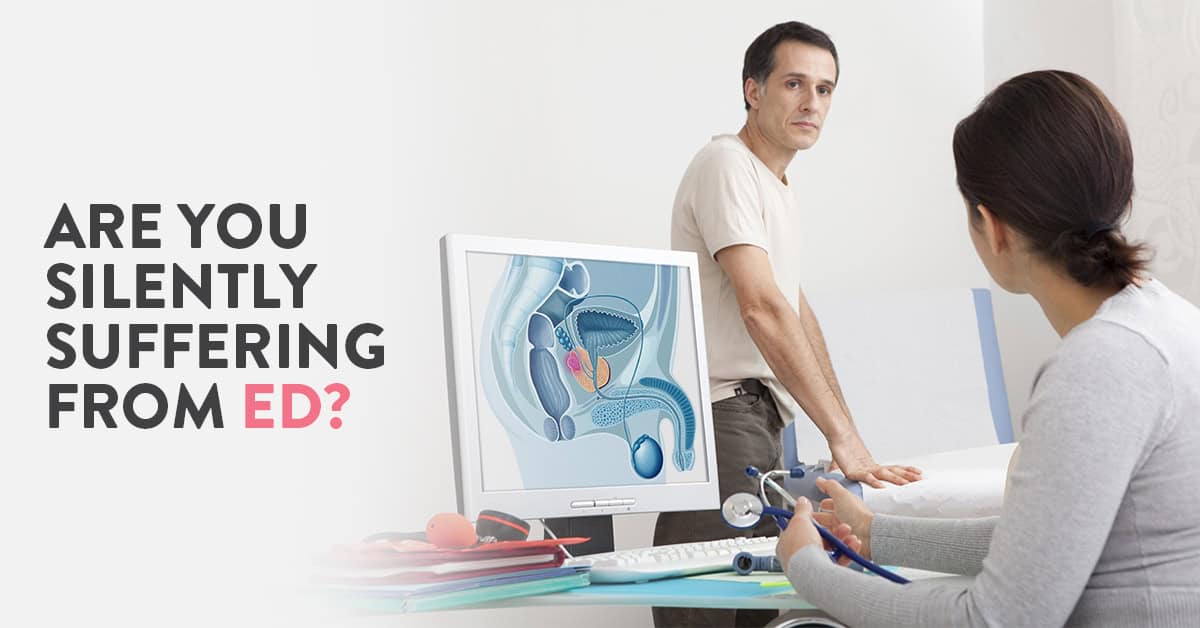
Many men may experience difficulty in getting aroused for sexual intercourse once in a while at some point of their lives. Having trouble with erection from time to time is not necessarily something to worry about. However, if the trouble persists, it might indicate erectile dysfunction or impotence.
Table of Contents
Erectile dysfunction or impotence is defined as the inability to get and keep an erection firm enough to engage in sexual intercourse. It can be a short or long-term problem. If you experience any of the following symptoms, you should get a medical consultation to rule out ED.
For a very long time, ED or impotence was brushed under the carpet as patients found it difficult and embarrassing to open up to their doctors, family or friends. “How to manage erectile dysfunction?” this is the burning question that many afflicted men find tough to ask. In this article, we answer some common questions about this sexual disorder and explore medical treatments and home remedies for erectile dysfunction.
Sexual arousal is an extremely complex process. Hormone levels, brain function, emotional state, muscle strength are just some of the factors which impact erections. Erectile dysfunction can be caused due to health conditions, lifestyle habits, psychological issues or a combination of the above.
In order to identify the cause of ED in any patient, the causes of ED are classified into two broad categories: Physiological causes of ED & Psychological causes of ED.
Read: Staying ahead of Urinary Tract Infections
Treatment protocol for erectile dysfunction involves treating the underlying cause of ED. Basically, erectile dysfunction cure depends on what is causing the condition in that particular individual. The cause is identified with the help of detailed medical examination, including the patient’s medical history and psychological analysis.
Depending on the cause of ED, the doctor can recommend any of the following treatments:
Kegel exercises are known to have a positive impact on erectile dysfunction. They help in strengthening pelvic floor muscles. In men, strengthening these muscles stimulates blood flow into the penis when sexually aroused, resulting in longer and more satisfying erections. Ideally, Kegel exercises should be performed multiple times every day.
Other exercises for ED include Pilates and aerobic exercises such as boxing, cycling, running etc. While Pilates strengthens your core muscles, aerobic exercises help in maintaining your cardiac health and improving blood circulation in the body.
Exercise also helps in regulating the mood. Hence, it is especially important for people facing problems with erections due to any psychological issues such as depression or anxiety.
“You are what you eat”, this stands true for all of us. Studies show that consuming more natural foods such as vegetables, fruits, fish, whole grains, etc and limiting your consumption of processed foods and meat can reduce the risk of having erectile dysfunction.
Being excessively sedentary, smoking, use of narcotic substances, consumption of too much alcohol etc are all lifestyle factors that can contribute towards increasing the likelihood of erectile dysfunction. If you consume tobacco in any form, quit now. If you are obese or overweight, getting your weight under control can also help in preventing erectile dysfunction. Follow a healthy balanced diet, exercise and keep your body physically fit to lead a healthy life.
For additional information about any men’s health condition, you can speak to our expert urologist & andrologist. Book your visit today at the CK Birla Hospital, Gurgaon.
Also, read: Premature ejaculation-causes and symptoms

वर्ष 2017 में माइक्रोसॉफ्ट के सीईओ (CEO) सत्या नडेला की किताब हिट रिफ्रेश प्रकाशित हुई थी जिसमे उन्होंने अपने सेरेब्रल पाल्सी से जूझ रहे बेटे से जुडी कई बातों का उल्लेख किया था।
हमारे आस पास भी बहुत से ऐसे बच्चे देखने मिल जाते हैं जो शारीरिक या मानसिक रूप से स्वस्थ नहीं होते या सेरेब्रल पाल्सी (Cerebral Palsy in Hindi) नामक बीमारी से जूझ रहे होते हैं।
सेरेब्रल पाल्सी एक न्यूरोलॉजिकल डिसऑर्डर होता है जो बच्चों की शारीरिक गति, चलने-फिरने की क्षमता को प्रभावित करता है। दरअसल, सेरेब्रल शब्द का अर्थ मस्तिष्क के दोनों भागों से होता है और पाल्सी शब्द का अर्थ शारीरिक गति की कमजोरी या समस्या से है। यह एक तरह की विकलांगता है जिसमे बच्चो को वस्तु पकड़ने और चलने में समस्या होती है।
यह रोग मस्तिष्क के किसी हिस्से में चोट लगने के कारण होता है। बच्चों में होने वाले इस रोग के विषय में और जानकारी देने के लिए हमारे बाल एवं अस्थि रोग विशेषज्ञ डॉ प्रवीण तित्तल ने इस बीमारी के विषय में कुछ खास जानकारी साझा की है, आइये जानते हैं क्या होती है यह बीमारी (Cerebral Palsy in Hindi) और किस प्रकार यह बच्चों के जीवन को प्रभावित कर सकती है।
Table of Contents
Cerebral Palsy in Hindi: सेरेब्रल पाल्सी बच्चो में उनके मस्तिष्क और मांसपेशियों से जुड़ी समस्या होती है जो लगभग तीन साल से अधिक उम्र के 1,000 में से लगभग 2 से 3 बच्चों को होती है।
एक सर्वे के अनुसार देश में करीब 500,000 बच्चे एवं वयस्क इस बीमारी से जूझ रहे हैं। यह बीमारी मस्तिष्क के कुछ भाग में चोट लगने के कारण होती है जो बच्चों में होने वाली मोटर डिजीज में से सबसे आम बीमारी है।
यह बीमारी संक्रामक नहीं होती है और न ही प्रोग्रेसिव होती है, जिसका मतलब होता है – समय के साथ इस बीमारी के लक्षण न हीं बढ़ते हैं और न ही बिगड़ते हैं। हालाँकि ये लक्षण सभी बच्चों में अलग अलग हो सकते हैं और लक्षणों और स्थिति की गंभीरता के आधार पर ही यह निर्धारित किया जा सकता है कि किस बच्चे को किस प्रकार की सहायता की आवश्यकता होगी।
उदाहरण के लिए, यदि किसी बच्चे की स्थिति गंभीर है तो उसे विशेष सहायता और उपकरणों की आवश्यकता हो सकती है वहीं कम लक्षण वाले बच्चे को अपेक्षाकृत कम सहायता की आवश्यकता होगी।
डॉ गौरव जंडियाल के अनुसार – अक्सर बहुत से माता पिता इस बीमारी के कारणों और कारकों को जानने के लिए, या यह बीमारी कभी सही हो सकती है या नहीं ऐसे ही बहुत से प्रश्नों के साथ आते हैं।
पहले, कुछ डॉक्टर्स ऐसा मानते थे कि सेरेब्रल पाल्सी होने का मुख्य कारण गर्भाशय में बच्चे को ऑक्सीजन की सही मात्रा न मिल पाना था किन्तु नई रिसर्च के अनुसार बहुत कम ऐसे मामले होते हैं जिनमे बच्चे को आक्सीजन न मिल पाने के कारण उन्हें सेरेब्रल पाल्सी की समस्या होती है।
सेरेब्रल पाल्सी से ग्रसित सभी बच्चे जन्म से ही इस बीमारी के साथ पैदा नहीं होते, कुछ मामलों में बच्चे जन्म के कुछ समय बाद उनके मस्तिष्क के विकास की अवधि के दौरान इस बीमारी से ग्रसित होते हैं। सेरेब्रल पाल्सी होने के कुछ कारण इस प्रकार हैं:
लक्षणों और मस्तिष्क के प्रभावित भाग के आधार पर सेरेब्रल पाल्सी को निम्न चार प्रकारों में विभाजित किया गया है:
यह प्रकार सबसे सामान्य प्रकार होता है जिस कारण अधिकांश मामलों में पाया जाता है। स्पासटीसिटी सेरेब्रल पाल्सी वाले लोगों की मांसपेशियाँ काफी टोंड और अधिक होती हैं जिस कारण उनकी माँसपेशियाँ काफी अकड़ी हुई रहती हैं जिससे उन्हें चलने फिरने में कठिनाई का समना करना पड़ता है। स्पासटीसिटी सेरेब्रल पाल्सी को भी प्रभावित अंगों के आधार पर पुनः 3 भागों में बांटा गया है:
यह सेरेब्रल पाल्सी का दूसरा सबसे आम प्रकार है और इसमें कई लक्षण शामिल हैं जैसे कि डायस्टोनिया (बार बार बोलना या बोलने में परेशानी होना), एस्थेटोसिस (राइटिंग मूवमेंट्स), और कोरिया (सही से चला न पाना)।
ऐसे लोगों के लिए चलने की गति या तो धीमी हो सकती है या बहुत झटकेदार और बहुत तेज भी हो सकती है। यहाँ तक की कुछ मामलो में रोगी का चेहरा और जीभ भी प्रभावित हो सकती है जिस कारण बात करने, खाने में समस्या हो सकती है।
यह प्रकार सबसे कम सामान्य है, जो पीड़ित को सामंजस्य में बाधा उत्पन्न करता है। इस प्रकार के सीपी से पीड़ित व्यक्ति को कचलने, बोलने, खाने, या अचानक गति करने में बहुत ही कठिनाई हो सकती है।
यह प्रकार उस समस्या को संदर्भित करता है जहां व्यक्ति के लक्षणों में दो या दो से अधिक लक्षणों का मिश्रण होता है।
बहुत से माता पिता अपने बच्चों की मांसपेशियों की गतिविधियों के बारे में चिंतित रहते हैं की कहीं उनके बच्चे को किसी प्रकार की विकृति तो नहीं है, इसका निदान करने का सबसे उत्तम तरीका है अपने बच्चे की मांसपेशियों का ध्यान रखना की किसी प्रकार का विकार या चलने फिरने में कोई परेशानी तो नहीं है। इसके अलावा कुछ चीजें जिनका विशेष ध्यान रखना चाहिए वो इस प्रकार हैं:
सेरेब्रल पाल्सी या CP के लिए कोई विशेष परीक्षण नहीं है, लेकिन कुछ टेस्ट्स ऐसे हैं जिनके द्वारा यह बताया जा सकता है कि आपके बच्चे को CP है या नहीं। टेस्टिंग के कुछ सामान्य तरीकों में निम्न शामिल हैं:
ब्रेन स्कैन- एमआरआई, क्रेनियल अल्ट्रासाउंड।
इलेक्ट्रोएन्सेफलोग्राम (ईईजी)- यह आमतौर पर उन बच्चों में किया जाता है जिनको दौरे पड़ते हैं, यह टेस्ट इलेक्ट्रोडों की एक श्रृंखला का उपयोग करके आपके बच्चे के मस्तिष्क की गतिविधियों को रिकॉर्ड करता है।
लेब टेस्ट- रक्त और मूत्र परीक्षण आनुवंशिक और चयापचय संबंधी समस्याएं को समाप्त करने में मदद कर सकते हैं।
विविध परीक्षण- यह बताने के लिए कि सेरेब्रल पाल्सी से शरीर का कौन सा अंग प्रभावित हुआ है, आपको उन विशेषज्ञों के पास भेजा जा सकता है जो आपको निम्नलिखित का निदान करने में मदद कर सकते हैं:
इसके अलावा, कोई भी परीक्षण बच्चे के गर्भ में होने के दौरान सेरेब्रल पाल्सी या मस्तिष्क पक्षाघात का पता नहीं लगा सकता।
यद्धपि सेरेब्रल पाल्सी का इलाज पूरी तरह से बच्चे को ठीक नहीं कर सकता है, लेकिन यह निश्चित रूप से कई तरीकों से बच्चे के विकास में मदद कर सकता है। दवाइयां, सर्जरी और थेरेपी इस बीमारी के इलाज का सबसे सामान्य रूप है। मांसपेशियों की रिहाई से संबंधित सर्जरी, टेंडन रिलीज, हिप डिस्लोकेशन, या स्कोलियोसिस इत्यादि कई मामलों में बच्चों के लिए सहायक हो सकती हैं। थेरेपी जैसे एक्वा, म्यूजिक, बिहेवियरल, फिजिकल और बॉवेल इत्यादि भी बच्चों में कमजोरी को कम करने में काफी सहायक होती हैं।
बोटोक्स और प्लास्टर जैसे गैर-पुरातन तरीके भी आज सेरेब्रल पाल्सी को सही करने में काफी योगदान देते हैं। बोटोक्स को मांसपेशियों में लगाने से यह मांसपेशियों की अकड़न को दूर करता है और उन्हें उन्हें सहनशक्ति प्रदान करता हैं। जबकि प्लास्टर बच्चे के निचले अंग के विस्तार तालमेल को रोकने में मदद कर सकता है जो बाद में सामान्य प्रकार से चलने फिरने में मदद कर सकता है और मांसपेशियों में तनाव को कम करता है।
सेरेब्रल पाल्सी से जुड़ा एक आम मिथक है कि यह बच्चे की जीवन प्रत्याशा को प्रभावित करता है। यहाँ हम आपको बता दें कि यह कथन असत्य है। बहुत से बच्चे जो सेरेब्रल पाल्सी से ग्रसित होते हैं उनकी जीवन प्र्तयशा भी सामान्य बच्चो के समान ही होती है। सेरेब्रल पाल्सी जीवन प्रत्याशा में बाधा नहीं डालता है बशर्ते आप अपने बच्चे को सर्वोत्तम चिकित्सा सहायता प्रदान करते हैं।
यदि आप अपने बच्चे को सेरेब्रल पाल्सी से ग्रस्त पते हैं तो आज ही सी के बिरला हॉस्पिटल आयें और हमारे बाल एवं अस्थि रोग विशेषज्ञ डॉ प्रवीण तित्तल से संपर्क करें।
इसके बारे में भी पढ़ें: क्या क्लबफुट का स्थायी इलाज संभव है?

“It’s your third trimester? Your baby is the size of a peach now!” Fruits and vegetables have been used to describe foetal growth throughout pregnancy for quite some time now. While this is a useful way to visualise the baby’s growth for most of us out there, your obstetrician has a more accurate form of measurement to monitor the growth of your little one. This measurement metric is called the “fundal height”.
According to the world health organisation, fundal height is a commonly practised method of foetal growth assessment. The measurement is defined as the distance in centimetres from your pubic bone to the top of your uterus. It is helpful in determining if the baby is small for its gestational age.
After completing 24 weeks of pregnancy, the fundal height should roughly be equal to the week of gestation, i.e. If you are 27 weeks pregnant, fundal height should ideally be 27 centimetres. As you approach your due date, your baby would begin descending into your pelvis in preparation for birth hence, the fundal height would start decreasing.
Your doctor would then compare your fundal height with the standard fundal height. If your fundal height measures smaller or larger than expected, or the rate of increase is more or less than expected, it could indicate
Based on what your doctor observes, you would be asked to undergo an ultrasound to determine the cause of the irregularities and would monitor your pregnancy closely.
It is important to remember that fundal height is not an exact science. Fundal height measurement is less accurate for women with a body mass index (BMI) greater than 30. Researchers are still trying to determine how effective it is in detecting intrauterine growth restriction.
Read: Is it safe to drink saffron (kesar) milk during pregnancy & will it influence my baby’s complexion?
Ques: What is my fundal height at 29 weeks of pregnancy?
After 20 weeks of pregnancy, your fundal height in centimetres should roughly be equal to the number of weeks you complete in your pregnancy. Basically, if you are in the 29th week of your pregnancy, your fundal height would be approximately 29 centimetres. As you get closer to your due date (37-40 weeks of pregnancy), your fundal height will start decreasing as your baby starts descending into your pelvis.
Ques: Is fundal height accurate?
Fundal height measurement is helpful in determining if the foetal growth is normal or not. However, it is not an exact science. Body mass index is also a factor in the accuracy of the fundal height. Your doctor might recommend an ultrasound to get a more accurate picture of foetal growth.
Ques: When can I start measuring my fundal height?
Your doctor would start recording your fundal height after you complete 20 weeks of pregnancy. In case there are any inconsistencies that are observed, your doctor would monitor your pregnancy more closely and recommend further diagnostic tests to determine the causes behind the irregularities.
Read: A guide to increasing baby weight when 9 months pregnant

