Filter :

Table of Contents
पित्ताशय की पथरी छोटे, कठोर जमाव होते हैं जो पित्ताशय में बनते हैं, जो यकृत के नीचे स्थित एक नाशपाती के आकार का अंग है। ये पत्थर आकार और संरचना में भिन्न हो सकते हैं, सबसे आम प्रकार कोलेस्ट्रॉल पत्थर और रंगद्रव्य पत्थर हैं। पित्त में पथरी उन पदार्थों में असंतुलन के कारण विकसित हो सकती है जो पित्त बनाते हैं, वसा के पाचन में सहायता के लिए यकृत द्वारा उत्पादित एक पाचन तरल पदार्थ।
ये भी पढ़े: पेट दर्द (Stomach Pain)के कारण, लक्षण और घरेलू इलाज
पित्त पथरी का निर्माण अक्सर पित्त के घटकों, अर्थात् कोलेस्ट्रॉल, पित्त लवण और बिलीरुबिन में असंतुलन के कारण होता है। कोलेस्ट्रॉल की पथरी तब बनती है जब पित्त में कोलेस्ट्रॉल की अधिकता हो जाती है, जो इसे तरल अवस्था में रहने से रोकती है।
दूसरी ओर, रंगद्रव्य की पथरी अतिरिक्त बिलीरुबिन के कारण बनती है, जो लाल रक्त कोशिकाओं के टूटने से उत्पन्न एक अपशिष्ट उत्पाद है। अन्य जोखिम कारकों में मोटापा, तेजी से वजन कम होना, गर्भावस्था, गतिहीन जीवन शैली और कुछ चिकित्सीय स्थितियाँ शामिल हैं।
पित्ताशय की पथरी से पीड़ित बहुत से व्यक्तियों में लक्षण नहीं होते हैं और उन्हें पता ही नहीं चलता कि उनमें यह पथरी है। हालाँकि, जब लक्षण प्रकट होते हैं, तो वे काफी असहज हो सकते हैं। पित्ताशय की पथरी से संबंधित लक्षणों में पेट के ऊपरी दाहिने हिस्से में या दाहिने कंधे के नीचे अचानक और तीव्र दर्द शामिल होता है, अक्सर वसायुक्त भोजन के बाद। यह दर्द, जिसे पित्त संबंधी शूल के रूप में जाना जाता है, मतली, उल्टी, सूजन और अपच के साथ हो सकता है। पीलिया, जो त्वचा और आंखों के पीलेपन के रूप में जाना जाता है, यह तब भी हो सकता है जब कोई पथरी पित्त नली को अवरुद्ध कर दे।
पित्ताशय की पथरी के लिए कुछ जोखिम कारक, जैसे आनुवंशिकी और कुछ चिकित्सीय स्थितियाँ, नियंत्रण से परे हैं, एक स्वस्थ जीवन शैली अपनाने से इसके गठन के जोखिम को कम करने में मदद मिल सकती है। संतृप्त वसा और कोलेस्ट्रॉल में कम संतुलित आहार बनाए रखने से पित्ताशय में अत्यधिक कोलेस्ट्रॉल के निर्माण को रोका जा सकता है।
धीरे-धीरे और टिकाऊ वजन घटाने, क्रैश डाइट से बचने से भी पित्त पथरी को रोकने में मदद मिल सकती है। नियमित शारीरिक गतिविधि फायदेमंद है, क्योंकि यह वजन प्रबंधन में सहायता कर सकती है और स्वस्थ पित्त कार्य को बढ़ावा दे सकती है।
यदि उपचार न किया जाए तो पित्ताशय की पथरी गंभीर जटिलताओं का कारण बन सकती है। एक सामान्य जटिलता कोलेसीस्टाइटिस है, जो तब होती है जब एक पत्थर सिस्टिक वाहिनी को अवरुद्ध कर देता है, जिससे पित्ताशय में सूजन और संक्रमण हो जाता है।
पित्ताशय की पथरी सामान्य पित्त नली में भी बाधा उत्पन्न कर सकती है, जिससे पीलिया हो सकता है और संभावित रूप से पित्तवाहिनीशोथ नामक अधिक गंभीर संक्रमण हो सकता है। अग्नाशयशोथ, अग्न्याशय की सूजन, विकसित हो सकती है यदि कोई पत्थर अग्न्याशय वाहिनी में बाधा डालता है। ये जटिलताएँ दर्दनाक हो सकती हैं और तत्काल चिकित्सा ध्यान देने की आवश्यकता होती है।
पित्ताशय की पथरी का उपचार दृष्टिकोण उनके आकार, संरचना और लक्षणों की उपस्थिति पर निर्भर करता है। स्पर्शोन्मुख पित्त पथरी को अक्सर उपचार की आवश्यकता नहीं होती है। हालाँकि, यदि लक्षण मौजूद हैं या जटिलताएँ उत्पन्न होती हैं, तो उपचार के विभिन्न विकल्प उपलब्ध हैं। लैप्रोस्कोपिक कोलेसिस्टेक्टोमी पित्ताशय और पथरी को हटाने के लिए एक सामान्य शल्य चिकित्सा प्रक्रिया है।
गैर-सर्जिकल तरीकों में कुछ प्रकार की पथरी को घोलने के लिए मौखिक दवाएं और लिथोट्रिप्सी शामिल हैं, जो पथरी को तोड़ने के लिए ध्वनि तरंगों का उपयोग करती हैं। जीवनशैली में बदलाव, जैसे कम वसा वाला आहार और वजन प्रबंधन, पुनरावृत्ति को रोकने के लिए उपचार के बाद महत्वपूर्ण हैं।
पित्त की पथरी हटाने के बाद, फाइबर, लीन प्रोटीन और स्वस्थ वसा से भरपूर संतुलित आहार पर धीरे-धीरे रिकवरी पर ध्यान केंद्रित करना चाहिए। हाइड्रेटेड रहें, नियमित रूप से व्यायाम करें और ऑपरेशन के बाद देखभाल के लिए अपने डॉक्टर के दिशानिर्देशों का पालन करें।
किसी भी संभावित जटिलताओं, जैसे दर्द, बुखार, या पाचन में परिवर्तन पर नज़र रखें। भविष्य में पित्त पथरी को रोकने के लिए अपने खाने की आदतों को समायोजित करें, उच्च वसा और तले हुए खाद्य पदार्थों से बचें। अपनी रिकवरी और समग्र स्वास्थ्य में सहायता के लिए नियमित चिकित्सा जांच को प्राथमिकता दें और एक स्वस्थ जीवन शैली बनाए रखें।
पित्त की पथरी कठोर जमाव है जो पित्त घटकों में असंतुलन के कारण पित्ताशय में बनती है। कुछ लोग लक्षण-मुक्त रह सकते हैं, पित्त की पथरी गंभीर दर्द, पाचन संबंधी समस्याएं और संभावित रूप से जीवन-घातक जटिलताओं का कारण बन सकती है। निवारक उपायों में स्वस्थ आहार अपनाना, स्वस्थ वजन बनाए रखना और शारीरिक रूप से सक्रिय रहना शामिल है।
अगर लक्षण या जटिलताएँ उत्पन्न होती हैं, तो उपचार के विकल्प में पित्ताशय की थैली को शल्य चिकित्सा से हटाने से लेकर गैर-आक्रामक तरीकों तक शामिल हैं, जिनका उद्देश्य पत्थरों को घोलना या तोड़ना है। पित्त पथरी से संबंधित मुद्दों को प्रभावी ढंग से प्रबंधित करने के लिए प्रारंभिक हस्तक्षेप और जीवनशैली में समायोजन महत्वपूर्ण हैं।
पित्त पथरी ठोस कण होते हैं जो पित्ताशय में बनते हैं, जो यकृत के नीचे एक छोटा अंग है। वे आकार और संरचना में भिन्न हो सकते हैं, जिनमें अक्सर कोलेस्ट्रॉल, बिलीरुबिन और अन्य पदार्थ शामिल होते हैं। पित्ताशय की पथरी पित्त नलिकाओं को अवरुद्ध कर सकती है, जिससे दर्द, सूजन और अन्य पाचन संबंधी जटिलताएँ हो सकती हैं।
पित्त का निर्माण करने वाले पदार्थों में असंतुलन के कारण पित्त पथरी विकसित हो सकती है। अतिरिक्त कोलेस्ट्रॉल, बिलीरुबिन, या कम पित्ताशय की गतिविधि उनके गठन का कारण बन सकती है। अन्य कारकों में मोटापा, तेजी से वजन कम होना, कुछ चिकित्सीय स्थितियाँ और आनुवंशिकी शामिल हैं।
उपचार के विकल्प लक्षणों की गंभीरता पर निर्भर करते हैं। हल्के मामलों में, वसा का सेवन कम करने के लिए आहार में बदलाव से मदद मिल सकती है। हालाँकि, अधिक गंभीर मामलों में दर्द को कम करने और अवरुद्ध पित्त नलिकाओं से जटिलताओं को रोकने के लिए लेप्रोस्कोपिक पित्ताशय की थैली को हटाने जैसे सर्जिकल हस्तक्षेप की आवश्यकता हो सकती है।
अत्यधिक दूध का सेवन पित्त की पथरी का प्रत्यक्ष कारण नहीं है। हालाँकि, संतृप्त वसा से भरपूर आहार योगदान दे सकता है। संतुलित आहार के हिस्से के रूप में दूध का मामूली सेवन आमतौर पर सुरक्षित होता है।
हाँ, 10 मिमी पित्त पथरी को बड़ा माना जाता है। पित्ताशय की पथरी आकार में भिन्न होती है; 8 मिमी से अधिक लंबाई वाले लोगों में रुकावट या जटिलताएं पैदा होने का खतरा अधिक हो सकता है।
छोटी पित्त की पथरी कभी-कभी दवा से घुल सकती है, लेकिन बड़ी पथरी को आमतौर पर सर्जरी की आवश्यकता होती है। गैर-सर्जिकल उपचार कुछ लोगों के लिए काम कर सकते हैं, लेकिन अवरुद्ध नलिकाओं या सूजन जैसी गंभीर जटिलताओं को रोकने के लिए अक्सर सर्जरी की सिफारिश की जाती है।

पीरियड एक प्राकृतिक जैविक प्रक्रिया है जो महिलाओं में उनके प्रजनन चक्र के एक भाग के रूप में होती है। हालाँकि, पीरियड के रक्तस्राव की तीव्रता और अवधि व्यक्ति से दूसरे व्यक्ति में व्यापक रूप से भिन्न हो सकती है। कुछ महिलाओं को पीरियड में रक्तस्राव कम हो सकता है, जिसे अक्सर हाइपोमेनोरिया कहा जाता है। हालाँकि यह एक सामान्य बदलाव हो सकता है, लेकिन यह अंतर्निहित स्वास्थ्य समस्याओं का भी संकेत दे सकता है।
ये भी पढ़े: पीरियड मिस होने के कारण और घरेलू उपाय
हार्मोन के स्तर में उतार-चढ़ाव, विशेष रूप से एस्ट्रोजन और प्रोजेस्टेरोन, पीरियड चक्र को विनियमित करने में महत्वपूर्ण भूमिका निभाते हैं। इन हार्मोनों में असंतुलन के परिणामस्वरूप गर्भाशय की परत में परिवर्तन हो सकता है और पीरियड के दौरान रक्तस्राव कम हो सकता है।
पीसीओएस एक सामान्य हार्मोनल विकार है जो अनियमित या अनुपस्थित पीरियड का कारण बन सकता है, जो कम रक्तस्राव के रूप में प्रकट हो सकता है। यह स्थिति अक्सर अन्य लक्षणों के साथ होती है जैसे वजन बढ़ना, मुंहासे और बालों का अधिक बढ़ना।
हाइपोथायरायडिज्म (अंडरएक्टिव थायरॉयड) और हाइपरथायरायडिज्म (अति सक्रिय थायरॉयड) दोनों सामान्य पीरियड पैटर्न को बाधित कर सकते हैं। थायराइड हार्मोन पीरियड चक्र को प्रभावित करते हैं, और उनके असंतुलन से हल्का या अनियमित रक्तस्राव हो सकता है।
तीव्र शारीरिक गतिविधि, विशेष रूप से जब अपर्याप्त कैलोरी सेवन के साथ संयुक्त होती है, तो हार्मोन उत्पादन बाधित हो सकता है और पीरियड में रक्तस्राव कम हो सकता है। यह आमतौर पर एथलीटों और खान-पान संबंधी विकार वाले लोगों में देखा जाता है।
दीर्घकालिक तनाव और मानसिक स्वास्थ्य स्थितियाँ हाइपोथैलेमस-पिट्यूटरी-अधिवृक्क अक्ष को प्रभावित कर सकती हैं, हार्मोनल संतुलन को बाधित कर सकती हैं और पीरियड के रक्तस्राव की नियमितता और तीव्रता को प्रभावित कर सकती हैं।
गर्भाशय के साथ संरचनात्मक समस्याएं, जैसे गर्भाशय फाइब्रॉएड, पॉलीप्स, या आसंजन, पीरियड प्रवाह को प्रभावित कर सकते हैं और परिणामस्वरूप रक्तस्राव कम हो सकता है।
कुछ दवाएं, जैसे हार्मोनल गर्भनिरोधक या अंतर्गर्भाशयी उपकरण (आईयूडी), दुष्प्रभाव के रूप में हल्के पीरियड का कारण बन सकती हैं।
जैसे-जैसे महिलाएं मेनोपॉज यानी रजोनिवृत्ति के करीब आती हैं, उनके हार्मोन के स्तर में स्वाभाविक रूप से उतार-चढ़ाव होता है, जिससे पीरियड के रक्तस्राव में बदलाव होता है। इसमें कम प्रवाह वाली अवधि शामिल हो सकती है।
ये भी पढ़े: पीरियड जल्दी लाने के घरेलु उपाय
यदि आप अपने पीरियड के रक्तस्राव पैटर्न में महत्वपूर्ण बदलाव देखती हैं, तो डॉक्टर से परामर्श करना महत्वपूर्ण है। वे अंतर्निहित कारण निर्धारित करने के लिए चिकित्सा इतिहास, शारीरिक परीक्षण और संभावित रूप से कुछ परीक्षणों सहित संपूर्ण मूल्यांकन कर सकते हैं।
हार्मोनल असंतुलन के मामलों में, पीरियड चक्र को विनियमित करने के लिए हार्मोन थेरेपी की सिफारिश की जा सकती है। इसमें जन्म नियंत्रण की गोलियाँ या अन्य हार्मोन-विनियमन करने वाली दवाओं का उपयोग शामिल हो सकता है।
अत्यधिक व्यायाम, खराब पोषण और तनाव जैसे कारकों को संबोधित करने से सामान्य हार्मोनल संतुलन को बहाल करने में मदद मिल सकती है। संतुलित आहार, तनाव प्रबंधन तकनीक और मध्यम व्यायाम स्वस्थ पीरियड पैटर्न में योगदान कर सकते हैं।
पीसीओएस, थायरॉइड डिसफंक्शन और गर्भाशय संबंधी असामान्यताएं जैसी स्थितियों के लिए व्यक्ति की आवश्यकताओं के अनुरूप विशिष्ट उपचार की आवश्यकता होती है। इन स्थितियों को प्रभावी ढंग से प्रबंधित करने से पीरियड के रक्तस्राव में सुधार हो सकता है।
गर्भाशय फाइब्रॉएड, पॉलीप्स या अन्य संरचनात्मक असामान्यताओं के मामलों में, समस्या को दूर करने या ठीक करने के लिए सर्जिकल प्रक्रियाओं पर विचार किया जा सकता है, जिससे संभावित रूप से पीरियड के रक्तस्राव में सुधार हो सकता है।
योग, ध्यान और माइंडफुलनेस जैसी तकनीकें तनाव को प्रबंधित करने और हार्मोनल संतुलन में सुधार करने में मदद कर सकती हैं, जो अप्रत्यक्ष रूप से पीरियड पैटर्न को प्रभावित करती हैं।
आपके पीरियड चक्र और रक्तस्राव के पैटर्न में किसी भी बदलाव पर नज़र रखने से आपको और आपके स्वास्थ्य सेवा प्रदाता को प्रगति की निगरानी करने और उपचार योजना में आवश्यक समायोजन करने में मदद मिल सकती है।
पीरियड में रक्तस्राव में कमी, या हाइपोमेनोरिया, हार्मोनल असंतुलन से लेकर चिकित्सीय स्थितियों तक, विभिन्न अंतर्निहित कारणों से हो सकता है। यह पहचानना महत्वपूर्ण है कि पीरियड प्रवाह में हल्के परिवर्तन सामान्य हो सकते हैं, लेकिन महत्वपूर्ण परिवर्तनों के लिए चिकित्सकीय ध्यान देने की आवश्यकता होती है।
एक डॉक्टर से परामर्श करना, अंतर्निहित कारण को समझना और एक अनुरूप प्रबंधन योजना का पालन करने से स्वस्थ पीरियड पैटर्न को बहाल करने और समग्र प्रजनन स्वास्थ्य सुनिश्चित करने में मदद मिल सकती है। प्रत्येक व्यक्ति का शरीर अद्वितीय है, इसलिए सर्वोत्तम परिणामों के लिए उपचार के तरीकों को वैयक्तिकृत किया जाना चाहिए।

उच्च रक्तचाप, जिसे हाई ब्लड प्रेशर के रूप में भी जाना जाता है, एक सामान्य चिकित्सा स्थिति है जो धमनियों में ऊंचे दबाव के कारण होती है। यह दिल के दौरे, स्ट्रोक और किडनी की समस्याओं सहित विभिन्न हृदय रोगों के लिए एक महत्वपूर्ण जोखिम कारक है। उच्च रक्तचाप के कारणों, लक्षणों और उपचार के विकल्पों को समझना स्वास्थ्य जोखिमों को कम करने के लिए महत्वपूर्ण है।
आप और अधिक पढ़ सकते हैं: डायबिटीज का आँखों पर प्रभाव और इसके बचाव
Table of Contents
अस्वस्थ जीवनशैली विकल्प उच्च रक्तचाप के विकास में महत्वपूर्ण योगदान दे सकते हैं। गतिहीन जीवनशैली, खराब आहार संबंधी आदतें, अत्यधिक शराब का सेवन और तंबाकू का सेवन जैसे कारक उच्च रक्तचाप का कारण बन सकते हैं।
अधिक वजन या मोटापा होने से हृदय और रक्त वाहिकाओं पर काम का बोझ बढ़ जाता है, जिससे रक्तचाप बढ़ जाता है।
आनुवंशिकी उच्च रक्तचाप में भूमिका निभा सकती है, जिन लोगों के परिवार में उच्च रक्तचाप का इतिहास रहा है, उनमें इस स्थिति के विकसित होने का खतरा अधिक होता है।
जैसे-जैसे लोगों की उम्र बढ़ती है, उच्च रक्तचाप विकसित होने का खतरा बढ़ जाता है। यह आंशिक रूप से समय के साथ रक्त वाहिकाओं और धमनियों में परिवर्तन के कारण होता है।
रक्तचाप को नियंत्रित करने में गुर्दे महत्वपूर्ण भूमिका निभाते हैं। जब क्रोनिक किडनी रोग जैसी स्थितियों के कारण उनमें समझौता हो जाता है, तो उच्च रक्तचाप का परिणाम हो सकता है।
दीर्घकालिक तनाव और उच्च स्तर की चिंता रक्तचाप में अस्थायी वृद्धि का कारण बन सकती है, और लंबे समय तक तनाव दीर्घकालिक उच्च रक्तचाप में योगदान कर सकता है।
कुछ दवाएं, जैसे नॉनस्टेरॉइडल एंटी-इंफ्लेमेटरी ड्रग्स (एनएसएआईडी) और जन्म नियंत्रण गोलियाँ, कुछ व्यक्तियों में रक्तचाप बढ़ा सकती हैं।
आप और अधिक पढ़ सकते हैं: हीमोग्लोबिन क्या है – कम होने का कारण, लक्षण और उपचार (Hemoglobin in Hindi)
उच्च रक्तचाप को अक्सर “साइलेंट किलर” के रूप में जाना जाता है क्योंकि यह आमतौर पर तब तक ध्यान देने योग्य लक्षण पैदा नहीं करता है जब तक कि यह खतरनाक स्तर तक नहीं पहुंच जाता है या जटिलताओं का कारण नहीं बनता है। उच्च रक्तचाप वाले कुछ व्यक्तियों को सिरदर्द, चक्कर आना या नाक से खून आना जैसे लक्षणों का अनुभव हो सकता है, लेकिन ये उच्च रक्तचाप के लिए विशिष्ट नहीं हैं और उन्हें विभिन्न अन्य स्थितियों के लिए जिम्मेदार ठहराया जा सकता है। अधिकांश लोगों को नियमित चिकित्सा जांच के दौरान पता चलता है कि उन्हें उच्च रक्तचाप है, जिससे शीघ्र पता लगाने और प्रबंधन के लिए नियमित रक्तचाप की निगरानी महत्वपूर्ण हो जाती है।
आप और अधिक पढ़ सकते हैं: बीपी (ब्लड प्रेशर) कम करने के 5 उपाय
जीवनशैली में बदलाव उच्च रक्तचाप प्रबंधन की आधारशिला है। इनमें निम्न शामिल हो सकते हैं:
यदि केवल जीवनशैली में बदलाव ही रक्तचाप को नियंत्रित करने में अपर्याप्त है, तो विशेषज्ञ दवाएं लिख सकते हैं। उच्च रक्तचाप के इलाज के लिए कई प्रकार की दवाओं का उपयोग किया जाता है, जिनमें शामिल हैं:
रक्तचाप की प्रगति को ट्रैक करने और यह सुनिश्चित करने के लिए कि उपचार प्रभावी हैं, रक्तचाप की लगातार निगरानी आवश्यक है। यह घर पर घरेलू रक्तचाप मॉनिटर का उपयोग करके या डॉक्टर के साथ नियमित जांच के दौरान किया जा सकता है।
ध्यान, गहरी सांस लेने के व्यायाम और योग जैसी विश्राम तकनीकों का अभ्यास करने से तनाव के स्तर को प्रबंधित करने और रक्तचाप को कम करने में मदद मिल सकती है।
कुछ लोगों को उच्च रक्तचाप को प्रबंधित करने में मदद के लिए एक्यूपंक्चर या बायोफीडबैक जैसी पूरक चिकित्सा से लाभ मिलता है, हालांकि उनकी प्रभावशीलता के वैज्ञानिक प्रमाण भिन्न हो सकते हैं।
ये भी पढ़े: गाइनेकोमैस्टिया क्या है – कारण, लक्षण और उपचार (Gynecomastia in Hindi)
उच्च रक्तचाप एक प्रचलित स्थिति है जिसे अगर नियंत्रित न किया जाए तो स्वास्थ्य पर गंभीर प्रभाव पड़ सकता है। स्वस्थ आहार, नियमित व्यायाम, तनाव प्रबंधन और, यदि आवश्यक हो, दवा जैसे जीवनशैली में संशोधन का संयोजन, उच्च रक्तचाप को प्रभावी ढंग से नियंत्रित कर सकता है। उच्च रक्तचाप का शीघ्र पता लगाने और सक्रिय प्रबंधन से जटिलताओं के जोखिम को काफी कम किया जा सकता है और समग्र हृदय स्वास्थ्य में सुधार हो सकता है। व्यक्तिगत उपचार योजना विकसित करने और बेहतर रक्तचाप नियंत्रण के लिए हृदय-स्वस्थ जीवनशैली अपनाने के लिए विशेषज्ञ से परामर्श करना आवश्यक है।
ये भी पढ़े: जानिए ओरल रिहाइड्रेशन सॉल्यूशन (ओआरएस) क्या है और इसके क्या फायदे हैं ?
उच्च रक्तचाप, तब होता है जब सिस्टोलिक दबाव (शीर्ष संख्या) लगातार 130 mmHg या अधिक होता है और डायस्टोलिक दबाव (निचली संख्या) लगातार 80 mmHg या अधिक होता है।
कुछ मामलों में, स्वस्थ आहार, नियमित व्यायाम और तनाव प्रबंधन जैसे जीवनशैली में बदलाव से हल्के उच्च रक्तचाप को प्रभावी ढंग से नियंत्रित किया जा सकता है। हालाँकि, कई लोगों को जीवनशैली में संशोधन और दवा के संयोजन की आवश्यकता हो सकती है।
पुरुषों को नियमित रूप से अपने रक्तचाप की जांच करनी चाहिए, खासकर यदि उनमें जोखिम कारक हों या उच्च रक्तचाप का पारिवारिक इतिहास हो। अधिकांश वयस्कों के लिए, वर्ष में कम से कम एक बार या किसी विशेषज्ञ की सलाह के अनुसार जांच कराने की सलाह दी जाती है।

अंडकोष में दर्द, जिसे वृषण दर्द या ऑर्कियलजिया के रूप में भी जाना जाता है, एक ऐसी स्थिति है जो प्रभावित व्यक्तियों के लिए असुविधा और चिंता पैदा कर सकती है। अंडकोष, जो पुरुषों में महत्वपूर्ण प्रजनन अंग हैं, विभिन्न अंतर्निहित कारकों के कारण दर्द का अनुभव कर सकते हैं। इस ब्लॉग का उद्देश्य अंडकोष में दर्द के सामान्य कारणों, इससे जुड़े लक्षणों और परेशानी को कम करने के लिए कुछ प्रभावी घरेलू उपचारों के बारे में बताना है। हालाँकि, यह ध्यान रखना महत्वपूर्ण है कि किसी भी लगातार या गंभीर वृषण दर्द के लिए उचित निदान और उपचार के लिए विशेषज्ञ के पास जाना चाहिए।
Table of Contents
वृषण मरोड़ एक चिकित्सीय आपातकाल है जो तब होता है जब शुक्राणु कॉर्ड मुड़ जाता है, जिससे अंडकोष में रक्त की आपूर्ति बंद हो जाती है। इस अचानक और गंभीर स्थिति से असहनीय दर्द हो सकता है और वृषण क्षति को रोकने के लिए तत्काल चिकित्सा ध्यान देने की आवश्यकता होती है।
एपिडिडीमाइटिस एपिडर्मिस की सूजन है, जो अंडकोष के पीछे स्थित एक कुंडलित ट्यूब है जो शुक्राणु को संग्रहीत और परिवहन करती है। यह स्थिति यौन संचारित संक्रमण (एसटीआई) या मूत्र पथ संक्रमण (यूटीआई) जैसे संक्रमणों के कारण हो सकती है।
ऑर्काइटिस एक या दोनों अंडकोष की सूजन है और अक्सर कण्ठमाला जैसे वायरल संक्रमण का परिणाम होता है। यह जीवाणु संक्रमण या ऑटोइम्यून विकारों जैसी अन्य स्थितियों के कारण भी हो सकता है।
वंक्षण हर्निया तब होता है जब आंत या पेट के ऊतकों का एक हिस्सा वंक्षण नलिका में फैल जाता है, जिससे अंडकोष पर दबाव पड़ता है और दर्द होता है
जब गुर्दे की पथरी मूत्र पथ से नीचे चली जाती है, तो वे कमर और वृषण क्षेत्र में दर्द का कारण बन सकती हैं।
अंडकोष में चोट, जैसे कि सीधे प्रहार या दुर्घटना से, दर्द और सूजन हो सकती है।
वैरिकोसेल अंडकोष के भीतर नसों का विस्तार है, जो पैरों में वैरिकोज नसों के समान है। इससे असुविधा हो सकती है और अंडकोष में खिंचाव महसूस हो सकता है।
हाइड्रोसील (Hydrocele) एक तरल पदार्थ से भरी थैली होती है जो अंडकोष के चारों ओर बन जाती है, जिससे दर्द और सूजन होती है।
अंडकोष में दर्द के लक्षण अंतर्निहित कारण के आधार पर भिन्न हो सकते हैं। कुछ सामान्य लक्षणों में शामिल हैं:
इस बात पर ज़ोर देना आवश्यक है कि घरेलू उपचार का उद्देश्य अंडकोष दर्द के हल्के मामलों में अस्थायी राहत प्रदान करना है। यदि आपको गंभीर या लगातार दर्द का अनुभव होता है, तो तत्काल चिकित्सा सहायता लेना महत्वपूर्ण है। यहां कुछ घरेलू उपचार दिए गए हैं जो असुविधा को कम करने में मदद कर सकते हैं:
पर्याप्त आराम करने और सहायक तकिये के साथ अंडकोष को ऊपर उठाने से सूजन को कम करने और दर्द को कम करने में मदद मिल सकती है।
प्रभावित क्षेत्र पर ठंडे पैक को पतले कपड़े में लपेटकर लगाने से सूजन को कम करने और दर्द को सुन्न करने में मदद मिल सकती है।
सपोर्टिव और आरामदायक अंडरवियर, जैसे ब्रीफ या एथलेटिक सपोर्टर पहनने से हल्का दबाव मिल सकता है और असुविधा कम हो सकती है।
गर्म पानी से नहाने से मांसपेशियों को आराम मिलता है और तनाव कम होता है, जिससे अंडकोष दर्द से राहत मिलती है।
इबुप्रोफेन या एसिटामिनोफेन जैसी नॉनस्टेरॉइडल एंटी-इंफ्लेमेटरी दवाएं (एनएसएआईडी) दर्द को प्रबंधित करने और सूजन को कम करने में मदद कर सकती हैं। हालाँकि, किसी भी दवा का उपयोग करने से पहले विशेषज्ञ से परामर्श लें, खासकर यदि आपको पहले से कोई चिकित्सा समस्या है या आप अन्य दवाएँ ले रहे हैं।
हाइड्रेटेड रहने से किसी भी संक्रमण पैदा करने वाले बैक्टीरिया को बाहर निकालने में मदद मिल सकती है और समग्र वृषण स्वास्थ्य को बढ़ावा मिल सकता है।
यदि आपका दर्द किसी गतिविधि या आदत से संबंधित है, जैसे साइकिल चलाना या लंबे समय तक बैठे रहना, तो दर्द कम होने तक इन ट्रिगर से बचने का प्रयास करें।
अंडकोष में दर्द कष्टकारी हो सकता है, और इसके कारण सौम्य समस्याओं से लेकर गंभीर चिकित्सा आपात स्थिति तक हो सकते हैं। सामान्य कारणों और लक्षणों को समझने से व्यक्तियों को यह पहचानने में मदद मिल सकती है कि उन्हें तत्काल चिकित्सा सहायता कब लेनी चाहिए। हालाँकि, कुछ घरेलू उपचार हल्के मामलों में अस्थायी राहत प्रदान कर सकते हैं, लेकिन उन्हें पेशेवर चिकित्सा सलाह का स्थान नहीं लेना चाहिए। उचित निदान और उचित उपचार के लिए विशेषज्ञ से परामर्श करना आवश्यक है। शीघ्र पता लगाने और शीघ्र उपचार से जटिलताओं को रोका जा सकता है और समग्र वृषण स्वास्थ्य सुनिश्चित किया जा सकता है।
वृषण दर्द विभिन्न कारकों के कारण हो सकता है, जिसमें वृषण मरोड़ (शुक्राणु कॉर्ड का मुड़ना), एपिडिडीमाइटिस (एपिडर्मिस की सूजन), ऑर्काइटिस (अंडकोष की सूजन), वंक्षण हर्निया, गुर्दे की पथरी, वृषण आघात, वैरिकोसेल (वृद्धि) शामिल हैं। अंडकोष में नसों का), और हाइड्रोसील (अंडकोष के चारों ओर तरल पदार्थ से भरी थैली)।
यदि आपको अचानक और गंभीर अंडकोष दर्द का अनुभव होता है, तो तत्काल चिकित्सा सहायता लेना आवश्यक है क्योंकि यह वृषण मरोड़ का संकेत हो सकता है, एक गंभीर स्थिति जिसके लिए तत्काल उपचार की आवश्यकता होती है। इसके अतिरिक्त, यदि आपको कोई सूजन, लालिमा, गांठ, बुखार या अन्य संबंधित लक्षण दिखाई देते हैं, या यदि दर्द कुछ घंटे से अधिक समय तक बना रहता है, तो उचित मूल्यांकन और निदान के लिए विशेषज्ञ से परामर्श लें।
हां, कुछ घरेलू उपचार हैं जो हल्के वृषण दर्द के लिए अस्थायी राहत प्रदान कर सकते हैं। अंडकोष को आराम देना और उसे ऊपर उठाना, ठंडी पट्टी लगाना, सहायक अंडरवियर पहनना, गर्म स्नान करना, हाइड्रेटेड रहना और इबुप्रोफेन या एसिटामिनोफेन जैसी ओवर-द-काउंटर दर्द निवारक दवाओं का उपयोग करने से असुविधा को कम करने में मदद मिल सकती है। हालाँकि, यह याद रखना महत्वपूर्ण है कि घरेलू उपचार चिकित्सा सलाह का विकल्प नहीं हैं, और यदि दर्द गंभीर है या बना रहता है, तो आपको उचित निदान और उपचार के लिए चिकित्सा सहायता लेनी चाहिए।
हाँ, वृषण दर्द कुछ यौन संचारित संक्रमणों (एसटीआई) का लक्षण हो सकता है। एपिडिडीमाइटिस और ऑर्काइटिस जैसी स्थितियां जीवाणु संक्रमण के कारण हो सकती हैं, जिनमें यौन संपर्क के माध्यम से फैलने वाले संक्रमण भी शामिल हैं। क्लैमाइडिया और गोनोरिया आम एसटीआई हैं जो वृषण दर्द का कारण बनते हैं। यदि आपको एसटीआई का संदेह है या पेशाब के दौरान डिस्चार्ज या जलन जैसे अन्य लक्षणों के साथ वृषण दर्द का अनुभव होता है, तो परीक्षण करवाना और उचित उपचार के लिए विशेषज्ञ से परामर्श करना आवश्यक है। लक्षण अनुभव होने के बाद भी उपचार में देरी करने से अन्य समस्याएं पैदा हो सकती हैं।

An urologist can improve the overall experience and result of important surgical operations by using robotic urologic surgery, also known as robot-assisted urologic surgery. It enables the operating surgeon to create a very tiny incision to access extremely remote and crucial areas. Moreover, precise movements and enhanced magnification are provided by the specialised technology.
Table of Contents
More control, flexibility, and accuracy may be achieved by doctors or surgeons performing a variety of complex operations with robotic-assisted urologic surgery than they could with traditional methods. Stated differently, robotic surgery is the only practical choice when more complex procedures requiring a higher level of accuracy are involved. Certain bodily sections, like the pelvic region, and diseases that affect those parts, such as prostate cancer, respond best to robotic surgery. Because robotic surgery is done laparoscopically, with tiny incisions made rather than larger skin incisions, it is a less invasive procedure. Doctors utilise surgical instruments and a camera for observation since it is a less invasive procedure that only requires minor skin incisions. Healing happens faster. Following surgery, patients can quickly resume their regular activities.
Among the typical urological surgical procedures that make use of robotic technology are:
Removing the entire prostate for men who have prostate cancer – results in improved recovery of erectile function and urine control after surgery.
For both men and women with bladder cancer, removal of the whole bladder, prostate, and lymph nodes reduces blood loss, reduces the need for blood transfusions, and speeds up recovery.
Removing just the cancer from the kidney and leaving the healthy kidney behind – allows our surgeons to treat more complex cancers with this minimally invasive technique.
For males with testicular cancer, eliminating the lymph nodes along the large blood veins in the belly lowers the likelihood that the disease may spread to this region.
For women who suffer from pelvic organ prolapse, which happens when any of the pelvic region’s organs—the bladder, uterus, vagina, or rectum—drop below their usual position, suturing the vagina’s end to the sacrum can assist support the vagina.
Recurrent persistent urine leaks in women, which may occur from the formation of a passageway between the vagina and the urinary system.
UPJ (ureteropelvic junction) obstruction repair refers to the treatment of an abnormal connection that results in a blockage of the kidney between the ureter and the kidney. It leads to fewer scars, less discomfort following surgery, and a shorter hospital stay.
Repair of a ureteric injury or scar tissue, which is the tube that joins the kidney and bladder. The bladder and ureter are rejoined. enables the minimum invasive completion of complicated rebuilding.
You Can Also Read: Prostate Cancer: Complete guide on symptoms, causes and treatments
Compared to traditional urological surgery, robotic urological surgery allows surgeons to execute more accurate surgeries. It has many benefits over conventional open surgery, including:
Because robotic technology is accurate and precise, surgeons prefer it over traditional techniques for delicate and complex urologic surgeries. They also favour robotic-assisted urological surgery because it is less invasive. With improved accuracy, dexterity, and control, it enables surgeons to perform even the most difficult treatments with ease. Patients and doctors are calling for robotic surgery at a much higher rate due to these advantages. Comparing robotic-assisted urological operations to open versions, favourable perioperative and postoperative results have been observed.
Depending on why you need surgery, your doctor could undertake any of the following:
Leading up to the procedure:
Urologists have efficiently presented the usage and benefits of robot-assisted surgery in urological operations in recent times. Below are some procedures which explain this further:
The most frequent robotic operation is robotic-assisted radical prostatectomy, often known as robotic prostatectomy. This has been successful in maintaining erectile function, resulting in nerve springs, and achieving complete continence. Within a day of their procedure, patients can also go from the hospital. Consequently, robotic prostatectomies have taken the role of previously employed open or laparoscopic procedures.
The process of robotic cystectomy using intracorporeal diversions is multifaceted and extended. Neobladders and ileal conduits are two intracorporal diversions that are implanted robotically to maintain urinary continence after the urinary bladder is removed. Compared to open surgery, this treatment results in less blood loss, a lower transfusion rate, a shorter hospital stay, and less pain for the patients.
Compared with open surgery, robotic partial or whole nephrectomy surgery involves a shorter hospital stay, less blood loss, and much less operating time. Since the kidneys are situated in the retroperitoneal area behind the abdominal cavity, highly qualified and specialised roboticists can also provide retroperitoneal robotic access. So, this method provides a more direct and safe path.
With its accuracy, less blood loss, decreased discomfort, and lower infection rates, urologic robotic surgery outperforms conventional surgery. It’s an appealing option because of the indisputable benefits in surgical results and patient recovery, even with possible greater expenses. In the end, medical practitioners assess the patient’s state, the state of technology at their disposal, and surgical proficiency to ascertain the best course of action. Promising better results and a more streamlined surgical experience, robotic-assisted surgery represents a breakthrough.
At the CK Birla Hospital, we guarantee that patients receive complete medical treatment, including compassionate care. In addition to encouraging quicker recovery, this patient-centred approach ensures that patients are aware of the necessary precautions. Our staff of highly skilled surgeons has completed over 6,000 procedures, and they are skilled in carrying out life-saving procedures.
Robot-assisted surgery is utilised in urology to treat a range of ailments, including non-cancerous disorders of the bladder, kidney, and prostate, as well as malignancies. Prostatectomies, or the removal of the prostate, are among the most common robotic urological treatments.
The recuperation period following robotically assisted prostate surgery is as short as two to three weeks. With or without the use of oral drugs, most patients may also anticipate a recovery of their potency, depending on their age and health.
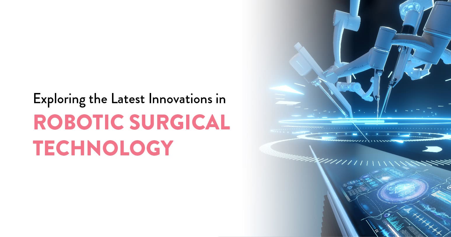
In the past ten years, the range of surgical procedures performed robotically has grown as a result of advancements in both hardware and software. Manufacturers are also building smaller, more specialised equipment that promises to reduce integration costs and increase access.
Today, gallbladders are routinely removed by a surgeon on one continent from a patient on another, using robotic telesurgery. Voice-activated robotic arms and haptic feedback provide surgeons with the control they need to perform surgery. It will be interesting to see how this technology continues to evolve and what we can expect in the next ten years.
Table of Contents
In four critical areas—adjustable stiffness, detectability, bendability, and controllability—newer colonoscopic surgical robots showed advances. It has been reported how a tiny robotic device called Endotics, which moves like a vacuum-based inchworm, can improve the precision of colonoscopic diagnostics and surgery.
Similarly, to get around these issues and improve control over the bending portion, a unique constrained tendon-driven serpentine mechanism (CTSM) has been developed.
An innovative soft robot, the NeoGuide colonoscopy system combines compliance, safe body interface, actuation and sensing, enhanced dexterity, and a larger workspace. This makes use of an entirely articulating computer-assisted insertion tube that resembles a snake. The tip of the scope determines the orientation of each subsequent segment, enabling the tube to assume various forms at various insertion depths. Additionally, it features variable stiffness control to avoid uncomfortable or insufficient endoscopy.
Based on a CTSM with SMA actuators, the Minimally Invasive Neurosurgical Intracranial Robot (MINIR) is another more recent robot utilised for brain tumour excision. The Flex system can perform several procedures with a smaller footprint than some other systems because it employs cables with varying tension to adjust the flexibility of the endoscope.
Like the Invendoscope, the Meshworm design mimics the movement of earthworms and is intended for colonoscopy. Based on the octopus arm, the STIFF-FLOP soft robot uses fluidic chambers with a modular stiffening and bending strategy.
The Israeli self-propelling colonoscope Aer-O-Scope pushes itself into the colon and up to the cecum using carbon dioxide without the assistance of an outside force.
With the removal of electromagnetic fields, remote endoscopic operation with MRI-based guiding permits high-scope detectability.
Noninvasive robots are useful in image-guided therapy, including the CyberKnife or image-guided radiosurgery. The use of non-invasive thermotherapy using high-intensity focused ultrasound (HIFU) coupled with robotic manipulators has been described.
Natural orifice transluminal endoscopic surgery (NOTES), where surgery can be truly non-invasive, is becoming closer thanks to these advancements, which also help to achieve greater stability of the instruments despite their flexibility, permit proper positioning of the instruments, greater dexterity, and higher-quality imaging.
An innovative strategy is to implant the whole MIS surgical platform into the peritoneal cavity using miniature in vivo robots. These robots have two multifunctional arms with various joints for infinite flexibility.
These provide scarless surgery with a small entry incision, tiny motors operating the arms, and great flexibility. Additionally, novice surgeons may utilise them under the guidance of more seasoned ones, greatly increasing patient access.
With the aid of these robots, surgeons may do procedures remotely or on-site while getting several views of the operative site. These are affordable and portable, making MIS accessible to nearly everyone at any time or location—from space exploration to distant medical emergencies or combat zones.
These are little endoscopes that are used for several medical procedures, medication administration, and diagnostic testing. They are incredibly tiny, resulting in less tissue damage and quick accessibility, and they can be moved by magnetic interactions, providing an untethered design with great flexibility of movement.
Even if they are still distant in the future, microbots are a possible innovation that might be put into circulation and transferred to a specific location without the need for any incisions.
In contrast to the previous master-slave systems, microrobots will have far more access, no physical link to the operator, and the ability to perform confined propulsion, consistent imaging, precise telemanipulation, and miniaturised functionality. Surgery might be revolutionised by microrobots once these problems are resolved.
Telemedicine first originated and developed in the US as a way to provide healthcare to those who lived in remote rural areas.
Both robotics and telemedicine have developed and improved over time, and they will do so in the future. How come? The numerous advantages that these two technological advancements may offer to patients, as well as to doctors, nurses, and other healthcare professionals in general, are evident to experts and researchers. They sincerely think that this may be the direction that modern medicine takes. As a result, there are various advantages that telemedicine and robotics-enabled healthcare practices are now providing.
AI has already been shown to be a huge help with patient monitoring and diagnoses in the medical field. The next natural step in integrating AI-based technologies and machine learning into medical treatment is to integrate AI into the operating room. This will greatly benefit patients and surgeons alike.
Surgical robotics is one of the next frontiers for optimising AI. Following are four ways robotic surgery can utilise AI to elevate the level of care, improve patient outcomes and reshape the healthcare industry:
Artificial intelligence-driven systems can process vast amounts of data in a matter of seconds. Developing “superhuman” advantages at the foundation of surgical robotics can maximise the possible use of information by optimising artificial intelligence. Artificial intelligence (AI) systems are not limited by time or memory. They can be given records of thousands of operations in a matter of seconds, and they can recall the first procedure they see with the same accuracy as the last.
Robotic surgery can thus rely on AI absorbing significantly higher volumes of data which can be utilised as a learning tool for surgeons at all stages of their careers. From educating physicians on different methodologies to centralising access for surgeons in developing regions, AI can reshape the way physicians learn, practice and perfect their surgical skills.
Artificial Intelligence (AI) offers doctors a fresh viewpoint and a new pair of eyes, which can bring innovative approaches to current surgical techniques and ultimately standardise methods. Through the collection of global data analytics, artificial intelligence (AI) can create complex visuals, identify minute variations, and establish novel patterns.
AI-based solutions can provide fresh, previously untested indications for the best surgical approaches by learning from thousands of surgical procedures. Understanding patterns and trends can help surgeons conduct some operations in new ways that improve patient and surgeon experiences. Standardised practices may result from this, as doctors all over the world execute AI-guided surgeries, using the same techniques to get optimal results.
AI can improve robotic surgery by reducing doctors’ stress levels, in addition to gathering a vast amount of data from which to learn and create new patterns. AI-based technologies may assist in guiding surgical processes and guaranteeing a more efficient process by identifying instruments, keeping an eye on operations, and sending alerts. Creating a plan of action based on the needs of each patient can help surgeons save valuable operating time and reduce cognitive strain, enabling them to do more surgeries at a higher level and with better results.
AI can also offer a fresh perspective on operating room ergonomics. The vast amount of “experience” that AI has amassed allows it to recognize and recommend ergonomically sound solutions, hence reducing the physical strain that many surgeons face while performing their procedures. Surgeons may prolong their careers and avoid compromising their physical health by utilising AI in conjunction with intelligent robotic surgical systems.
With the use of artificial intelligence (AI), more doctors will be able to learn from the top models in their area, which will enable more medical professionals to undertake surgery. Surgeons may benefit from and employ AI-based surgical robotics to reach a larger patient population, regardless of their location or the resources at their disposal. By diversifying into more sub-specialities, surgeons who now only handle a single kind of surgery may be able to impact a larger range of sub-specialities with their newfound ability to handle them.
Robotic surgery and artificial intelligence go hand in hand. It is essential to incorporate AI-based technologies into medical technology to enhance the experience of patients and doctors alike. Artificial intelligence (AI) has the potential to revolutionise surgical robots, propel the healthcare sector to unprecedented heights, and pave the path for automated care by empowering doctors at every stage of their careers and improving the quality of treatment.
Since its start, robotic surgery has advanced significantly. Thanks to technical developments, it is now a practical choice for many surgical operations. With several firms investing in research and development to advance the technology, the future of robotic surgery appears bright. This includes improvements in haptic feedback, artificial intelligence, and robotic arm motion. It follows that in the upcoming years, robotic surgery should become more widely available, reasonably priced, and effective.
At the CK Birla Hospital, we guarantee that patients receive complete medical treatment, including compassionate care. In addition to encouraging quicker recovery, this patient-centred approach ensures that patients are aware of the necessary precautions. Our staff of highly skilled surgeons has completed over 6,000 procedures, and they are skilled in carrying out life-saving procedures.
Modern surgical robots have tiny cameras built into them, giving the operator a visual aid to help increase precision. Cutting-edge technologies may even combine images from CT, MRI, and ultrasound scans, enabling a surgeon to see concealed surgical sites.
Medical practitioners may perform a wide range of difficult procedures with more control, accuracy, and flexibility thanks to robotic or automated surgery than is possible with traditional techniques. A lot of the time, minimally invasive surgeries involving tiny incisions are associated with mechanical surgery.
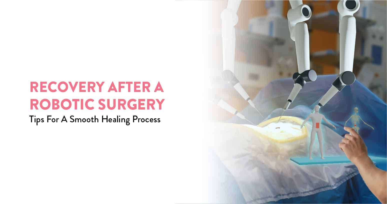
Many patients postpone their treatments as they are too busy to stay in bed for many days at home after spending a week in the hospital. Robotic surgery offers a surgical option with a recuperation period that is usually much shorter than with traditional surgery. Speak with your surgeon as soon as possible so they may assess the progress of your recuperation and address any concerns you may have. Let’s take an example of a hysterectomy in this article to show you what recovery or post-robotic surgery care looks like.
Table of Contents
While recovery times vary, most patients spend no more than one night in the hospital.
You’ll be taking painkillers. As soon as you feel like it, your healthcare staff will urge you to exercise.
After your robotic hysterectomy, some vaginal bleeding is normal for a few days to a few weeks.
Compared to an abdominal hysterectomy, the recovery period following a robotic hysterectomy is quicker and less painful. It might take three to four weeks for a full recovery.
Even if you feel better, wait six weeks following surgery to engage in vaginal sex or lift anything heavy (more than 20 pounds, or 9.1 kilograms).
Should you have nausea, vomiting, or bleeding that is more intense than usual during your menstrual cycle, get in touch with your doctor.
You may experience relief following a hysterectomy if you are no longer experiencing severe pelvic pain or bleeding.
After a hysterectomy, most women have no change in their ability to conceive. However, some women report greater sexual satisfaction following a hysterectomy, possibly as a result of the discomfort they no longer experience during sexual activity.
It’s common to have feelings of loss and sadness following a hysterectomy. If you’re young and expecting to get pregnant in the future, you may also be depressed about losing your fertility. See your doctor if being depressed or experiencing other unpleasant emotions prevents you from enjoying daily life.
You will be monitored in the recovery room following surgery while you come out of the anaesthesia. Most hospital stays last several days. Following your hospital stay, the following might occur:
Upon returning home, you must adhere to your surgeon’s recommendations and schedule your follow-up visits. As prescribed, take any medicine. Early recovery is common, with some discomfort. Find out from your surgeon which painkillers are recommended.
You can anticipate the following when recovering at home:
During the healing process, inform your surgeon of any of the following:
You could have emotional symptoms following this operation in addition to the physical ones associated with healing. Following a hysterectomy, your menstrual cycle will end and you will no longer be able to become pregnant. A certain amount of women feel depressed because of these losses.
Menopause symptoms, such as hot flashes and dry vagina, may be apparent if you have surgery to remove your ovaries. Hormone treatment may be beneficial for certain women following a hysterectomy. Talk about this with your doctor.
Compared to conventional, open hysterectomies, most patients will recover from robotic hysterectomy faster and with less discomfort.
You may get relief from bothersome symptoms like pain or irregular bleeding, depending on the reason for your hysterectomy. On the other hand, some individuals have minor menopausal symptoms following a hysterectomy.
Feelings differing from one another on the surgery are common. After having a hysterectomy, some people experience loss or despair since they are unable to conceive and no longer have their menstrual period.
Following your robotic hysterectomy, you will get a list of activities to avoid from your surgeon. Here are a few examples:
Following a robotic hysterectomy, a complete recovery typically takes four to six weeks. After surgery, you can resume mild, routine activities in 24 to 28 hours. Having someone help you at home during this period is beneficial since you might not be able to do much and you could be exhausted and hurting. Typically, you can resume driving after 72 hours, however, this can vary according to the type of painkiller you’re using. After around two weeks, most people can resume their jobs, however, this varies according to the nature of the employment.
You Can Also Read: Hysterectomy or uterus removal surgery
Compared to traditional surgery and even laparoscopic surgery, recovery with robotic surgery is far shorter. It is also far more accurate and minimally invasive, requiring less anaesthesia, which facilitates patient recovery. You may need to modify your diet, refrain from doing strenuous exercise, or take longer time off than usual following the procedure, depending on the type of surgery. Additionally, your surgeon will probably schedule a follow-up session to assess your progress and address any concerns you may have.
At the CK Birla Hospital, we guarantee that patients receive complete medical treatment, including compassionate care. In addition to encouraging quicker recovery, this patient-centred approach ensures that patients are aware of the necessary precautions. Our staff of highly skilled surgeons has completed over 6,000 procedures, and they are skilled in carrying out life-saving procedures.
There is no requirement for bed rest at home following robotic surgery. Although patients may experience some discomfort and increased weariness, they should make an effort to remain active by walking often and carefully, as well as for as long as they can. To hasten recovery, patients should consume enough water and maintain a balanced diet.
Traditional open surgery can require several days of hospitalisation and recovery time can last several months. While every case is unique, the return to normal activities (except for lifting heavy objects and strenuous exercise) following robotic-assisted surgery can occur in as little as two to three weeks.
Any surgical treatment carried out with the aid of robotic devices is referred to as robotic surgery or robot-assisted surgery. Compared to current minimally invasive surgical procedures, it gives the surgeon improved control over the surgical instruments and a clearer view of the operational site. Physicians are also relieved from the physical strain of having to stand the entire time during long procedures.
In addition to having superior control over the laparoscopic camera placement—which in this instance is also notably steadier with fewer inadvertent movements than human assistance—the robot’s computer program also filters out natural vibrations. The finest element is that the robot won’t burn out when employed for several rotating surgical teams.
Table of Contents
Through the use of specialised technology, robotic surgery improves the hands of your surgeon. Small incisions allow surgeons to do procedures in hard-to-reach areas. Additionally, the specialised technology allows for accurate motions and improved magnification.
The Technology Includes:
During the past ten years, surgical robots have radically changed how doctors practise medicine and how rapidly patients heal. Even for major surgery, robotic surgery only necessitates tiny incisions and allows doctors to perform complicated treatments with unparalleled accuracy.
During a robotic surgery, the patient has three or four robotic arms introduced through tiny incisions. A camera is attached to one arm, two arms serve as the surgeon’s hands, and a fourth arm can be utilised to clear a blockage. There is a surgical team on hand next to the patient, and the surgeon uses a nearby console to control all four arms.
Certain treatments that would ordinarily need open surgery can now be completed with minimally invasive robotic-assisted surgery. This is significant because patients who have minimally invasive surgery typically experience fewer side effects.
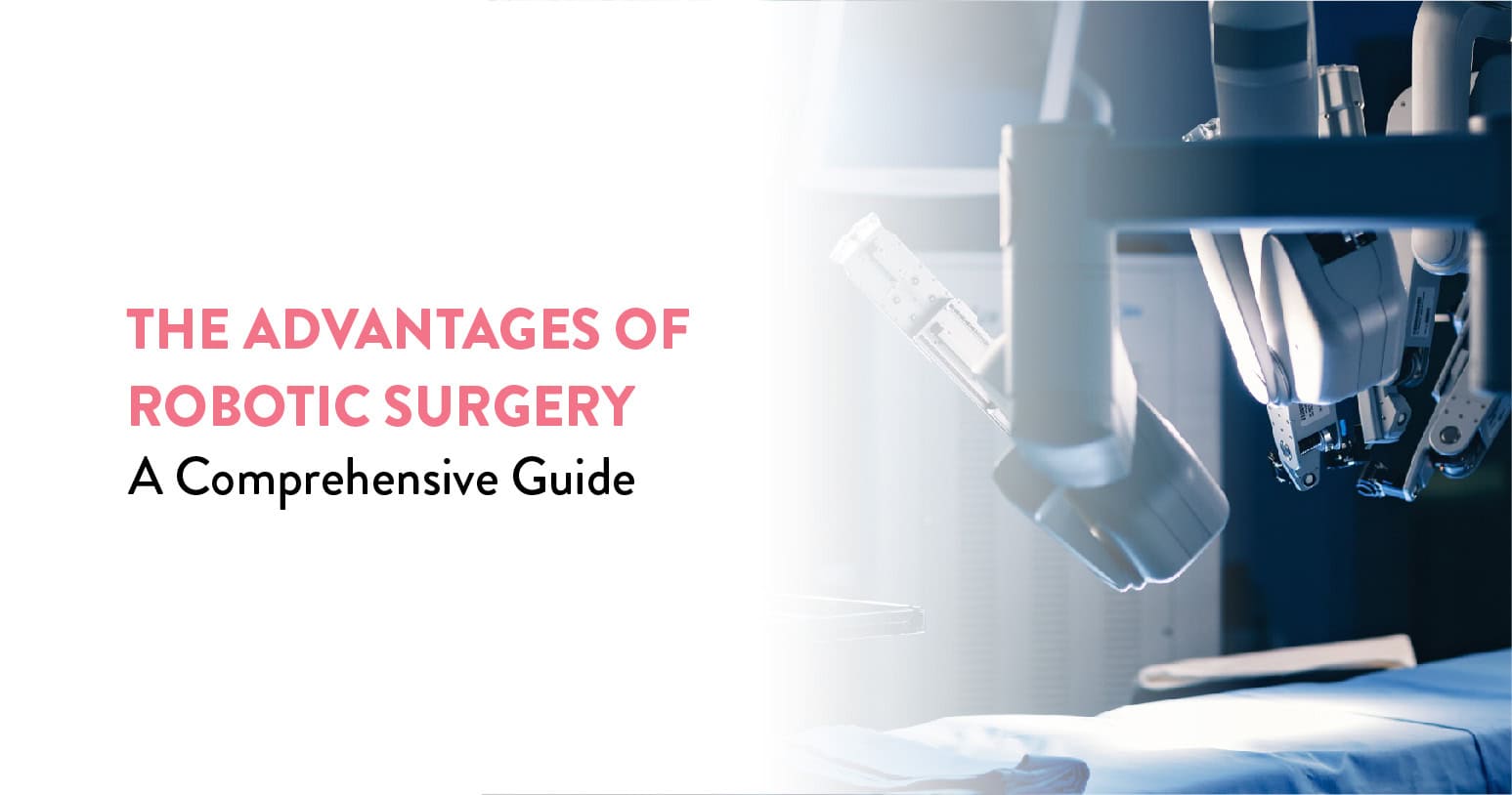
Although robotic-assisted surgery might not be something you’ve thought about before, there are many good reasons to talk to your doctor about robotic surgery options if you need a medical procedure.
Let’s examine the benefits of robotic surgery:
The incisions are smaller than in traditional surgery since the doctors do not have to use their hands to directly reach the body. To lessen the possibility of unintentional cuts or punctures that might result in bleeding and infections, the robotic arms also filter out hand tremors.
When an operation is required in a part of the body that is difficult to access, robotic surgery is a fantastic alternative. Major blood arteries or other important organs may be close by in these places, which increases the danger of the procedure. To have more room for the surgeon to see exactly what they’re doing to operate, surgery on these locations typically requires a wider incision.
Rapid healing is one of the main benefits of robot surgery. Compared to traditional surgery, robotic surgery is minimally invasive, thus your body will probably heal faster.
Since each person is unique, the length of time it takes to recover varies depending on the situation. Nonetheless, following robotically assisted surgery, the majority of patients are back to their regular activities, including work, in a few weeks.
You’ll be able to return to your regular life sooner and avoid the often expensive expenditures associated with an inpatient hospital stay thanks to a possible quicker healing period. You’ll recuperate with less scarring and blood flow since there is less damage to the body.
You can Also Read: Future Of Robotics Surgery – Revolutionizing Treatments
You’ll feel less pain during and after surgery if there are fewer incisions and more accuracy. Additionally, you won’t need as many painkillers while in recovery, which lowers the possibility of addiction.
Due to their ability to manoeuvre in constricted areas, robotic arms also make far smaller incisions and cuts than traditional surgical methods, which helps to minimise blood loss. In addition, the robot keeps the active blood vessels cauterised to prevent unnecessary bleeding and triggers instantaneous blood coagulation in the operating area of the body. Due to this, patients do not need blood transfusions, which otherwise carry the hazards of infection. Blood loss following other procedures may result in problems and even longer recovery periods.
Any surgical operation has the danger of infection, which can impede your recovery and lengthen your stay in the hospital. Certain body sections are more susceptible to infection than others, especially when lengthy incisions expose a considerable portion of the body. However, because robotic surgery is minimally invasive, there is a lower chance of infection, which spares you from any potential consequences that may arise from an infection.
Enhanced accuracy is one of the main benefits of robotic surgery. A robot’s arms need less room and can even operate an instrument in a small place, but a human hand needs a specific amount of room to grasp the instrument and control the affected region. It makes surgery more accurate and necessitates smaller, less incisions.
The robot can readily access confined locations because of the more substantial mobility of its robotic arms. It also aids with the patient’s ability to undergo surgery, maintain composure, and prevent panic attacks.
Occasionally, surgical sites are small, cramped areas, which makes it challenging for medical professionals to complete the procedure. The surgical site is seen in three dimensions on the associated computer console thanks to the robot arms that are connected to the camera. It makes the procedure considerably more precise for the doctor to undertake. For a better surgical experience, the camera provides a clearer view of the operative region.
Procedures using robot assistance take less time to operate and recuperate from. Patients stay in the hospital for shorter periods as a result. On the other hand, conventional operations often need more time since the surgeon must first mark, prepare, and open the patient’s chest and cardiovascular regions. Nonetheless, because of the robot’s accuracy and precision, these procedures are finished far more quickly in robotic surgery.
Moreover, fewer problems arise after surgery using robotic surgery. In addition, it lessens the chance of wound discomfort, heart attacks and strokes, which can happen if a patient is not up to the demands of traditional surgery. Robotic surgery reduces the danger of problems while enabling surgeons to achieve accurate and consistent results by doing away with the need for manual intervention.
An improved prognosis for the patient as a whole is another benefit of robotic surgery. A better patient outcome is a result of less blood, shorter operating times, fewer problems following surgery, etc. Robotic surgery is particularly helpful in high-risk situations when maximum precision, less operating risk, and a quicker recovery period are necessary.
You can Also Read: Robotic Surgery vs. Traditional Surgery: Which Is Right for You?
In India, robotic or robot-assisted surgery is improving minimum access surgery with several advantages for the patient and the doctor. It has increased the safety and precision of both simple and complex surgeries and is now a standard component of the majority of surgical procedures. The outcome has proven beneficial to both the patient and the surgeon.
At the CK Birla Hospital, we guarantee that patients receive complete medical treatment, including compassionate care. In addition to encouraging quicker recovery, this patient-centred approach ensures that patients are aware of the necessary precautions. Our staff of highly skilled surgeons has completed over 6,000 procedures, and they are skilled in carrying out life-saving procedures.
There are intrinsic advantages to robotic surgery; compared to traditional surgical techniques, this technology is often safe and successful.
There is less pain and physical scarring than with open procedures. Robotic-assisted surgery is more advantageous than traditional surgery and provides cancer patients with a better, less painful, less hazardous, and faster recovery period.
Robotic surgery technology lacks self-moving capabilities. Surgeons are always in command. Safety measures are in place to guarantee that the surgeon maintains control over the robot at all times.
The benefits of robot-assisted technology for surgeons include improved vision, increased clinical accuracy, and less tiredness. Patients also benefit from shorter recovery times and minimal to no scarring.
As each patient is different, you should discuss your recuperation with your doctor. Patients often spend one or two nights in the hospital before going home. After surgery, most patients discover that they have healed in six weeks.

The phrase “robotic surgery” is familiar to many, but what does it mean? This topic comes up when people have to choose between seeing a surgeon who suggests traditional open surgery or one who chooses this new and more complex style of surgery. Thankfully, robotic surgery is not as scary as it seems and generally has many benefits.
If you need surgery, think about asking your surgeon if robotic surgery is the best option for you. Here is more information about this novel surgical technique.
Table of Contents
Robotic surgery — or robotic-assisted surgery — is a minimally invasive type of surgery in which surgeons use a console equipped with controllers that manoeuvre robotic arms. This console also has a 3-D high-definition camera that provides a clear and magnified view of the target area. Using the console’s tiny and precise instruments, the surgeon can create small incisions, cauterise, staple, grasp and perform other actions — just as they would with their hands.
Despite the word “robotic surgery” suggesting that a robot is carrying out the operation, the surgeon has complete control over how all of the devices move.
One type of surgery that is becoming more and more common is robotic surgery. It doesn’t mean that robots are doing your operation, unlike what the general public believes. Rather, a surgeon uses extremely precise robotic arms.
Because robotic surgery only requires a little incision, it is regarded as minimally invasive surgery. Comparing a smaller incision to typical open surgery can frequently result in significant reductions in risk factors and recovery durations.
During robotic surgery, the surgeon uses a computer to control the robotic arms that perform the procedure. Robotic surgery gives him/her unparalleled flexibility and precision during surgery.
Robotic surgery can frequently be used for the following common procedures:
With less blood loss, discomfort, and recovery time than more conventional surgical techniques, robotic surgery provides consistent, dependable results.
Robotic surgery is not a suitable solution for many procedures and medical problems. The surgeon provides the finest conventional surgical methods, such as laparoscopic surgery, in these situations.
Because laparoscopic surgery involves a significantly smaller incision than standard open surgical approaches, it is a very popular surgical alternative. Laparoscopic procedure incisions are typically half an inch long.
The doctor makes an incision, inserts a plastic tube, and performs the procedure using a camera and tiny surgical equipment. Upon completion of the surgery, she uses a few stitches to seal the wound.
Laparoscopic surgery is similar to robotic surgery in that both minimise the incision size and can reduce risks associated with surgery. Patients may experience less discomfort, along with faster recovery times when they undergo laparoscopic or robotic surgery.
The surgeon could suggest open surgery in specific circumstances. Typically, open surgery necessitates a wider incision, and the surgical team uses their hands rather than tiny instruments to move muscle and tissue.
The best kind of surgery for you will depend on your particular medical situation. When providing a personalised surgical strategy, we examine your injuries, health, and medical history.
All surgeries come with a set of risks, but your healthcare provider can help you navigate surgery and recover as quickly as possible.
Each kind of surgery has advantages. Research shows that patients can recover from robotic surgical operations more quickly and with less discomfort. For instance, robust urologic operations benefit greatly from the accuracy of robotic technology. One of them is a prostatectomy, in which the target location is surrounded by nerves that govern erectile function and urine control and are firmly contained. By employing a robotic device, surgeons can precisely minimise harm to neighbouring nerves, speed up recuperation, and facilitate a return to regular activities.
Although statistics have demonstrated the statistical benefits of robotic surgery, you are not a number. You are an individual with a distinct anatomy, suffering, and medical issues. In certain cases, conventional surgery is the superior option.
When you feel uneasy, having robotic surgery is one of those situations. It’s normal for some people to find the concept of robot-assisted surgery repulsive. We allow our patients to select the orthopaedic surgery that best suits their needs, even if it is painful. This way, you may still receive the necessary treatment to end your chronic pain and restore function to your leg and hip.
If you have a lot of scar tissue around your hip or have certain health conditions, you and your surgeon may decide that traditional surgery is a better option for you.
With its accuracy, less blood loss, decreased discomfort, and lower infection rates, robotic-assisted surgery outperforms conventional surgery. It’s an appealing option because of the indisputable benefits in surgical results and patient recovery, even with possible greater expenses. In the end, medical practitioners assess the patient’s state, the state of technology at their disposal, and surgical proficiency to ascertain the best course of action. Promising better results and a more streamlined surgical experience, robotic-assisted surgery represents a breakthrough.
At the CK Birla Hospital, we guarantee that patients receive complete medical treatment, including compassionate care. In addition to encouraging quicker recovery, this patient-centred approach ensures that patients are aware of the necessary precautions. Our staff of highly skilled surgeons has completed over 6,000 procedures, and they are skilled in carrying out life-saving procedures.
Robotic surgery has a recovery period of as little as two to three weeks, whereas conventional surgery necessitates a lengthy hospital stay and several months of recuperation. This results from decreased blood loss, discomfort, and infection as well as from minimal invasion.
Because robotic-assisted surgery uses smaller surgical equipment and smaller incisions than traditional surgery, it is less intrusive than traditional surgery. This lowers the chance of infection and minimises blood loss.
For patients, the main advantage of robotic surgery is a quicker recovery. Patients can resume their regular activities sooner than they could after open or laparoscopic surgery. Robotic surgery also leaves smaller, less obvious scars and has fewer surgical problems.
| Robotic Surgery vs. | Traditional Surgery | Conclusion |