Filter :

Your body’s like a little furnace. It puts out heat all the time. It results from your body working to keep you alive. When it puts out a lot less or a lot more heat than usual, it is trying to tell you there’s a problem.
A normal body temperature can vary depending on your age and other factors. Whether you take it in the armpit, rectally or orally also can impact your temperature reading. A healthy body is normally pretty good at keeping its temperature at a comfortable level, however, if the body goes into some sort of illness or trauma, the temperature can become high or low depending upon the trigger. In such cases, do not hesitate to reach out to a doctor. When and how do you know you need help? Read on to know more…
Table of Contents
The average person most likely learned as a child that the body’s normal temperature is 98.6 degrees Fahrenheit (or 37 degrees Celsius). A mid-1800s research is where that frequently used figure came from.
The average person nowadays probably runs a little bit cooler than that, between 97.5 F (36.4 C) and 97.9 F (36.6 C), according to a more recent study.
The reality is that although it often stays within a range, your body temperature can fluctuate up, down, and all about.
Your normal body temperature changes throughout your life. It frequently increases from youth through adulthood before declining in one’s senior years. It looks like this as per stages:
A temperature that is higher than 100.4 F (or 38 C) is considered a fever, and it is normally something you should bring to your doctor’s attention, especially if it lingers for more than two days.
Oftentimes, a fever is your body’s reaction to a virus or an infection (like influenza). A fever itself does not require any specific treatment, other than trying to bring the temperature down for your comfort.
Persistent high-grade or low-grade fevers could signal that something else is going on in your body. Some medical conditions, including hyperthyroidism and other endocrine disorders, can raise your body’s core temperature.
Young kids usually tend to push thermometer readings higher than adults. Their bodies have not yet mastered the art of regulating their body temperature, so they are also more likely to spike fevers and severe ones, at that.
If an infant younger than 3 months develops a fever or if your child’s fever does not come down with fever reducers, call your paediatrician.
According to studies, as people age, their core body temperature drops. An underactive thyroid (hypothyroidism) can also slow down metabolism, which can lead to a drop in body temperature.
If your core body temperature dips down to 95 F (35 C) or lower, that is considered hypothermia. It is often caused by exposure to cold weather, but other factors can put you at risk for hypothermia, such as certain medications and age.
Hypothermia is a medical emergency, so call an ambulance or take the patient to the emergency room if you suspect someone has hypothermia.
During the 19th century, German doctor Carl Wunderlich identified the average body temperature of 98.6°F (37°C). However, many studies have since determined that that is not always the case. The average body temperature was determined to be 97.86°F (36.59°C) in a 2019 research. That is a little lower than initially thought so many years ago.
However, it is best to take this information with a grain of salt since no single number defines your average body temperature. Instead, it is best to look at a temperature range that might be lower or higher than the average.
Here are some of the factors that affect body temperature:
If you take your temperature with 3 different kinds of thermometers, you may get 3 different results.
A thermometer that goes under the tongue will likely give you a slightly higher reading than the one that goes under your armpit or a forehead thermometer (But those kinds might be easier to use with young kids).
Which is the most accurate? A thermometer that you place beneath your tongue would be that. A rectal thermometer, usually used with young children, would be more accurate for the same reason. A thermometer used rectally should never be used orally to avoid spreading bacteria. Keep thermometers used orally and rectally separate and clearly marked.
Usually, you go to the medical cupboard to get your thermometer because someone at home is feeling under the weather.
As a result of the crucial information the reading gives, consider the optimum moment to measure your temperature. Temperature is one of your vital signs, and it is an important indicator of your health.
It is also important to note that body temperatures usually run slightly higher in the afternoon and a little lower in the morning. Additionally, it may vary with menstrual cycles.
A human body’s temperature is an important indicator of its overall health. Your body can normally maintain its temperature at a comfortable level, however, if you have any concerns or questions about your temperature, then it is always advisable to seek medical help from an experienced general physician. Timely care and help can ensure an optimum body temperature for the best possible health.
At the CK Birla Hospital, we ensure patients get timely medical support which includes treatment in a hands-on yet compassionate environment. This patient-first approach not only helps patients heal better but also ensures they are aware of the preventive measures at hand. In case you need to consult a general physician, reach out to us, or book a direct appointment at the CK Birla Hospital.
A fever of 103°F or higher typically requires a doctor’s consultation. If your fever reaches 105°F, go straight to an emergency room.
People might feel hot for many reasons other than a fever. Some causes might be easy to identify and temporary, like experiencing anxiety and stress, being in a humid environment or eating spicy foods. However, some people might feel hot frequently for no apparent reason.
Typically, the following thermometer readings signify a fever: temperature of at least 100.4 (38 C) in the temporal, ear, or rectal arteries, and at least 99 F (37.2 C) in the armpit and oral temperature of at least 100 F (37.8 C).

A bone is a rigid organ that constitutes part of your skeleton. Bones come in a variety of sizes and shapes and have complex external and internal structures. They have a variety of uses and are tough but lightweight.
Occasionally cracking or popping your knuckles or other joints is normally not harmful and very common. It does not cause arthritis contrary to the old wives’ tale. It can give you greater motion in a joint and provide a feeling of relief. Joint cracking might become more noticeable as you age, as some of your cartilage wears away. If it is accompanied by swelling or pain or follows an injury, it might be caused by an underlying condition. Then it is best to contact a health professional to determine the cause.
Table of Contents
In sports medicine and orthopaedic medicine, a clicking, popping or crackling sound in a joint (also called bone cracking by some) might mean that air is moving in the joint, which is normally harmless.
People often notice this in their knees in the form of knee cracking, but it can also happen in other joints such as the neck, elbow or shoulder.
Bone cracking with pain can be a sign of injury or wear and tear. If it is painful, you should consult a doctor.
Bone cracking and joint sounds/voices can be a normal part of the movement. Joint popping is a common problem, especially as individuals age. You might notice:
There are different causes for joint cracking. It is common and is normally not an indication of a bone health condition. Exactly what causes the popping or cracking noise is the subject of many studies, but it is still not completely understood.
Some natural causes of joint cracking are:
Why people experience popping or cracking noises in some parts of their body is something which is not fully known. A traditional explanation is that pressure on a joint creates tiny bubbles in the synovial fluid, which pop when they form quickly. Your bones are protected from rubbing against one another by synovial fluid, which is composed of carbon dioxide, nitrogen, and oxygen.
Cracking your knuckles or other joints is not bad, but it might be annoying to the people around you if you do it frequently. In rare instances, you may hurt yourself by squeezing a nerve or straining a muscle if you are cracking a joint too forcefully, such as in your back.
According to a small 2011 study, the cracking process can give you a physical feeling of relief from pressure, whether you are having a chiropractor manipulate a bone or doing it yourself.
The common myth that you will get arthritis in your hands if you crack your knuckles has proven to be just a myth by another 2011 study. Knuckle cracking does not decrease cartilage and is not likely to cause osteoarthritis, according to studies.
Many causes of joint cracking and popping (crepitus) improve with home remedies, such as using the RICE method (rest, ice, compression and elevation) or taking anti-inflammatory medications. Other causes or effects may require a doctor’s help.
Your doctor will discuss several treatment options with you, such as:
Sometimes, a splint or brace can help align the elbow, shoulder or knee so an injury can heal.
Our physical therapy teams tailor treatment plans to your goals, activities and condition.
Orthotics (special shoe inserts that stabilise the knee and foot), can relieve pain and help you stay active.
Handled correctly, pain relief methods can eliminate inflammation and discomfort and might let you get back to your favourite activities safely.
Some causes of bone cracking may require surgical treatment. You may consider:
Arthroscopy is a minimally invasive procedure that accesses the joint by inserting small tools through extremely small incisions. For Patellofemoral pain syndrome, your surgeon can remove bits of damaged tissue or reposition tendons to provide more knee movement.
Some types of arthritis and patellofemoral pain syndrome improve with debridement. The surgeon can smooth damaged cartilage via this minimally invasive procedure to lessen friction.
When joint damage or arthritis is advanced, you may consider joint replacement such as hip replacement, elbow replacement, knee replacement . The goal of joint replacement surgery is to replace a damaged joint with an artificial one.
Popping or cracking the joints is quite common and usually not harmful. If you notice
any pain, swelling or injury, then it is always advisable to seek medical help from an experienced orthopaedist. Timely care and help can ensure an appropriate diagnosis and treatment of your condition (if any).
At the CK Birla Hospital, we ensure patients get holistic medical support which includes treatment in a compassionate environment. This patient-centric approach not only helps patients heal better but also ensures they are aware of the preventive measures as well. In case you need to consult an orthopaedist, reach out to us, or book a direct appointment at the CK Birla Hospital.
There is no evidence that knuckle cracking is either advantageous or detrimental. Knuckle cracking particularly does not cause arthritis. Joint cracking can result from a negative pressure pulling nitrogen gas temporarily into the joint, like when knuckles are cracked. This is not harmful.
Vitamin D deficiency is a contributing factor to fatigue and joint pain. Additionally, vitamin D helps strengthen your bones if you are getting enough of it.
Some foods which are good for your joints are cherries, red peppers, turmeric and walnuts.
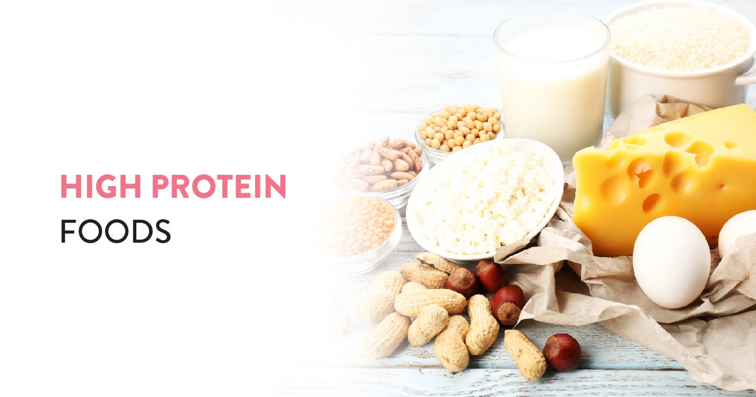
Do you know the secret to the gorgeous hair and lean physiques of most celebrities? It is simply a high-protein diet. Protein is a nutrient needed by the human body for maintenance & growth, as well as the most prevalent type of molecule in our body after water. They act as a fuel source and building block of our bodily tissue.
Proteins are a large category of molecules that impact our cell structures, hormone synthesis, immunity and much more. They are all made up of tiny building blocks called amino acids. Not only is protein essential for your health but consuming it can keep you feeling satisfied and full, which supports a healthy body weight.
Nine types of amino acids are essential, meaning your body needs them but can not make them on its own, so you need to get them in your diet. The recommended dietary allowance (RDA) for protein is 0.36 grams (g) per pound of body weight (0.8 g per kilogram). Keep in mind that this is the very minimum amount of protein your body requires. Fortunately, there are many high-protein foods to choose from, including plant-based and animal sources.
Table of Contents
An individual’s protein goals might vary depending on medical history and physical activity level. Some people follow a high-protein diet when trying to reduce weight because protein encourages a feeling of fullness. The following are some of the best high-protein foods that a person can consume and include in their chart/list:
Whole eggs are a good source of protein that is easy to absorb, and they are also an excellent source of antioxidants, healthy fats, minerals and vitamins.
Remember that egg whites are almost pure protein, but whole eggs that include the yolk provide many more nutrients, including healthy fats, antioxidants, minerals and vitamins. 50 g of big eggs provide 6.3 g of protein.
Almonds are a nutritious tree nut rich in essential nutrients such as magnesium, manganese, vitamin E and fibre. They are also high in plant-based protein.
Eating almonds might benefit your health in several ways, including lowering heart disease risk factors like high blood pressure and high LDL (bad) cholesterol.
Almonds have 6 g of protein per ounce (28.35 g). Other high-protein nuts include pistachios, which deliver 5.73 g per 1-ounce (28.35 g) serving and cashews, which contain 4.34 g of protein per 1-ounce (28.35 g) serving.
Chicken breast is an excellent choice if you are trying to increase your protein intake. Chicken offers a range of B vitamins, as well as minerals like selenium and zinc, in addition to protein. 26.7 g of protein is included in an 86 g portion of chicken breast.
You Can Also Read: Vitamin D Foods and its Benefits
Cottage cheese is a type of cheese that is high in protein and low in fat and calories. It is rich in riboflavin (vitamin B2), vitamin B12, selenium, phosphorus and calcium.
Cottage cheese has 28 g of protein per cup (226 g).
Milk has a small amount of almost every nutrient your body requires. It is a good source of high-quality protein, and it is high in minerals and vitamins, like riboflavin (vitamin B2), phosphorus and calcium.
Many people with lactose intolerance can not tolerate milk and other dairy products, and they avoid many dairy-containing foods. One cup (246 mL) of dairy milk provides 8.32 g of protein.
Lentils are among the richest sources of plant-based protein, making them an excellent choice if you follow a vegan or vegetarian diet.
Plus, they are loaded with other nutrients too, including manganese, copper, iron, potassium, magnesium, folate and fibre.
Studies show that people who regularly consume lentils and other legumes have a lower risk of developing health conditions like fatty liver disease and heart disease.
The amount of protein in 100 g (or half a cup) of cooked lentils is 9.02 g. Chickpeas, which have 7.05 g of protein per 100 g of cooked weight, and black beans, which have 8.86 g of protein per 100 g of cooked weight, are two more high-protein legumes.
Fish is an excellent source of protein and provides several important minerals and vitamins, like vitamin B12, selenium and iodine.
People who include a lot of fish in their diet tend to have a lower risk of health conditions like type 2 diabetes and heart disease. Some fatty fish are also high in omega-3 fats, which have powerful benefits for your overall health, including supporting heart health.
Protein powder can come in handy when you are pressed for time and unable to prepare a meal. You can easily add protein powders like pea protein and whey to yoghurt, energy balls, smoothies and shakes to increase the protein and fullness factor.
Whey protein powder provides about 16.6 g of protein per scoop (28.6 g), while pea protein provides 15 g of protein per scoop (20 g). Note that the protein content per scoop differs between products, even when the scoop size is the same.
Pumpkin seeds are a great source of minerals such as zinc, magnesium, phosphorus and iron. Plus, they are loaded with fibre and plant-based protein.
Try adding pumpkin seeds to yoghurt, oatmeal, baked goods, and salads or mix them with unsweetened dried fruit and almonds for a convenient snack.
8.8 g of protein is included in 1/4 cup (29.5 g) of pumpkin seeds. Other high-protein seeds include sunflower seeds, which provide 7.25 g per 1/4-cup (35-g) serving, and flax seeds, which provide 7.5 g of protein per 1/4-cup (42-g) serving.
Peanut butter and peanuts are packed with nutrients like vitamin E, magnesium, folate and protein.
Eating peanut butter and peanuts might help make you feel full due to their high protein content. Studies show that adding peanut butter to a high-carb meal might help reduce blood sugar spikes after the meal.
A 1-ounce (28.35-g) serving of peanuts provides 7.31 g of protein, while a 2-tablespoon (32-g) serving of smooth peanut butter provides 7.2 g of protein.
You Can Also Read: High-Fibre Foods You Should Eat
Protein deficit happens when you don’t consume enough of it in your diet. However, protein deficiency might occur in people with special requirements, such as people following strict vegan or vegetarian diets and older people.
Symptoms of protein deficiency include:
Protein is a vital nutrient required for your body’s growth and maintenance. If you are unable to decide or meet your body’s protein requirements with your daily meal routine, then it is always advisable to seek medical help from an experienced dietitian. Timely care and help can ensure the inclusion of sufficient protein in your regular diet.
At the CK Birla Hospital, we ensure patients get holistic medical support which includes looking after their pathological and dietary needs. We believe that any treatment yields the healthiest outcomes in a stress-free and compassionate environment. Our patient-first approach not only helps patients heal better but also ensures they are aware of the preventive measures to maintain their health in the long run. In case you need to consult a dietitian, reach out to us, or book a direct appointment with Dr. Prachi Jain at the CK Birla Hospital.
The healthiest protein sources that you may eat daily are those that come from plants, such as soy, lentils, beans, seeds, and nuts; lean meats, including skinless, white-meat chicken; a range of fish and shellfish; egg whites; or low-fat dairy.
High-protein foods include eggs, nuts, seeds, cheese, milk, low-fat yoghurt, lentils, beans, fish and lean chicken.
Guava is one of the fruits with the greatest protein content. Every cup contains a massive 4.2 grams of the substance. Additionally, this tropical fruit is rich in fibre and vitamin C.
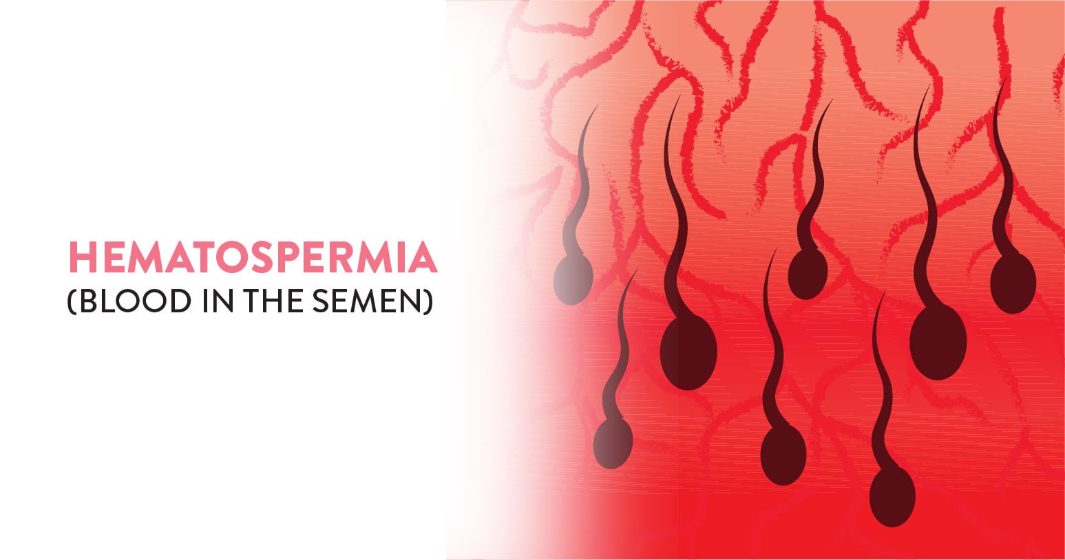
If you suddenly spot blood in your ejaculated semen, don’t be frightened. You might be experiencing a condition called Hematospermia. While initially it might seem relatively frightening or distressing, it so happens that this condition is commonly experienced by a majority of men, but not openly discussed.
This may be a one-time occurrence or a recurring episode. Whether the blood condition is old or fresh, Hematospermia is most often caused by infectious or inflammatory disorders and follows a self‐limiting, benign course.
Although major dysfunctions are not readily associated with this condition, in males over the age of 40, a more serious pathology may be present, necessitating further testing. Irrespective of the persistence of the condition, one must always seek medical advice to rule out any chances of prostate cancer. Before you visit your doctor for an examination or any necessary tests, let us understand the condition better, in order to voice your worries and receive reassurance.
Table of Contents
Semen is your organic bodily fluid which contains spermatozoa. It is secreted by a male’s sexual glands (gonads) and organs and can fertilise the female’s ovum.
Hematospermia (or Hemospermia) is the presence of blood in your semen. It may naturally worry men who encounter it, but it does not mean that it is a likely indication of a serious health issue.
It is not clear how common the symptom of blood in semen is because men typically do not examine their semen after ejaculation. Men of any age can have blood in their semen, although men in their 30s and 40s seem to have it more frequently.
Although it is frightening to see blood in your semen, it is rarely a symptom of a major health problem. A single episode of blood in your semen is normally not a sign of cancer. However, you might still want to see your healthcare provider to be reassured, to fully discuss this symptom and get an exam and perhaps other tests if your provider thinks they are needed.
Your semen may include as little as a single drop of blood or as much as is necessary to give it the appearance of blood. The reason for your bleeding will determine how much blood is in your semen. You may also experience:
You Can Also Read: Factors that affect male infertility
On its route to the urethra for ejaculation, the semen travels via many channels. Blood vessels along this tract might rupture for any variety of reasons, causing blood to spill into the semen.
In many instances, the precise reason for blood in the semen remains unknown. If you’re under 40, most incidences of blood in the semen are not dangerous. Some of the possible causes of bloody semen that your doctor may investigate are below:
Bloody semen frequently results from inflammation of the seminal vesicles. Inflammation of any organ, tube, duct or gland involved in the male genitals can cause blood in your semen. This includes:
Additionally, irritation from calculi (stones) in the prostate, urethra, bladder, or seminal vesicles can result in inflammation.
Just as with inflammation, infections in any organ, tube, duct or gland involved in the male genitals can cause blood in the semen.
Sexually transmitted infections (STIs), like herpes, gonorrhoea or chlamydia can also cause blood in semen. Infections caused by fungi, bacteria and viruses can also lead to this condition.
Blockages in ducts like the ejaculatory duct might cause the blood vessels in the area to enlarge and rupture. When your urethra is under strain from an enlarged prostate, bloody semen may result.
Malignant tumours or benign polyps in the seminal vesicles, epididymis, testicles or prostate could lead to blood in your semen.
The blood you’ve noticed in your semen may be due to vascular anomalies in the male genitals, such as vascular cysts.
Semen blood might result from illnesses that impact your entire body. These include haemophilia (a disorder that leads to easy and excessive bleeding) and hypertension (high blood pressure). Other possibilities include chronic liver disease and leukaemia.
Blood in your semen might result from physical damage, such as getting struck in the testicles while participating in sports. Trauma can cause blood vessels to leak, and that blood might leave your body in semen. A medical procedure such as a vasectomy, biopsy or prostate exam can cause blood in your semen.
You Can Also Read: Premature ejaculation-causes, symptoms and treatment
Your healthcare provider will:
Your healthcare provider might order one or more of these tests:
The results of your tests and exam might not show the cause of blood in your semen. Your provider might refer you to a urologist (a doctor who specialises in the male reproductive organs and urinary tract) if your urinalysis and initial evaluation are not normal or if the blood in your semen is present for longer than a month. Your urologist might order some or all of the following tests:
You may be able to treat yourself at home depending on the cause of the blood in your semen.
If you have blood in your semen as a result of a trauma, simply resting and allowing your body to heal might help. If your groin is also swollen, you should apply ice to it for no more than 10 to 20 minutes at a time.
Most cases of Hematospermia are cured on their own. Keep an eye on your symptoms and alert your doctor if they persist for longer than one month or get worse.
Your doctor will recommend antibiotics if an infection is the reason for the blood in your semen. If the only problem is swelling, anti-inflammatory drugs are readily available.
If the blood in your semen is caused by a blockage in your genitourinary tract, surgery might be necessary. Potential surgeries include the removal of a bladder stone that’s obstructing the urinary tract or the removal of tumours.
Your doctor will likely recommend an oncologist to you if cancer is the cause of the blood in your semen, who will then decide on the most effective course of action.
Blood in your semen might look scary, but it is a common symptom for many men. You can treat this issue at home, but if it continues for a long duration or gets worse, then it is always advisable to seek medical help from an experienced urologist. Timely care and help can ensure an appropriate diagnosis and treatment of your condition.
At the CK Birla Hospital, we ensure patients get holistic medical support which includes treatment in a compassionate and judgement-free environment. This patient-centric approach not only helps patients heal better but also ensures they are aware of the preventive measures in place to make an informed decision about their health. In case you need to consult a urologist, reach out to us, or Book a direct appointment with Dr. Kumar Saurav at the CK Birla Hospital.
Although it is worrisome to see blood in your semen, it is typically not a symptom of a serious health problem. See your healthcare provider to get reassurance, discuss your concerns and get any needed tests or exams.
The majority of patients experience several episodes spread out over weeks or months. Blood in the ejaculate that lasts for more than 10 ejaculations necessitates additional assessment, although there is no universally agreed-upon definition of chronic Hematospermia.
Urine is the liquid waste product from your digestion process. It travels from your kidneys to the bladder, after which it exits the body when you urinate.
Urinary tract infections, or UTIs, are a catch-all phrase that refers to both infections of the lower urinary tract, which may also include the bladder, as well as infections of the upper urinary system, which may include the kidneys. The term UTI is most commonly used interchangeably with those infections involving the lower urinary tract, which generally present as causing moderate to mild discomfort or pain.
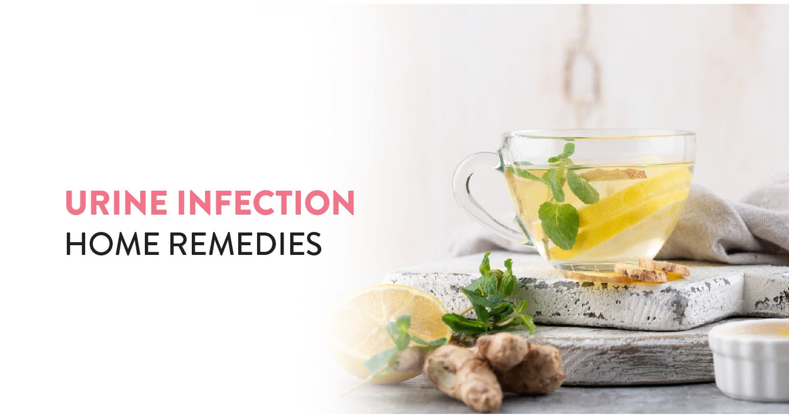
UTIs are a frustrating and common problem, especially if they keep recurring. While medications can effectively cure UTIs, many individuals also find relief from their UTI symptoms with natural solutions. Let’s examine a few of the most popular all-natural remedies for UTIs.
Table of Contents
UTIs (urinary tract infections) are very common. UTIs happen when bacteria, typically from the rectum or skin, enter the urethra. Any section of the urinary tract can get infected, but bladder infections are the most frequent.
Although UTIs can affect anybody, those who are born as females are more likely to have them. That is because the female urethra, the tube that carries urine out of the bladder, is shorter than the male urethra.
Bacteria might enter the bladder more easily due to the shorter distance (in women). Another factor is the urethra’s closeness to the rectum and vagina, both of which are bacterial breeding grounds. Additional UTI risk factors include:
Bacteria cause almost 95% of UTIs, but fungi can also cause infection.
Though antibiotics usually treat UTIs, there are various natural ways to reduce the risk of recurrence and help manage infections.
For mild UTIs, home remedies might help alleviate symptoms, and/or prevent infections from developing. The following are some of the most popular DIY treatments for UTIs:
Cranberry juice may help against urinary tract infections. It has a long history of usage in the management of UTIs. The use of cranberry juice in UTIs is due to its potential antibacterial properties.
Cranberry juice has many bioactive compounds (organic acids, anthocyanins, flavonoids etc.) which may result in the excretion of hippuric acid. Hippuric acid, a strong antibacterial agent, may therefore contribute to reducing the number of germs responsible for UTIs.
It is known that cinnamon may have an action against the bacteria causing UTIs in long-term catheter users. This activity may be due to its potential effect of preventing the bacteria from coating the urinary tract. Therefore, cinnamon may be an effective natural remedy for UTIs.
Probiotics are gut-friendly bacteria that may be helpful against UTIs as well. They mostly include Lactobacillus bacteria, which cling to the walls of the urinary system and appear to function as a barrier against the bacteria that cause UTIs.
Coconut water has multiple benefits in many different forms. Drinking coconut water regularly may be helpful for those suffering from UTIs and enhance urinary health in general. It facilitates urine formation and urination and may help in flushing the kidneys too.
Your doctor may recommend that you increase the intake of fluids, especially water. This habit may be helpful in urinary tract infections as it might help fight against bacterial infection. Therefore, you should be drinking the daily recommendation of 6 to 8 glasses of water.
According to some research, boosting your vitamin C consumption may help prevent UTIs. According to theory, vitamin C works by making urine more acidic, which kills the infection-causing bacteria.
Fruits and vegetables are a good way to increase your intake of vitamin C and are especially high in it. Kiwifruit, oranges and red peppers all contain the full recommended daily amount of vitamin C in just one serving.
Preventing UTIs starts with practising a few good hygiene and bathroom habits.
First, it’s important not to hold your urine for too long as it can lead to a buildup of bacteria, resulting in infection. By limiting the transmission of germs, urinating after intercourse has long been associated with a decreased incidence of UTIs.
Those who are prone to UTIs should also refrain from using spermicide because it has been associated with an increase in UTIs.
Finally, when using the toilet, especially if you have a female urethra, make sure you wipe front to back. Wiping from back to front increases the risk of UTIs by allowing germs to enter the urinary system.
Some other tips for managing UTIs are:
You Can Also Read: Urinary Tract Infections – A common issue in the corporate crowd
If you have symptoms of a UTI, it is important to talk with a doctor. While natural remedies might help, a medical professional can diagnose a UTI and prescribe antibiotics to treat the infection.
UTIs have a risk of severe complications, including spreading to your kidneys if they are left untreated. Signs of a severe infection should not be ignored. If you encounter any of the following, get in touch with a medical expert:
Urinary tract infections are quite common and can be irritating. You can use home remedies in most cases for relief, however, if they don’t work then medications might be required. It is always advisable to seek medical help from an experienced urologist. Timely care and help can ensure an appropriate diagnosis and treatment of your condition.
At the CK Birla Hospital, we ensure patients get holistic medical support which includes treatment in a compassionate environment. This patient-centric approach not only helps patients heal better but also ensures they are aware of the preventive measures as well. In case you need to consult a urologist, reach out to us, or book a direct appointment with Dr. Shalabh Agarwal at the CK Birla Hospital.
You can try home remedies such as taking probiotics, vitamin C supplements or drinking cranberry juice to reduce and prevent the recurrence of UTIs. You should also be aware that you might still have a UTI even if your symptoms go away, so it is advisable to speak with a doctor about the best UTI treatment for yourself.
Water is a fantastic drink which helps get rid of a urinary tract infection faster by flushing out the bacteria in your bladder. It also dilutes your urine so that urinating might be less painful.
Some foods and drinks which should be avoided in case of a urinary tract infection are fizzy drinks, coffee, alcohol, Inflammatory and spicy food, citrus fruits, refined carbohydrates and sugar.
Vaginal discharge is perfectly natural. It is the body’s way of protecting the vagina from infections and keeping it clean. Discharge from the vagina can also range in amount and colour, depending on things like where you are in your menstrual cycle.
While brown discharge might look alarming, it is not always a reason to worry. Brown discharge can also be related to hormonal changes, periods or certain conditions affecting the ovaries and uterus. You may see this colour throughout your cycle, typically around the time of menstruation.
Getting to know your body can help you notice when something might be a cause for concern. Try to take note of what your vaginal discharge is normally like and speak with a healthcare professional if you notice any significant changes or any worrying symptoms.
Table of Contents
Brown discharge is common among most females, especially around 2 to 3 days before the period. The brown discharge might be a sign of a period coming In such cases. Some women get brown discharge following their period. This happens when the uterus cleans itself after the period and there might be very light or no bleeding in such a case. Several days after the menstruation has ended, there may be extremely mild discharge.
Use of Hormonal Birth Control or Contraceptives can also cause brown vaginal discharge. Using hormonal birth control causes the uterine lining to thin. This condition might result in mild brown discharge even when there is no period. In this situation, brown discharge develops as the body tries to adjust to the altered hormone levels.
A female might experience brown discharge as a sign of ovulation in rare cases. When the mature egg breaks open the ovary, there might be slight bleeding. The colour of the vaginal discharge can be brown to reddish pink. Sometimes, it also looks like brown discharge mixed with clear discharge. The female can also experience cramps, lower back pain and abdominal pain in such situations.
The brown discharge might also be a result of breakthrough bleeding and there is mild light brown discharge in this case. A female might experience mild brown discharge between period cycles.
Some women might have mild brown discharge after sexual intercourse. This might be the effect of intense sexual activity. In such cases, brown discharge occurs due to slight bleeding of the vaginal canal (cervix). Certain medical conditions can also cause unusual or abnormal brown discharge.
There are many possible causes of brown discharge. Brown discharge is nothing to be concerned about in most cases. However, brown discharge can indicate a possible health issue if it is accompanied by other symptoms, such as changes to your menstrual cycle, a strong odour, pain or vaginal itching. There are several reasons why brown discharge could occur suddenly, including the following:
If you are in the early stages of pregnancy, you may have brown spotting or light bleeding. As many as 30% of pregnant people have brown spotting or light bleeding during their first trimester. This is normal, but you should still call your healthcare provider or doctor to check that everything is alright.
Some contraceptive methods like implants or IUDs release the progestin hormone into your body to prevent you from getting pregnant. As your body adjusts to the new form of birth control, you may experience side effects like brown discharge, breakthrough bleeding, spotting and irregular menstruation.
PID is an infection of the uterus and cervix that can sometimes result in brown discharge. It’s usually caused by an untreated STI (sexually transmitted infection) like chlamydia or gonorrhoea. Other PID symptoms include heavy discharge with a bad smell, painful urination, fever, pain during sex and pain in the pelvis and lower abdomen. PID is a severe medical illness that requires immediate diagnosis and care.
Some STIs(Sexually Transmitted Infections), like gonorrhoea or chlamydia, can cause you to have spotting or brown discharge when you do not have your period. Other symptoms include vaginal discharge with an unpleasant odour, a burning sensation when urinating and pain during sex.
One in ten persons with a vagina of reproductive age (15–49) have PCOS, a very prevalent hormonal disorder. Its exact cause is unknown but likely has to do with excess insulin in the body and genetics. People with PCOS have an imbalance of reproductive hormones; their bodies produce higher levels of hormones called androgens, resulting in missed or irregular periods. Brown discharge in place of your menstruation is one indication of PCOS. Other symptoms include dark patches on the skin, ovarian cysts, infertility, obesity, excessive hair growth, acne and irregular menstrual cycles.
Endometriosis is a chronic condition that occurs when the tissue that is usually in your uterine lining starts growing in other areas, like your bowels, fallopian tubes or ovaries. Heavy periods and irregular bleeding are common in endometriosis patients, and they occasionally have internal bleeding that results in brown discharge.
It is normal to experience some spotting or light bleeding after a vaginal exam or a pap smear test. Gynaecologists take great care in these cases, but occasionally their instruments might produce a minor irritant inside the walls of your vagina or cervical lining. If you experience this without any other symptoms, you do not normally need to worry about it.
Vaginal irritation can cause light bleeding if you have recently had vigorous sex. It might take a few days for the blood to leave your body, and during this time the blood turns brown as a result of oxidation.
Occasionally after unprotected sex, a fertilised egg will attach itself outside your uterine cavity, causing an ectopic pregnancy. This is rare, but when it happens, it is serious and requires medical care as soon as possible to prevent life-threatening complications. Signs of ectopic pregnancy include spotting and light bleeding, lightheadedness, vomiting and nausea and severe abdominal cramps.
In extremely rare cases, brown discharge could be a sign of cervical cancer if it’s accompanied by symptoms including weakness, unusual weight loss, bleeding between periods, prolonged or heavy periods and painful intercourse. Gynecologic care and regular pap smears can help detect and prevent any early signs of cervical cancer. Between the ages of 21 and 65, everybody with a vagina should have these tests to be proactive.
A female might experience abdominal cramps and light bleeding in her early pregnancy. Brown discharge might be a cause of implantation bleeding, as an early sign of pregnancy. Implantation bleeding happens when the fertilised egg implants in the inner lining of the uterus. There might be slight bleeding that may be brown at this point.
This kind of brown discharge typically appears when a woman is about to start menstruating. Brown discharge instead of a missed period in a female is typically an indication of pregnancy. Consult your gynaecologist as soon as possible in such a case.
The abnormal brown discharge accompanied by several other symptoms and complications might be something you need to consult with your gynaecologist
There might be certain infections that cause abnormal brown discharge. If you observe brown discharge for several days and it does not stop on its own, it is wise to visit a gynaecologist and get help. Certain diseases or infections that cause brown discharge are:
There are other symptoms associated with brown discharge that result from any of the aforementioned problems. In cases of severe illness, the following other symptoms may also appear along with brown discharge:
Because they may worsen over time, you shouldn’t treat these symptoms lightly. So, waste no time and consult a gynaecologist as soon as you observe pain or discomfort accompanied by brown discharge during pregnancy.
Brown vaginal discharge may not necessarily indicate cause for concern. If you are concerned about your condition and feel that it might be serious, then it is always advisable to seek medical help from an experienced gynaecologist. Timely care and help can ensure appropriate treatment as per the cause of your condition.
At the CK Birla Hospital, we ensure patients get holistic medical support which includes treatment in a compassionate environment. This patient-centric approach not only helps patients heal better but also ensures they are aware of the preventive measures as well. In case you need to consult a gynaecologist, reach out to us, or book a direct appointment with Dr. Seema Sehgal at the CK Birla Hospital.
A few days before and following a period, a person frequently has brown vaginal discharge. When it occurs before, it is likely an early, light flow. Brownish discharge after a period is the result of extra menstrual blood leaking out of the vagina.
Spotting or brown vaginal discharge can sometimes be an early sign of pregnancy. This is known as implantation bleeding.
Yes, it is normal. Sometimes your uterus has less tissue to clear than others, and when this happens, you will experience brown discharge instead of a full period. It is usually nothing to worry about, but if you find this happening month over month, talk to your Abnormal Vaginal Discharge doctor.

You must obtain iron from food since it is a necessary nutrient. It aids in the body’s transportation of oxygen.
An adult human’s body has 4 grams (0.005% of body weight) of iron, largely in the form of myoglobin and haemoglobin. Oxygen transport by blood and oxygen storage in muscles are the essential roles played in vertebrate metabolism by these two proteins respectively. Human iron metabolism requires a minimum of iron in the diet to maintain the necessary levels.
Iron is both potentially toxic and necessary to the body. Controlling iron levels in the body is a critically important part of many aspects of human disease and health. A deficiency can occur if your iron intake is too low to replace the amount you lose daily as it can lead to symptoms like fatigue and cause anaemia. Fortunately, you may satisfy your daily needs for iron by eating a variety of healthy food items.
Iron is a mineral that serves various important functions, its main one being to carry oxygen throughout your body as a part of red blood cells.
For non-pregnant adults, the daily value for iron is 8-18 mg. If your intake is insufficient to make up for the quantity you lose each day, a deficiency may result.
It’s interesting to note that your body’s ability to absorb iron depends in part on how much you have stored. Some foods which are high in iron are listed below:
You Can Also Read: High-Fibre Foods You Should Eat
You Can Also Read: Potassium Rich Foods
Iron plays a crucial role in oxygen transportation in your body. If you are unable to decide or meet your body’s iron requirements, then it is always advisable to seek medical help from an experienced dietitian. Timely care and help can ensure the inclusion of sufficient iron in your regular diet chart.
At the CK Birla Hospital, we ensure patients get holistic medical support which includes treatment in a compassionate environment. This patient-centric approach not only helps patients heal better but also ensures they are aware of the preventive measures as well. In case you need to consult a dietitian, reach out to us, or book a direct appointment at the CK Birla Hospital.
Yes, almonds are a good source of iron, offering 5.3 mg per cup whole (66% of the recommended dietary allowance).
Iron content in milk and milk substitutes is low. Milk hinders the body’s capacity to absorb iron from meals and supplements.
Apples are known for their high iron content and vitamin C content, both of which are essential for reversing and preventing anaemia. Increasing your iron consumption will help you overcome anaemia, which is a haemoglobin deficit in the blood.
OA (Osteoarthritis) is one of the most common types of knee arthritis which can result in disability and pain. Daily activities which you like can become challenging and symptoms may worsen with weight-bearing.
A high tibial osteotomy (HTO) is a surgical procedure that realigns your knee joint. For some patients who have knee arthritis, this surgery can prevent or delay the need for a total or partial knee replacement by preserving damaged joint tissue.
People with knee issues, particularly when combined with bowleggedness, should talk to their orthopaedic team about HTO as it is recommended to patients on a case-to-case basis after considering the pros and cons.
Table of Contents
An orthopaedic surgical treatment called a high tibial osteotomy treats compartmental osteoarthritis and a varus deformity. Since the procedure’s inception, improvements in technique, fixation technology, and patient selection have allowed HTO to gain popularity among younger, more active patients looking to treat arthritis. Realigning the weight-bearing line of the knee is the surgery’s objective. By realigning the knee, the arthritic medial compartment receives less force from weight-bearing and more comes from the lateral compartment, which is healthy. Delaying the onset or progression of osteoarthritis in the medial compartment of the knee due to this reduction in force or load in the diseased portion of the knee joint also reduces knee discomfort.
The International Society of Arthroscopy, Knee Surgery, and Orthopaedic Sports Medicine (ISAKOS) created the recognized methodology for patient selection in 2004. An ideal patient with this procedure is:
Contraindications specified by ISAKOS are:
You Can Also Read: Common Knee Injuries You Should Be Aware Of
The general surgical technique includes either performing HTO alone or performing HTO in combination with ligament reconstruction. When deciding which treatment avenue to take, one must consider patient demographics, their predominant symptoms, and which ligaments, if any are involved. When ligaments are involved, but the ACL deficiency is chronic and pain is due to arthritis and malalignment, HTO alone should be sufficient. However, if instability is the predominant symptom, for example, an acute ACL deficiency, HTO in combination with ACL reconstruction may be performed to protect the ACL graft that was constructed.
The two most common surgical techniques used in HTO are lateral close wedge osteotomy and medial open wedge osteotomy.
The initial cut is made between the posteromedial border of the tibia and the medial aspect of the tibial tubercle. The medial collateral ligament (MCL) is exposed by cutting the sartorius fascia and pulling it medially. MCL is then removed from its insertion medially. Two K-wires are placed towards the lateral cortex, about 4 cm below the joint line. The osteotomy is done below the K-wires and parallel to the joint line.
The advantages of the medial open wedge method include less risk of peroneal nerve injury compared to the lateral close wedge method, no limb shortening, no bone loss, and the use of a single cut with no need to detach muscles.
Starting at the anterolateral aspect about 1 cm below the knee joint line, an L-shaped cut is made to the lateral edge of the tibial tubercle and anterior tibial crest. To expose the bone, the fascia of the anterior compartment is cut near the anterior tibial crest and the anterior tibialis is elevated. Osteotomy starts 15 mm below the joint line, just above the tibial tubercle, and is directed parallel to the joint line, medially.
Some of the advantages of the lateral close wedge method are faster healing with less morbidity, greater potential for healing, and no need for bone grafting, unlike the medial open wedge method.
Two main types of fixation plates are used: spacer plates and plate fixators. Spacer plates are lower-profile implants that require a smaller incision. The disadvantage of using a spacer plate is the decreased rigidity associated with increased rates of delayed union or nonunion. Because of this, spacer plates require a longer period of staying off the leg that was operated on. Plate fixators give a stronger fixation, allowing for earlier weight-bearing and initiation of therapy. A couple of studies attempted to compare these two methods but found no differences in reliability.
After part of the bone is removed, there is a space that may need to be filled. Some prefer using a graft or bone substitute, which will hopefully increase stability and decrease healing time. Bone can also be taken from the hip of the patient to use as a graft. This has a lower complication rate so is considered in someone who is at risk of the bone not healing, like a smoker or obese patient.
The most common complications are the same as those occurring for any orthopaedic procedure performed on a lower limb. These are:
The complications specific to the HTO are rare and include the failure of the bone to heal, common peroneal nerve palsy, decreased range of motion, a low-lying knee cap, and a fracture inside the knee joint.
Knee replacement surgery can treat this problem when the joint deterioration is beyond repair or recovery. However, in certain cases, a high tibial osteotomy can realign the knee by wedge-opening the top part of the tibia to reposition the knee joint, relieving pressure from the injured side. Then, the burden of bearing shifts from the unhealthy tissue to the injured or worn tissue.
This sort of osteotomy is normally thought of as a technique to delay the need for a knee replacement because these advantages typically disappear after 8 to 10 years. Younger individuals who experience discomfort from instability and misalignment are often the only ones who benefit from this therapy. To allow cartilage repair tissue to develop without being put under too much strain, an osteotomy may also be carried out in combination with other joint preservation techniques.
An HTO (high tibial osteotomy) is a surgical technique that realigns the knee joint. For individuals with knee arthritis, it is a blessing since it can postpone or eliminate the need for a partial or full knee replacement. It is always advisable to seek medical help from an experienced orthopaedist. Timely care and help can ensure an appropriate diagnosis and treatment of your condition.
At the CK Birla Hospital, we ensure patients get holistic medical support which includes treatment in a compassionate environment. This patient-centric approach not only helps patients heal better but also ensures they are aware of the preventive measures as well. In case you need to consult an orthopaedist, reach out to us, or book a direct appointment at the CK Birla Hospital.
A high tibial osteotomy (HTO) is a major surgical procedure that improves the condition of the knee joint. It is also less of an intervention compared to a total knee replacement.
Following surgery, you must utilise crutches for 12 weeks while wearing your brace for 6 weeks. You progressively put more weight on your leg during the second six weeks while continuing to use crutches as protection. Before progressively increasing the weight you bear on your leg, an x-ray is done at six weeks to monitor bone repair.

फैटी लीवर को हेपेटिक स्टीटोसिस के रूप में भी जाना जाता है। यह लीवर की एक सामान्य स्थिति है जब लीवर कोशिकाओं में अत्यधिक वसा जमा हो जाती है। इस स्थिति को मोटे तौर पर दो प्रकारों में बांटा जा सकता है: अल्कोहलिक फैटी लीवर रोग (एएफएलडी) और गैर-अल्कोहल फैटी लीवर रोग (एनएएफएलडी)। फैटी लीवर वसा के सौम्य संचय से लेकर अधिक गंभीर रूपों तक हो सकता है जिससे लीवर में सूजन, घाव और लीवर की बीमारी हो सकती है।
फैटी लिवर की समस्या कई कारणों से होती है जिसमें मुख्य रूप से निम्न शामिल हो सकते हैं:
अल्कोहलिक फैटी लीवर मुख्य रूप से लंबे समय तक अत्यधिक शराब के सेवन के कारण होता है। लीवर अल्कोहल को संसाधित करता है, और लंबे समय तक शराब के सेवन से लीवर कोशिकाओं के भीतर वसा का संचय हो सकता है।
गैर-अल्कोहल फैटी लीवर रोग अधिक आम है और इसके कई योगदान कारक हैं, जिनमें शामिल हैं:
मोटापा एनएएफएलडी के लिए एक प्रमुख जोखिम कारक है। शरीर में अतिरिक्त वसा लीवर में फैटी एसिड के प्रवाह को बढ़ा सकती है, जिससे वसा जमा हो जाती है।
इंसुलिन प्रतिरोध, जो अक्सर टाइप 2 मधुमेह से जुड़ा होता है, यकृत में वसा के भंडारण को बढ़ा सकता है।
ऊंचा रक्त शर्करा स्तर, जैसा कि अनियंत्रित मधुमेह में देखा जाता है, फैटी लीवर के विकास में योगदान कर सकता है।
ट्राइग्लिसराइड्स और अन्य रक्त लिपिड का ऊंचा स्तर एनएएफएलडी के खतरे को बढ़ा सकता है।
भारी वजन घटाने या कुपोषण के कारण लीवर रक्तप्रवाह में संग्रहित वसा को छोड़ सकता है, जो लीवर की कोशिकाओं में जमा हो सकता है।
कुछ दवाएँ, जैसे कॉर्टिकोस्टेरॉइड्स, टैमोक्सीफेन और एंटीरेट्रोवायरल दवाएं, फैटी लीवर में योगदान कर सकती हैं।
ये भी पढ़े: Neutrophils in Hindi (White Blood Cell Kaise Badhaye)
फैटी लीवर के लक्षण उनकी गंभीरता में व्यापक रूप से भिन्न हो सकते हैं, और कई मामलों में, व्यक्तियों को किसी भी ध्यान देने योग्य लक्षण का अनुभव नहीं हो सकता है। जब लक्षण प्रकट होते हैं, तो वे हल्के और गैर-विशिष्ट होते हैं। थकान एक आम शिकायत है, जिसमें व्यक्ति लगातार थकान और ऊर्जा की कमी महसूस करते हैं। कुछ लोग पेट के ऊपरी दाहिने हिस्से में हल्की असुविधा या दर्द की भी शिकायत कर सकते हैं, जो बढ़े हुए लीवर से जुड़ा हो सकता है। कुछ मामलों में बिना कारण वजन कम हो सकता है। इस बात पर ध्यान देना जरूरी है कि ये लक्षण फैटी लीवर के लिए विशिष्ट नहीं हैं और विभिन्न अन्य चिकित्सीय स्थितियों के कारण हो सकते हैं।
और अधिक पढ़ें: हाई कोलेस्ट्रॉल के लक्षण(High Cholesterol Symptoms in Hindi)
फैटी लीवर के प्रबंधन (Managing Fatty Liver) में मुख्य रूप से जीवनशैली में बदलाव और अंतर्निहित जोखिम कारकों को संबोधित करना शामिल है। विशिष्ट दृष्टिकोण स्थिति की गंभीरता, जटिलताओं की उपस्थिति और व्यक्तिगत रोगी कारकों के आधार पर भिन्न हो सकता है। फैटी लीवर के इलाज के कुछ प्रमुख पहलू नीचे दिए गए हैं:
मोटापे से ग्रस्त व्यक्तियों के लिए, संतुलित आहार और नियमित शारीरिक गतिविधि के संयोजन के माध्यम से वजन कम करना महत्वपूर्ण है। धीरे-धीरे, स्थायी वजन घटाने से लीवर में वसा कम हो सकती है और लीवर की कार्यप्रणाली में सुधार हो सकता है।
ऐसा आहार जिसमें संतृप्त वसा, शर्करा और परिष्कृत कार्बोहाइड्रेट कम हों, फैटी लीवर को प्रबंधित करने में मदद कर सकते हैं। अधिक फल, सब्जियाँ, साबुत अनाज और लीन प्रोटीन आदि का सेवन करें। प्रसंस्कृत खाद्य पदार्थों और शर्करा युक्त पेय पदार्थों के अत्यधिक सेवन से बचें।
इंसुलिन संवेदनशीलता में सुधार और लीवर में वसा कम करने के लिए नियमित शारीरिक गतिविधि आवश्यक है। रोजाना सुबह या शाम में हल्का-फुल्का व्यायाम करने की कोशिश करें।
यदि आपको मधुमेह या प्रीडायबिटीज है, तो दवा, आहार और व्यायाम के माध्यम से ब्लड शुगर के स्तर को प्रबंधित करना महत्वपूर्ण है। इससे लीवर की और अधिक क्षति को रोकने में मदद मिल सकती है।
कुछ मामलों में, डॉक्टर फैटी लीवर रोग के विशिष्ट पहलुओं को प्रबंधित करने के लिए दवाएं लिख सकते हैं, जैसे इंसुलिन-संवेदनशील दवाएं या लिपिड-कम करने वाली दवाएं।
यदि अल्कोहलिक फैटी लीवर रोग का निदान किया जाता है, तो शराब पीना पूरी तरह से बंद करना महत्वपूर्ण है। यह लीवर की क्षति की प्रगति को रोक सकता है और प्रारंभिक चरण में स्थिति को उलट भी सकता है।
इन सबके अलावा, अगर फैटी लीवर अधिक गंभीर रूप में बढ़ता है जिसे गैर-अल्कोहल स्टीटोहेपेटाइटिस (एनएएसएच) के रूप में जाना जाता है, जिससे लीवर फाइब्रोसिस और सिरोसिस हो सकता है, तो मेडिकल हस्तक्षेप आवश्यक हो सकता है।