Filter :
Your body has cartilage, which is a supple and durable kind of connective tissue. It protects and envelops the ends of long bones at the joints. Blood vessels and nerves are absent from cartilage.
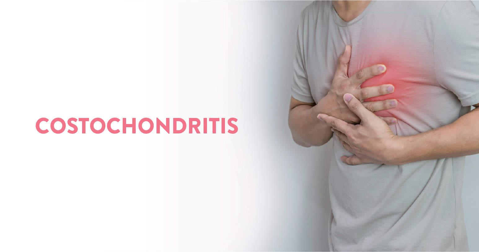
The harmless inflammation (swelling) of cartilage in your chest is called costochondritis. Costochondritis feels like an aching or sharp pain, which can develop slowly or start suddenly and spread across your chest.
Costochondritis that is not severe may go away in a few days. The majority of instances don’t last more than a year, however chronic cases might linger for weeks or longer. If you suffer chest discomfort while engaging in activities such as manual labour or high-impact exercises, get medical help right away.
Table of Contents
Costochondritis is the medical term for cartilage inflammation in the rib cage. It might happen due to an illness, an accident, or other health problems. This ailment often affects the cartilage at the costosternal joint or costosternal junction, which is where the upper ribs join the breastbone, commonly known as the sternum.
Costochondritis can result in mild to severe chest discomfort. Mild instances may simply result in a mild tenderness or soreness when you press on the affected area in your chest.
Deep breathing and certain motions may make more severe instances worse. The problem often goes away within a few weeks or months, although in certain situations, treatment may be necessary.
Costochondritis frequently manifests as chest pain in the upper and middle rib area, on either side of the breastbone. This discomfort may worsen over time gradually or unexpectedly.
Additional symptoms can include:
It’s crucial to remember that signs like tightness in the chest and radiating pain might signify a variety of illnesses, including a heart attack. If your chest discomfort is severe and persistent, get medical help right away.
You Can Also Read: 10 tips to prevent back pain during work from home
In the majority of cases, the actual cause of costochondritis is unknown. However, the following circumstances might lead to it:
According to some studies, women, particularly athletes, are more likely than males to develop costochondritis. You may also be more susceptible to developing this illness if you:
While there is no specific test to identify costochondritis, your doctor will almost certainly do some tests and ask you several questions to identify the cause of your chest discomfort.
However, depending on your unique medical history, your doctor may order certain tests to rule out other possible causes of your chest discomfort, such as pneumonia or coronary heart disease. Typically, lab tests are not required to identify costochondritis.
To be sure your lungs aren’t emitting anything odd, your doctor could ask you to obtain an X-ray.
Your X-ray ought to be normal if you have costochondritis. To make sure your heart isn’t the source of your chest pain, they can also advise getting an electrocardiogram (ECG).
When diagnosing costochondritis, it is frequently necessary to rule out other potential, more serious causes.
Usually, costochondritis resolves on its own, however, it may persist for a few weeks or longer. The therapy aims to make people feel less uncomfortable.
Your health care provider might recommend:
Physical therapy treatments might include:
Injecting numbing medicine and a corticosteroid straight into the affected joint is another possibility if conservative procedures don’t relieve the pain.
You Can Also Read: Causes of stomach pain after eating
Normally, treatment of the inflammation and pain causes costochondritis to eventually go away on its own.
Even with therapy, the pain from chronic costochondritis may come back or linger when you exercise or do particular activities. In these circumstances, it may be necessary to seek long-term care to prevent costochondritis from impairing your quality of life and capacity for everyday tasks.
Costochondritis-related pains might be a sign of other problems. When you have chest pain, you should seek medical attention right away to rule out pneumonia or a heart attack. Chest discomfort is frequently a sign of heart or lung problems.
Costochondritis-related chest discomfort may be a sign of fibromyalgia. You could also have pain in your chest if you have fibromyalgia in addition to:
Inquire with your doctor about a fibromyalgia test if you also suffer chest pains in addition to these other symptoms. Understanding this disease will make it easier for you to deal with the symptoms and prevent it from interfering with your everyday activities.
Costochondritis is a benign inflammation (swelling) of the cartilage in your chest. The majority of cases last no more than a year, however, chronic cases might persist for weeks or longer. It is always advisable to seek medical help from an experienced rheumatologist. Timely care and help can ensure an appropriate diagnosis and treatment of your condition.
At the CK Birla Hospital, we ensure patients get holistic medical support which includes treatment in a compassionate environment. This patient-centric approach not only helps patients heal better but also ensures they are aware of the preventive measures as well. In case you need to consult a rheumatologist, reach out to us, or book a direct appointment with Dr. Chirag Arora at the CK Birla Hospital.
Typically, the front left side of your breastbone is where discomfort first manifests. It frequently affects more than one rib. It might impact your arms and shoulders in addition to the rest of your chest.
Any action that puts stress on your chest area, such as rigorous exercise or even basic movements like reaching up to a high cabinet, might cause costochondritis. You should refrain from engaging in any activities that aggravate your chest pain until the cartilage and rib inflammation have subsided.
Your knee is the largest joint in your body and it joins your leg with the thigh. Each knee has 2 joints, one between the femur and patella (patellofemoral joint) and one between the femur and tibia (tibiofemoral joint).
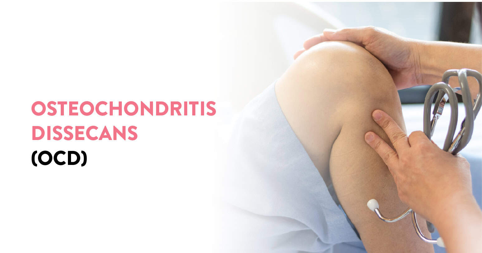
The joint condition which occurs when your bone separates from cartilage and starts to die is known as osteochondritis dissecans (OCD). It is typically due to a lack of blood flow to the bone. When small pieces of the separated cartilage and bone start to break loose, it can reduce your range of motion in the affected area and cause pain.
Although the majority of patients recover completely, having OCD can raise your chance of subsequently getting osteoarthritis in the affected joint. OCD usually requires you to rest the affected area for several weeks or might even require surgery in some cases (if the symptoms do not improve after 4 to 6 months). Your doctor will also likely recommend surgery if you have loose cartilage or bone fragments in your joints.
Table of Contents
Osteochondritis dissecans is a disorder where the interior of the joints (where the ends of one bone meet the ends of another bone) become mushy due to a lack of blood flow. As a result, a tiny portion of the bone degenerates and splits away from the larger bone. The bone portion may then split and come free, along with the cartilage that covers and shields the bone.
The cartilage and loose bone may stay where they are or they may slide inside the joint, making the joint unstable. Where the bone and cartilage part ways, a lesion results from the disorder. The entire process may take many months or even years, and symptoms may not manifest for a long period.
Osteochondritis dissecans often affects the knee, elbow, ankle, and thigh bone (femur) area. Other joints, such as the hip and shoulder, are also susceptible to the illness.
Osteochondritis dissecans frequently affects just one joint. The condition, known medically as sporadic osteochondritis dissecans, presents as a single lesion in a single joint.
You May Also Read: Arthritis of Knee: Symptoms & treatment
Depending on the affected joint, the following osteochondritis dissecans signs and symptoms may be present:
Most of the time, there is no recognized aetiology for sporadic osteochondritis dissecans. One reason for the syndrome is that repetitive damage or stress to a joint over time, such as from participating in sports, can cause it.
The ACAN gene, which produces the aggrecan protein, is the source of the hereditary mutations (changes) that lead to familial osteochondritis dissecans. The protein cannot properly produce cartilage as a result of the mutation, leaving the cartilage weak and disorganized. It is unclear, nevertheless, how the brittle and disorderly cartilage contributes to lesions and bone separation.
Sporadic osteochondritis dissecans in children and young adolescents will often go away on their own as they age. Rest and a break from physically demanding activities like jogging and jumping might help them reduce discomfort and swelling. The doctor could suggest using an over-the-counter painkiller or anti-inflammatory drug.
The damaged joint will resume normal function within six to twelve weeks. The young athlete should ease back into sports activity with mild workouts (yoga, swimming, cycling, or stretching).
The doctor may advise using crutches or may apply a brace, splint, or cast to the joint if the recovery is taking too long. A recommendation for physical treatment from the doctor is also possible.
For patients with osteochondritis dissecans, the doctor could advise surgery if:
A camera and small equipment are frequently used during arthroscopic surgery, which is done through very small incisions. There are three types of surgery:
Following surgery, the patient will use crutches for approximately six weeks before beginning physical therapy for two to four months to strengthen the body and regain the range of motion in the joint. Four to five months following surgery, the patient might be ready to start engaging in strenuous physical activity again.
You May Also Read: Chondromalacia Patella: Causes, Symptoms and Treatment
Since its origins are unknown, osteochondritis dissecans can be challenging to prevent. By wearing pads and other protective gear, for example, young athletes can take precautions to safeguard their joints. They should also practise the required physical skills for their sport, stretch and warm up before engaging in strenuous exercise, and stretch and cool down afterwards.
Osteochondritis dissecans (OCD) is a disorder of the joints in which the bone begins to detach from the cartilage and die. It might make the afflicted region more painful and limit your range of motion. If you don’t feel better after resting, you could need surgery since OCD increases your risk of developing osteoarthritis later on. It is always advisable to seek medical help from an experienced orthopaedist. Timely care and help can ensure an appropriate diagnosis and treatment of your condition.
At the CK Birla Hospital, we ensure patients get holistic medical support which includes treatment in a compassionate environment. This patient-centric approach not only helps patients heal better but also ensures they are aware of the preventive measures as well. In case you need to consult an orthopaedist, reach out to us, or book a direct appointment with Dr. Praveen Tittal at the CK Birla Hospital.
Pain frequently becomes worse with exercise. If left untreated, the problem may result in arthritis and cause discomfort, swelling, catching or locking of the joint.
Once the patient heals, osteochondritis dissecans often don’t reoccur. But occasionally the illness just looks to get better when the symptoms disappear. In such circumstances, symptoms may eventually reappear.
Your knee is the largest joint in your body and it joins your leg with the thigh. Each knee has 2 joints, one between the femur and patella (patellofemoral joint) and one between the femur and tibia (tibiofemoral joint).
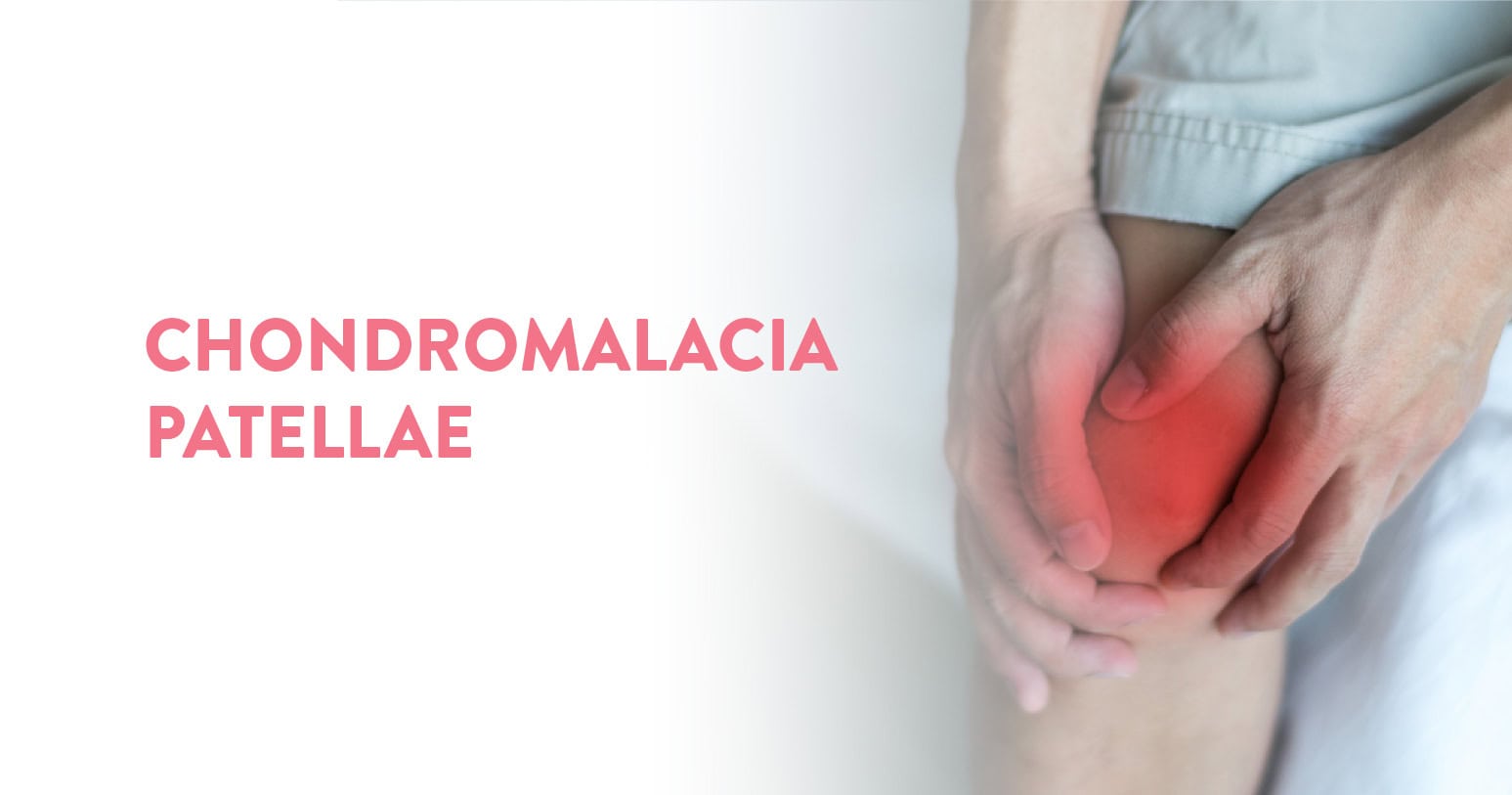
Chondromalacia patellae (or “runner’s knee”) is caused by the softening of your kneecap cartilage. Common among young athletes, it might also affect older adults with knee arthritis.
Chondromalacia is mostly seen as an overuse injury in sports, and sometimes taking some days off from training can produce good results. In other cases, the cause is improper knee alignment, and simply resting does not provide relief. A conversation with your healthcare provider will help you get clarity for further courses of tests or treatments.
Table of Contents
The deterioration or softening of the cartilage on the bottom of the kneecap is known as chondromalacia patellae. It is comparable to patellofemoral pain syndrome (runner’s knee), in which the kneecap and surrounding area are painful.
This condition is common among young athletes but may also occur in older adults who have arthritis of the knee.
A smooth (articular) cartilage layer covers the top of the thigh bone and the bottom of the kneecap, allowing the two bones to easily glide over one another. When the knee is bent or stretched, the injured cartilage can cause the joint’s surface to become rough and easily inflamed. The pain might range from mild to severe depending on the severity of the injury.
A dull discomfort under or around the kneecap that worsens when going downstairs is the most typical sign of chondromalacia patellae. Additionally, there could be pain while getting out of a chair or ascending steps.
When moving the knee, a person with chondromalacia frequently complains of a grinding or cracking sensation. When doing activities that put a lot of strain on the knees, such as exercising, or after sitting or standing still for a long time, the discomfort is frequently severe. Additionally typical are kneecap swelling and irritation.
Normally, your kneecap sits on top of your knee joint. The back of your kneecap slides across the femur, or thigh bone, at the knee as you bend your knee. Your kneecap is joined to your shinbone and thigh muscle via ligaments and tendons. Your kneecap may bump against your thigh bone if one of these parts isn’t functioning properly. This unnatural rubbing can cause the patella to deteriorate, which causes chondromalacia patellae, often known as runner’s knee.
Improper kneecap movement may result from:
Several things might make you more likely to get chondromalacia patellae.
Inflammation of the joint and surrounding tissue is a sign of arthritis, which can also cause a runner’s knee. The kneecap may become dysfunctional due to inflammation.
A high level of activity or regular exercise that stresses your knee joints might raise your chance of developing knee issues.
Your chance of getting a runner’s knee might rise if you have previously suffered from a kneecap injury, such as a dislocation.
The knee joints may experience more stress from flat feet than from higher arches.
Since women normally have less muscle mass than men do, they are more susceptible to acquiring a runner’s knee. This may result in improper knee alignment and increased lateral (side) kneecap pressure.
Young adults and adolescents are particularly vulnerable to this illness. Rapid bone and muscle development during growth spurts may cause temporary muscular imbalances.
Your kneecap and joint are under pressure, thus the treatment aims to lessen it. The first course of therapy could involve resting, stabilising, and applying ice to the joint. Resting frequently helps to heal the cartilage damage that results in a runner’s knee.
To minimise inflammation around the joint, your doctor may recommend anti-inflammatory medicine for many weeks. The following treatments should be considered if swelling, soreness, and discomfort continue.
Quadriceps, hamstrings, adductors, and abductors-specific physical treatment can help you gain more muscular strength and balance. The balance of muscles will aid in preventing knee misalignment.
Non-weight-bearing workouts like swimming or using a stationary bike are frequently advised. Furthermore, isometric workouts that require you to contract and release your muscles can support the maintenance of muscular mass.
To inspect the joint and establish whether the knee is misaligned, arthroscopic surgery may be required. This procedure entails making a small incision to put a camera into your joint. A surgical surgery might resolve the issue. A lateral release is one typical method. To relieve stress and promote a greater range of motion, this procedure entails cutting a few of your ligaments.
Other surgical procedures can include repositioning the insertion of the thigh muscle, implanting a cartilage graft, or smoothing the rear of the kneecap.
By heeding the advice below, you can lessen your chance of acquiring runner’s knee:
Finally, carrying extra weight may put a strain on your knees. It might assist in relieving pressure on the knees and other joints by maintaining a healthy body weight. By consuming less sugar and fat, eating more fruits, vegetables, and whole grains, and engaging in at least 30 minutes of exercise five days a week, you may take measures to reduce weight.
The weakening of the cartilage in your kneecap is what causes chondromalacia patellae, also known as “runner’s knee”. Sometimes, resting is insufficient for recovery and in bringing comfort, so it is always advisable to seek medical help from an experienced orthopaedist. Timely care and help can ensure an appropriate diagnosis and treatment of your condition.
At the CK Birla Hospital, we ensure patients get holistic medical support which includes treatment in a compassionate environment. This patient-centric approach not only helps patients heal better but also ensures they are aware of the preventive measures as well. In case you need to Consult an Orthopaedist, reach out to us, or book a direct appointment with Dr. Ashwani Maichand at the CK Birla Hospital.
FAQs
You can remain active while you have chondromalacia patellae, as long as you stick to activities that don’t put any stress on your knee. Low-impact activities are best, such as walking on flat surfaces.
The problem can sometimes get better with rest and anti-inflammatory medications. In many situations, the issue becomes worse with activity and gets better with rest since the kneecap has been out of alignment for the whole person’s life. A lot of individuals decide to get surgery to fix this issue.
Cartilage is a smooth and resilient type of connective tissue in your body. At the joints, it shields and covers the ends of your lengthy bones. Cartilage does not contain nerves or blood vessels.
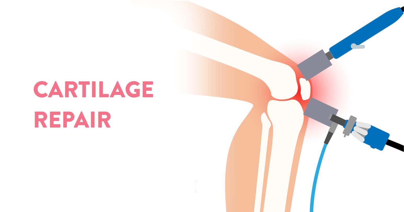
Knees contain 2 types of cartilage: Meniscus and Articular. Articular cartilage helps the knee move freely. Meniscus cartilage serves as a shock absorber or cushion between the bones. Years of normal wear and tear, accidents and sports injuries can damage both types of cartilage in the knee, making it necessary to replace or repair the lost cartilage. Osteoarthritis is a condition where the cartilage begins to wear down.
In this blog, we review newly emerging and traditional approaches to knee cartilage repair and replacement.
Table of Contents
Thanks to advancements in orthopaedic surgery, there are several alternatives for treating knee problems. Some traditional methods include knee joint replacement or surgery to restore broken cartilage. In addition to this, there are currently minimally invasive (keyhole) procedures that use cartilage harvested from other parts of the body or tissue grown from a patient’s cells.
The several options for the replacement and repair of knee cartilage are:
If the loss of knee cartilage and symptoms are minor, you might be able to put off surgery or other interventions through physical therapy.
Physical therapy’s primary objective is to increase the knee joint’s surrounding muscles’ range of motion and strength to lessen the strain on the joint itself. Physical therapy helped reduce pain, not just in cases of mild knee osteoarthritis, but in people dealing with moderate pain, too.
Physical therapy had less of an impact on persons with severe arthritis pain, suggesting that pain severity may be a key consideration when determining whether to seek physical therapy as a therapeutic option.
Weight loss achieved through diet and exercise can often help ease knee arthritis symptoms in people who are overweight or obese.
Microfracture is one method of stimulating the creation of new cartilage. To promote better blood flow and the release of cells that build new cartilage, small holes are drilled in one or more of the knee joint’s bones.
Microfracture is best suited for people who:
For elderly people with severe osteoarthritis or to treat extensive lesions, microfracture is not advised.
After the procedure, you will need to keep weight off your knee for about six weeks and use a CPM (continuous passive motion) machine several hours a day to straighten and bend the knee. Resuming sports or other strenuous activities might take up to nine months.
The comparatively low incidence of infections or other problems following microfracture surgery is an additional benefit. However, occasionally the operation does not produce enough new cartilage, necessitating the potential long-term need for a different kind of therapy.
This procedure to grow new cartilage, also known as MACI (matrix-induced autologous chondrocyte implantation), is a two-step process. An arthroscopic cartilage biopsy of the knee is the first step. The cartilage cells from the biopsy tissue are then stimulated to begin growing in a lab. When the fresh cartilage is ready for implantation, the surgeon cuts and moulds it to suit the missing piece of natural cartilage.
The following individuals are the best MACI candidates:
MACI is an effective and safe approach to cartilage transplant for most people.
MACI is typically an outpatient procedure, though you can expect to wear a knee brace for about six weeks while your knee regains stability and strength.
Aside from the need for 2 separate operations, the other main downside to MACI is that there is a risk of cartilage overgrowth, which can require a third surgery to treat.
The main justifications for MACI are that it works well for repairing tiny patches of cartilage and that it makes use of the patient’s cells, lowering the likelihood of rejection by the body.
This procedure, also known as a mosaicplasty, substitutes worn-out cartilage for good cartilage from another area of the knee. Osteochondral allograft, a similar surgery, uses donor tissue.
During the procedure, a surgeon removes the damaged cartilage and a portion of the underlying bone. The hole is then filled up with a replacement core consisting of bone and cartilage from another section of the body or a donor’s knee. Typically, no screws or other mechanical fasteners are required to hold the new tissue in place.
Like other knee procedures, an allograft or an osteochondral autograft requires about four to six weeks of recovery before the knee can start to bear weight. With the help of rehabilitation, a complete return to sports or other activities should be possible within six to nine months.
There are some downsides, which include the limited availability of donor tissue and the risk that the body may reject the implant.
The rehabilitation period is typically longer with an allograft than with an autograft, and an autograft is normally less expensive and carries a smaller risk of complications. Research suggests that autografts provide satisfactory outcomes for at least ten years among 72% of people who undergo the procedure.
Young people are the best candidates for this treatment since they have more regenerative cells available for transplant. In addition, an allograft is usually used to repair areas of worn cartilage that are no larger than a dime.
However, an autograft is a more invasive surgical treatment since it requires the removal of healthy tissue from another part of the body. Additionally, some people are reluctant to take the risk – even a small one – of issues arising from the removal of healthy bone and cartilage.
You Can Also Read: Cracking of Bones and Joints
When other less invasive approaches have not improved knee pain and movement, severe knee arthritis might require a knee replacement.
The procedure begins with an incision from above the knee down past the knee joint. All or part of the damaged knee joint is then removed and replaced with a prosthetic knee joint that duplicates the movement and function of a natural knee.
To create new gliding surfaces, worn-out knee surfaces that were once covered in cartilage are also resurfaced with ceramic, plastic, and metal materials.
By participating in physical therapy following the operation, most people can resume everyday activities within six weeks, though a full recovery can take several months. On the bright side, knee replacement can ultimately give your knee joint full functionality again and eliminate the discomfort that might make standing or even walking uncomfortable.
As with any surgery, knee replacement carries the potential risk of infection or other complications. Because the knee is a complicated joint, there is a chance that the prosthetic knee might not completely meet your expectations. As a result, you might need to consider a second operation or adjust to life with the new knee as it is.
Knee cartilage can get significantly worn down or damaged to the point that it no longer offers cushioning between the bones or smooth bone movement inside the joint, which can result in decreased mobility and ongoing discomfort. It is not a condition that will improve on its own.
Replacing or repairing damaged knee cartilage can:
Also Watch: Cauda Equina Syndrome treated by Endoscopic Spine Surgery
Damaged knee cartilage can either be repaired or replaced as per your existing condition. It is always advisable to seek medical help from an experienced orthopaedist. Timely care and help can ensure an appropriate diagnosis and treatment of your condition.
At the CK Birla Hospital, we ensure patients get holistic medical support which includes treatment in a compassionate environment. This patient-centric approach not only helps patients heal better but also ensures they are aware of the preventive measures as well. In case you need to consult an orthopaedist, reach out to us, or book a direct appointment with Dr.Reetadyuti Mukhopadhyay at the CK Birla Hospital.
With knee cartilage injuries, locking and pain of the knee can lead to difficulty with running, walking and other activities. The most visible indicator of knee cartilage degradation is pain, but repeated knee swelling is also a common warning sign.
Most meniscal tears improve with physiotherapy and time. Physiotherapy will include load modification e.g. reducing your walking and running and reduction or avoidance of the activities and movements that make your swelling or pain worse.
Meniscus Replacement Procedure (MRP) is a prosthetic, joint-preserving orthopedic implant surgery using a manufactured, non-biological medical device. It does not involve transplantation, human tissue, donor material, or biological grafts, and is not governed by transplant or tissue laws. MRP is clinically distinct from partial or total knee replacement, as it preserves native joint surfaces and bone and is intended for selected patients with early compartment-specific degeneration. Clinical outcomes may vary; no assurance, guarantee, or claim of superiority over other treatments is made, and future knee replacement may still be required. Final treatment decisions are made by patient through informed consent upon considering the available treatment modalities advised by the treating surgeon following individualized clinical assessment. This information is provided solely for patient education and professional clarification.
Your knee is the largest joint in your body and it joins your leg with the thigh. Each knee has 2 joints, one between the femur and patella (patellofemoral joint) and one between the femur and tibia (tibiofemoral joint).
One of the most common knee injuries in children and adolescents, as well as in some adults is patellar subluxation. Surgery is normally not required for the first occurrence, however, if it is needed, then several new techniques make it likely that you will regain all or most of your previous activity and strength. If you are unable to straighten or bend your knee and put weight on your leg or walk, then you should consult your healthcare provider for further advice.
Table of Contents
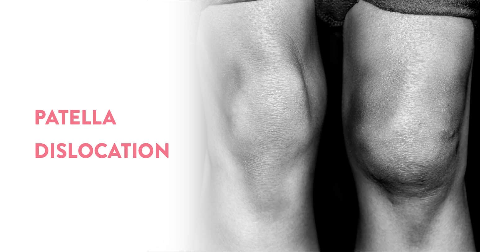
Another term for a bone’s partial dislocation is subluxation. Patellar subluxation is a partial dislocation of the patella (kneecap). It is also known as kneecap instability or patellar instability.
The kneecap is a small protective bone that attaches near the femur (bottom of your thigh bone). The trochlea, a groove at the bottom of the thigh, is where your kneecap glides up and down when you straighten and bend your knee.
Several groups of ligaments and muscles hold your kneecap in place. When these become injured, your kneecap might move out of the groove, causing difficulty and pain in flexing the knee.
The extent of the dislocation determines whether it is called a dislocation or a patellar subluxation.
The majority of injuries force the kneecap to move toward the outside of the knee. This can also damage the ligament on the inside of the knee, known as the MPFL (medial patellofemoral ligament). A second dislocation may occur if the MPFL does not heal adequately.
You might experience the following symptoms with patellar subluxation:
Although you may be able to self-diagnose, you will need to see a doctor for treatment.
You Can Also Read: Arthritis of Knee: Symptoms & treatment
Any contact sport or extreme activity can cause a patellar subluxation.
Dislocations and patellar subluxations mainly affect active and young people, especially between the ages of ten to twenty years. Most first-time injuries occur during sports.
The chances of a second dislocation are very high after an initial injury.
To diagnose a patellar subluxation, your doctor will straighten and bend the injured knee and feel the area around the kneecap.
X-rays might be used to see how the kneecap fits into the groove at the bottom of the patella and to identify any other possible bone injuries.
MRI (magnetic resonance imaging) might be used to visualise the ligaments and other soft tissue around the patella. Adolescents and children are sometimes not aware that they have had a patellar dislocation. The MRI can help confirm it.
There are 2 types of treatment for patellar subluxation – surgical and non-surgical.
Nonsurgical treatment is recommended for the majority of people with a first-time patellar dislocation or subluxation.
Nonsurgical treatment includes:
You have about a 33% chance of recurrence after a patellar subluxation.
The majority of first-time patellar subluxation instances don’t require surgery and are managed conservatively. Surgical treatment is recommended in special cases or if you have a repeat episode.
Some common types of surgery for repeat episodes of patellar dislocation or subluxation are:
The MPFL (Medial patellofemoral ligament) pulls the kneecap toward the inside of the leg. When the ligament is damaged or weak, the kneecap can dislocate toward the outside of the leg.
MPFL reconstruction is an arthroscopic surgery involving 2 small incisions. In this procedure, the ligament is rebuilt using a little piece of tendon taken from either your or a donor’s hamstring muscle. It takes about 1 hour. You normally return home the same day wearing a brace to stabilise your knee.
The brace keeps your leg straight while walking. It is worn for 6 weeks. After 6 weeks, you begin physical therapy. Most people can resume sports and play activities 4 to 7 months after MPFL reconstruction.
Your shin bone is also known as your tibia. The tibial tuberosity is a bulge or an oblong elevation, in the tibia just below your knee.
The tibial tuberosity receives an attachment from the tendon that controls the motion of your kneecap in the trochlear groove. An injury that has caused the kneecap to dislocate might have damaged the connection point for this tendon.
Tibial tubercle transfer operation requires an incision about 3 inches long above the shin bone. In this procedure, your doctor moves a small portion of the tibial tuberosity to enhance the tendon’s adhesion. This then facilitates correct kneecap movement inside its groove.
The surgeon will place 1 or 2 screws inside your leg to secure the piece of bone that is transferred. The operation takes about 1 hour.
You will be given crutches to use for 6 weeks following surgery. After that, physical therapy begins. Most people can return to school or work 2 weeks after surgery. It takes about 9 months before you can return to sports.
Until about ten years ago, lateral release was the standard surgical treatment for patellar subluxation, but it is rare nowadays because it increases the risk of recurrence of instability in the kneecap.
To stop them from dragging the kneecap to one side during this treatment, ligaments on the outside of the knee are partially severed.
Patellar subluxation is a common knee injury. Surgery is not required initially as you can try non-surgical treatment, however, if you still face difficulties in knee movement, then it is always advisable to seek medical help from an experienced orthopaedist. Timely care and help can ensure an appropriate diagnosis and treatment of your condition.
At the CK Birla Hospital, we ensure patients get holistic medical support which includes treatment in a compassionate environment. This patient-centric approach not only helps patients heal better but also ensures they are aware of the preventive measures as well. In case you need to consult an orthopaedist, reach out to us, or book a direct appointment with Dr. Praveen Chawla at the CK Birla Hospital.
With chronic patellar subluxations, the pain might be less severe than in a traumatic injury. Patients might complain of pain underneath the kneecap, especially with activities that involve deep knee bending.
A minor subluxation of the patella, especially if it is not a first-time subluxation, might recover fairly quickly with full recovery in a few days to a few weeks. Ongoing physical therapy and a kneecap stabilising brace might be required for a full recovery and to prevent a recurrence of the injury.
The ankle is the area where your leg and the foot meet. The main bones of the ankle region are the tibia and fibula (in the leg) and talus (in the foot).
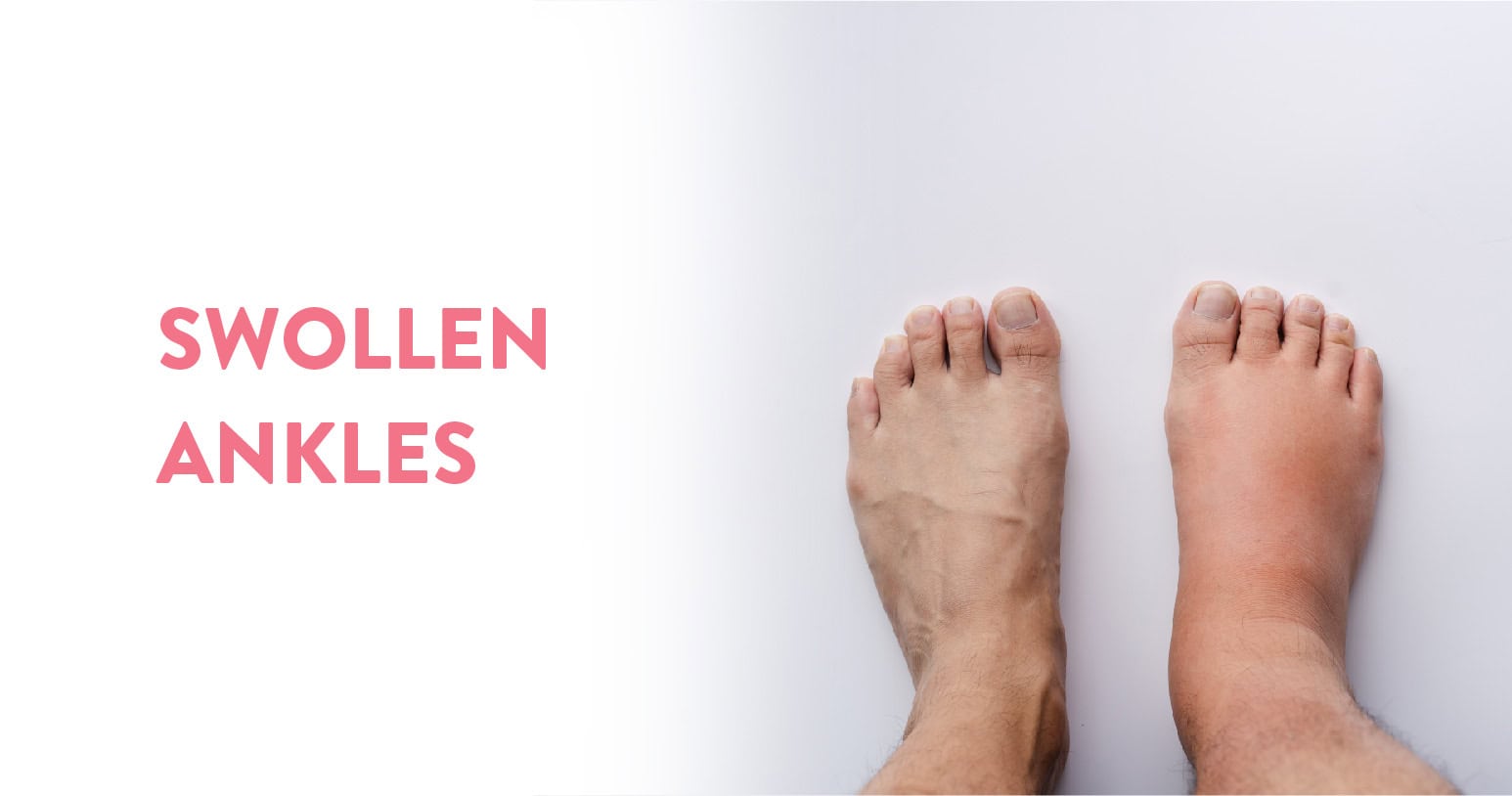
Inflammation and injuries can cause swelling in your lower extremities. Swollen ankles are common and normally not a cause for concern, particularly if you have been walking a lot or standing. It often goes away on its own. There are many reasons for a swollen ankle, however, it is usually the result of your body’s response to an infection or injury or due to fluid buildup.
Swollen ankles that persist or come with additional symptoms may be an indication of a serious medical condition. Additionally, you can have fluid retention as a result of a medically important underlying health issue. See a doctor if it does not get better in a few days; they can run tests to determine a diagnosis and treatment plan.
Table of Contents
The legs, ankles and feet are common sites of swelling because of gravity’s effect on the fluids in the human body. A swollen ankle or leg might have other causes besides fluid retention from gravity. Injuries and subsequent inflammation can also cause swelling and fluid retention.
A swollen ankle can cause the lower part of the leg to appear larger than normal. It could be challenging to walk due to the swelling. It might be painful, with the skin over your leg feeling stretched out and tight.
This swelling is typically temporary and not cause for concern. But you will still want to take measures to reduce swelling. This way, you can reduce any pain you are experiencing and resume your daily activities.
If parts of your lower leg remain swollen or you have other symptoms, it could signal that you have an underlying health condition. It might be easier to rule out a more serious issue if you know what’s causing your swelling.
Some possible causes of a swollen ankle and what you can do to reduce swelling are:
During pregnancy, some ankle and foot swelling is typical. Excessive or sudden swelling, however, might be a sign of preeclampsia, a serious condition in which protein and high blood pressure in the urine develop after the 20th week of pregnancy. Call your doctor right away if you develop severe oedema or swelling coupled with other symptoms including headaches, nausea, vomiting, vision changes, or stomach discomfort.
An injury to the ankle or foot can lead to swelling. The most common is a sprained ankle, which occurs when a misstep or an injury causes the ligaments that hold the ankle in place to be stretched beyond their normal range. To reduce the swelling from an ankle or foot injury, elevate the foot on a stool or pillow, wrap the foot or ankle with a compression bandage, and use ice packs and rest to avoid walking on the injured ankle or foot. If pain and swelling are severe or do not improve with home treatment, see your doctor.
This is a buildup of lymphatic fluid in the tissues that can occur when lymph veins are absent, have difficulties or after the excision of lymph nodes. Lymph is a protein-rich fluid that usually travels along an extensive network of capillaries and vessels. The lymph nodes, which catch and eliminate undesirable elements like germs, filter it. However, the fluid might get obstructed when there is an issue with the lymph nodes or veins. Untreated, lymph buildup can impair wound healing and lead to deformity and infection. Lymphedema is common following radiation therapy or removal of the lymph nodes in patients with cancer. See your doctor right away if you’ve had cancer treatment and are experiencing swelling.
Swelling of the feet and ankles is often an early symptom of venous insufficiency, a condition in which blood inadequately moves up the veins from the feet and legs up to the heart. Usually, the veins keep blood flowing upward with one-way valves. When these valves become weakened or damaged, the blood leaks back down the vessels and fluid is retained in the soft tissue of the lower legs, especially the feet and ankles. Chronic venous insufficiency can lead to infection, skin ulcers and skin changes. Visit your doctor if you exhibit symptoms of venous insufficiency.
Swelling in the ankles and feet can be a sign of infection. People who have diabetic neuropathy or other foot nerve issues are more likely to get foot infections If you have diabetes, it is important to inspect your feet daily for sores and blisters because nerve damage can blunt the pain sensation and foot problems can progress quickly. If you notice a blister or swollen foot that appears to be infected, contact your doctor right away.
Leg vein blood clots can prevent blood from returning to the heart from the legs and result in swelling in the feet and ankles. Blood clots can be deep (deep vein thrombosis) or superficial (occurring in the veins just below the skin).
Large leg veins may become partially or completely blocked by deep clots. These blood clots can be life-threatening if they break loose and travel to the lungs and heart. If you have swelling in one leg, along with pain, low-grade fever, and possibly a change in colour of the affected leg, call your doctor immediately. Treatment with blood thinners might be necessary.
Sometimes swelling can indicate a problem such as kidney, liver or heart disease. Ankles that swell in the evening could be a sign of retaining water and salt because of right-sided heart failure. Kidney disease can also cause ankle and foot swelling. Fluid can accumulate in the body when the kidneys are not working correctly. Albumin is a protein that the liver makes that prevents blood from spilling out of blood vessels and into the surrounding tissues. Liver illness can change how much albumin the liver produces. A lack of albumin production may cause fluid leakage. Fluid can build up in the chest and belly as well as the ankles and feet, where it tends to collect more due to gravity. If your swelling is accompanied by other symptoms, including weight gain, loss of appetite and fatigue see your doctor right away.
In many cases, you can treat a swollen ankle at home. Depending on the cause, home management will vary.
If your swelling is the result of fluid buildup, the following home tips might help relieve swelling:
Keep R.I.C.E (rest, ice, compression, elevation) in mind if you have swelling from an injury but consult with a doctor regarding how much activity your leg should get.
Swollen ankles are quite common and usually not a cause for concern. It normally gets cured on its own, however, if the swelling stays or accompanies other problems, then it is always advisable to seek medical help from an experienced orthopaedist. Timely care and help can ensure an appropriate diagnosis and treatment of your condition.
At the CK Birla Hospital, we ensure patients get holistic medical support which includes treatment in a compassionate environment. This patient-centric approach not only helps patients heal better but also ensures they are aware of the preventive measures as well. In case you need to consult an orthopaedist, reach out to us, or book a direct appointment with Dr. Ashwani Maichand at the CK Birla Hospital.
If one or both of your ankles are swollen and the condition worsens or does not get better after a few days of home treatment, consult a doctor.
People with heart failure tend to retain fluid. Due to an accumulation of extra fluid, this manifests as swollen ankles and legs. You might notice that your shoes do not fit and socks appear tight or leave a prominent indent above the ankle.
Swollen ankles (also known as oedema) can be harmless, but might also signal a serious health condition or injury.
A testicle (or testis) is the reproductive gland or gonad in males. They are small, egg-shaped reproductive organs which rest inside the scrotum (a thin pouch of skin behind your penis). Its function is to produce both androgens (primarily testosterone) and sperm.
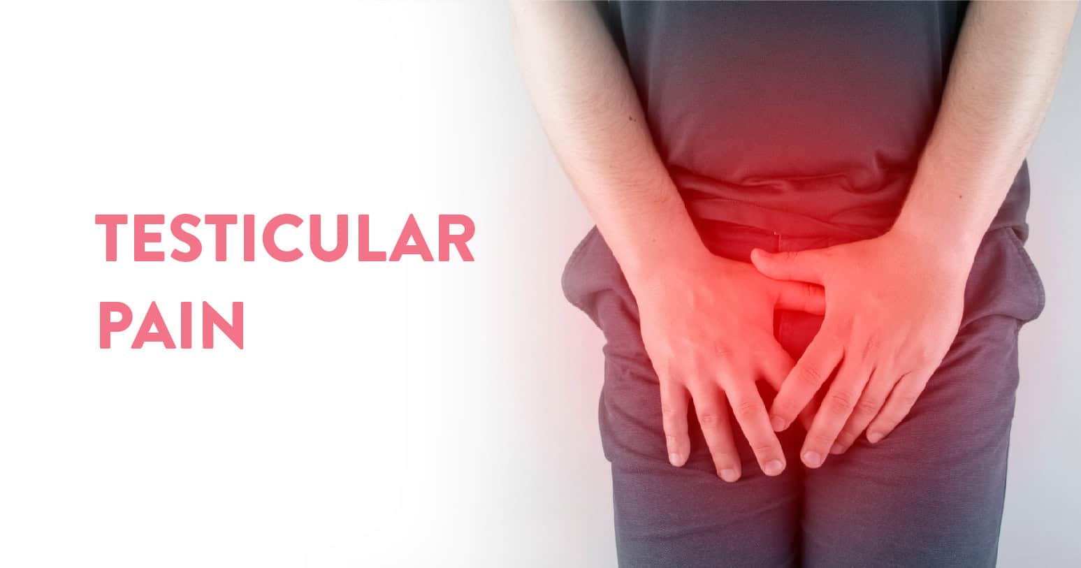
Pain in the testicles can affect anyone at any age. You may feel it in one or both testicles, however, the pain might come from another part of your body, like your groin or stomach (referred pain). Testicular pain can be chronic or acute. Acute refers to fast onset, a sharp ascent, and brief duration. Chronic refers to a long-lasting, gradually worsening form of pain.
There are a lot of delicate nerves in your testicles, which can make testicular discomfort quite painful. Reach out to a healthcare provider if you have testicular pain that lasts for more than an hour. Go to an emergency room if you have intense testicular pain as it could be a sign of testicular torsion, which is a serious medical emergency.
Table of Contents
The reproductive organs known as testicles are situated in the scrotum. Minor injuries to the region may result in testicular pain. However, if you are experiencing pain in the testicle, you need to have your symptoms evaluated.
Pain in the scrotum can be the result of serious conditions like an STI (Sexually Transmitted Infection) or testicular torsion. Ignoring the discomfort might harm the testicles and scrotum permanently. Ignoring the pain might cause irreversible damage to the scrotum and testicles.
Often, problems with the testicles cause groin or abdominal pain before pain in the testicle develops. Unexplained groin or abdominal pain should also be evaluated by your doctor.
There are various common causes of testicular pain. The cause might be obvious if you have had an accident or a recent injury while exercising or playing a sport. But in other cases, it might not be obvious why you have pain.
Some other common causes of testicular pain might include:
Other symptoms that might occur alongside testicular pain include:
The following techniques can be used to alleviate pain at home that does not require medical attention:
You must consult your doctor for treatment if the pain is more severe. Your doctor will complete a physical exam of your scrotum, groin and abdomen to determine what is causing your pain and will also ask you about your current health conditions and any other symptoms.
To accurately diagnose your condition, your doctor might need to order additional tests, including:
Your doctor will be able to treat you once they have determined the source of your discomfort. The treatment might include:
Pain in the testicles can affect anyone and can be acute or chronic. If the pain is
severe and persists for a longer duration, then it is always advisable to seek medical help from an experienced urologist. Timely care and help can ensure an appropriate diagnosis and treatment of your condition.
At the CK Birla Hospital, we ensure patients get holistic medical support which includes treatment in a compassionate environment. This patient-centric approach not only helps patients heal better but also ensures they are aware of the preventive measures as well. In case you need to consult a urologist, reach out to us, or book a direct appointment with Dr. Kumar Saurav at the CK Birla Hospital.
It is advisable to rest and protect your testicles and groin. Stop, change, or take a break from any activity that might be causing your soreness or pain. Put a cold pack or ice on the area for 10 to 20 minutes at a time. Put a thin piece of fabric between your skin and the ice.
Sometimes there is no need for treatment since testicular discomfort will go away on its own. Depending on the cause of your testicle pain, your condition might take up to four weeks to heal. Limit your activity until your pain decreases. Get more rest while you heal.
Yes. Blue balls can develop after a protracted time of arousal without ejaculation; this condition is referred to medically as epididymal hypertension. Sperm accumulation might give your testicles a faint blue tint.
The circulatory system’s blood arteries are used by the heart, a muscular organ, to pump blood. The pumped blood carries nutrients and oxygen to the body while carrying metabolic waste such as carbon dioxide to the lungs.
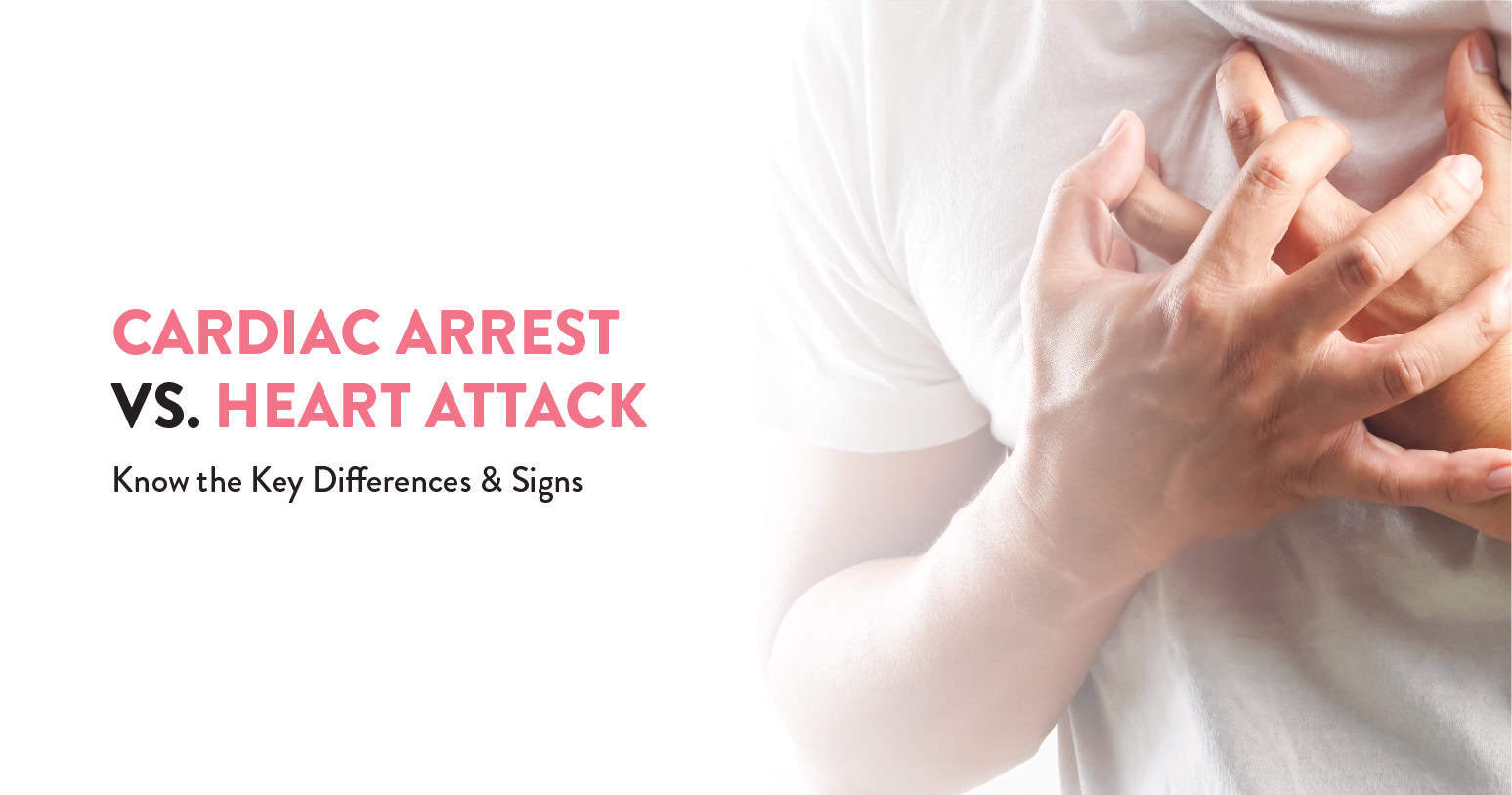
Cardiovascular diseases are the most common cause of death worldwide as of 2008, accounting for 30% of deaths (including cardiac arrest and heart attack). Circulation issues in the body that obstruct blood flow to the heart are what lead to a heart attack. A cardiac arrest is an electrical systemic failure of the heart.
It can be easy to forget the differences between a cardiac arrest and a heart attack, but the most important thing to remember is that both are serious, life-threatening conditions. If you believe you or someone you know is suffering from either illness, get medical attention right away. A healthcare team and doctor can quickly assess your symptoms and begin treatment. Your recovery and outlook will depend on how quickly you can get to a healthcare professional or doctor.
Table of Contents
A heart attack happens when a clogged artery stops oxygen-rich blood from getting to a particular area of the heart. The area of the heart ordinarily supplied by the blocked artery starts to perish if it is not rapidly unblocked. Damage increases with time and duration of non-treatment.
Sudden cardiac arrest is caused abruptly and frequently without notice. When the heart suffers from an electrical problem, it results in an Arrhythmia or irregular heartbeat. With its pumping action disrupted, the heart can not pump blood to the lungs, brain and other organs. When this occurs, a person has no pulse and loses consciousness. If the person doesn’t get help, they’ll die in a matter of minutes.
A heart attack, which is often referred to as a myocardial infarction, happens when blood that usually flows to the heart is cut off or blocked. Without enough oxygen-rich blood flowing to the heart, one of the body’s most vital organs may suffer injury, and the heart muscle may start to deteriorate.
On the other hand, abrupt cardiac death refers to a cardiac arrest. The word “arrest” means to bring to a halt or stop. The heart stops beating during cardiac arrest, which is a very dangerous medical condition. Cardiac arrest can cause disability or near-immediate death.
Although acute chest pain is a common sign of a heart attack, it can also start gradually with moderate discomfort that comes and goes over many hours. Symptoms of a heart attack can vary, and if you have had one heart attack, your symptoms might be different if you experience another one.
Symptoms can also vary between females and males, but for both sexes, the most common symptom of a heart attack is chest pain. However, women are somewhat more likely than men to experience other symptoms, particularly jaw or back pain, vomiting, nausea and shortness of breath.
Cardiac arrests can often happen to people who did not know they had a heart problem. A person experiencing cardiac arrest might lose consciousness and collapse. They might experience difficulty breathing or stop breathing.
Heart attacks are normally caused by coronary heart disease, which starves your heart of oxygen. Most of the time, people know they are at risk of a heart attack because they are being treated for heart disease.
The risk factors for a heart attack can include:
Certain health factors and heart conditions can increase your risk of cardiac arrest, including coronary heart disease. Other factors that are not always known could also put you at risk such as:
By doing a physical examination and prescribing an electrocardiogram to examine the electrical activity of your heart, doctors can determine whether you are having a heart attack.
A doctor might order a cardiac catheterization or an echocardiogram to determine the vitality and strength of your heart. It is also common to have a sample of your blood taken to check for signs of heart muscle damage.
A cardiac arrest means your heart has stopped. Without immediate resuscitation, it is fatal.
If a doctor is successful at restarting your heart and getting the blood flowing again, they will then perform diagnostic tests to determine the cause of your cardiac arrest. These tests might include a chest X-ray, blood tests and an echocardiogram to look for other signs of disease in your heart.
If you have had a heart attack, a doctor might recommend any number of procedures depending on the severity of the emergency to prevent another heart attack, help relieve pain, or both.
Some of these treatments or procedures can include:
It is also common for doctors to prescribe medications, such as beta-blockers and pain relievers, to help prevent another heart attack or to help with recovery.
Treatment for cardiac arrest nearly always starts with a defibrillator or CPR to get the heart started again. A doctor will probably begin one or more therapies after someone has survived a cardiac arrest to help reduce the likelihood of it happening again. These often include:
Cardiovascular diseases account for about one-third of all deaths globally. You can forget the differences between a heart attack and a cardiac arrest, however, both are life-threatening conditions. It is always advisable to seek medical help from an experienced cardiologist. Timely care and help can ensure an appropriate diagnosis and treatment of your condition.
At the CK Birla Hospital, we ensure patients get holistic medical support which includes treatment in a compassionate environment. This patient-centric approach not only helps patients heal better but also ensures they are aware of the preventive measures as well. In case you need to consult a cardiologist, reach out to us, or book a direct appointment with Dr. Sanjeeva Kumar Gupta at the CK Birla Hospital.
Both a heart attack and a sudden cardiac arrest are significant medical situations that can be fatal. Cardiac arrest is usually more severe, as it can result in death within minutes if proper medical care is not provided. Even when a heart attack doesn’t happen right away, it can still be quite hazardous if untreated.
Quick action increases a person’s chance of survival during a heart attack or sudden cardiac arrest. For a heart attack, seek medical attention immediately by calling an ambulance. In case of a cardiac arrest, give CPR (cardiopulmonary resuscitation) first immediately and seek professional emergency medical services.
In females, the period is the regular discharge of mucosal tissue and blood from the inner lining of the uterus through the vagina. It is triggered by falling progesterone levels and is an indicator that pregnancy has not occurred.
A light period or a period that stops before you expect might happen for a variety of reasons. Light periods might not be a sign that you have something to worry about. Even a period as short as 2 to 3 days is considered normal. Changes in medication, age or weight can impact your period.
A missed period or spotting might indicate a pregnancy or an underlying medical condition. In such a case, you can take a pregnancy test. Keep track of your menstrual cycles and consult your doctor.
Table of Contents
About 2-3 teaspoons of blood make up the majority of menstrual cycles. It might be very challenging to estimate how much blood a person is losing because there is a large difference across individuals.
A person should make a note if their periods are lighter than they normally are. They can accomplish this by monitoring their pad or tampon usage or the amount of blood a menstrual cup gathers.
The following might indicate a light period:
Sometimes, a light period might also cause a reduction in symptoms of premenstrual syndrome, like mood shifts, uterine cramping or reduced back pain.
Several factors can lead to light periods. These include:
Your period can vary in flow and length if you are in your teenage years. On the other hand, if you are going through menopause, you can have irregular, light-flowing periods. Hormonal imbalances are the cause of these events.
Body fat percentage and body weight can affect your period. Because your hormones are not functioning regularly, being extremely underweight might cause your menstruation to become irregular. Additionally, gaining or losing an extreme amount of weight can cause irregularities with your period.
It is unlikely that you will have a period if you are pregnant. You might notice some spotting and think it is your period, but it might be implantation bleeding. This can occur when a fertilised egg attaches to the lining of the uterus. Implantation bleeding typically lasts for 2 days or less.
Your periods might not come back immediately after you give birth if you are breastfeeding. The milk hormone delays the onset of your menstruation and inhibits ovulation. You might get your period months after giving birth if you are breastfeeding.
Even if your period hasn’t yet come back, you can still become pregnant while breastfeeding. That is because you will ovulate 2 weeks before your first postnatal period. If you have had unprotected sex while breastfeeding and are experiencing spotting, it is a good idea to take a pregnancy test to confirm that the spotting was not caused by implantation bleeding.
Hormonal birth control might be the cause of a light period. Some methods of birth control stop an egg from releasing into your body. These techniques can take many different shapes, including:
Your uterus does not create a thick lining when your body does not release an egg. This may lead to missing periods or periods that are completely lighter.
You might also experience irregular periods if you have stopped or started taking birth control recently.
Your brain can alter the menstrual cycle hormones if you are stressed. You might experience lighter or skipped periods because of it. Your periods should return to normal once a stressful event passes.
Women who exercise frequently might experience changes in their period. Athletes use a lot of body energy, have low body weight and can be under stress. This can result in altered periods.
Bulimia and anorexia nervosa are types of eating disorders that might cause irregular periods. Eating disorders that result in low body weight may have an impact on the hormones that regulate your menstrual cycle.
If you have stopped menstruating or you are experiencing irregular periods, it could be the result of PCOS. Your body experiences a hormonal alteration as a result of which your eggs cease developing. This hormonal change might also:
An ultrasound can be used by your doctor to determine if you have PCOS. That is because PCOS causes cysts to form in your ovaries. If you have PCOS, your doctor will likely recommend that you take contraceptive pills and lose weight to help your period return to normal.
You Can Also Read: Living with PCOS
Irregular or unusual periods might be a sign of a more serious health condition. Your body is in good shape if you get regular periods. A light period could indicate a medical illness or issues with hormone levels. Issues with reproductive organs and polycystic ovary syndrome can lead to irregular periods.
Discussing symptoms with your doctor might help you determine the cause of lighter-than-normal periods.
Sometimes, people with no known risk factors can have light periods. Light periods are, nevertheless, more likely due to specific circumstances.
The following are some potential risk factors for a light period:
A person might wish to talk with a doctor about individual risk factors that might affect the severity of their periods.
Your light period might be caused by one of many factors. It might be a one-time occurrence. If you experience any troubling symptoms or your light periods persist, you might need further treatment.
Your doctor will discuss possible reasons for your light periods and test you for various conditions to determine an appropriate treatment plan.
Problematic and persistent light periods might be treated with changes to your medications and lifestyle. Hormonal birth control might occasionally make your periods more regular. Treatment options might include further therapies or other drugs if your light periods are an indication of anything more serious.
Less bleeding during your periods may have several reasons. It usually is not a sign of something worrying, however, if you suspect a missed period or spotting as a symptom of an underlying medical condition, then it is always advisable to seek medical help from an experienced gynaecologist. Timely care and help can ensure an appropriate diagnosis and treatment of your condition.
At the CK Birla Hospital, we ensure patients get holistic medical support which includes treatment in a compassionate and judgement-free environment. This patient-centric approach not only helps patients heal better but also ensures they are aware of the preventive measures as well. In case you need to consult a gynaecologist, reach out to us, or book a direct appointment with Dr. Astha Dayal at the CK Birla Hospital.
Light periods can be a sign of pregnancy. Spotting in early pregnancy is often caused by implantation bleeding, which might just seem like a light period. Lighter than normal period blood can be brownish, red or pink. It may or may not involve period pain and cramps.
Light periods may also result from a drop in oestrogen levels. Oestrogen plumps up the endometrium, so if oestrogen is low, that lining does not plump up and results in less bleeding.