Filter :
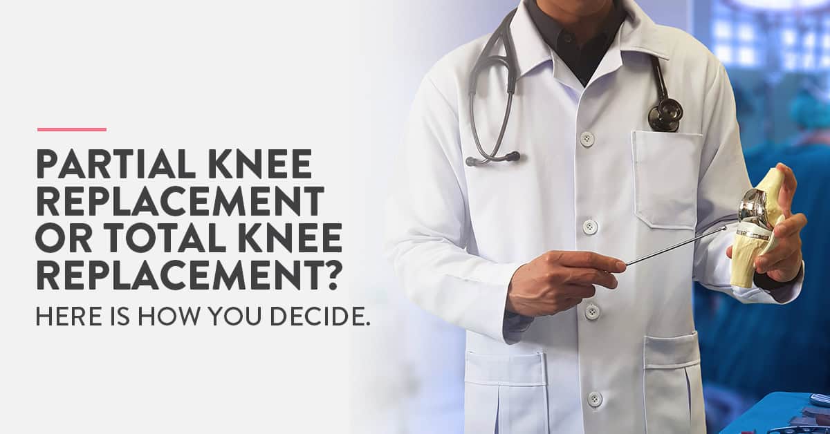
Joint replacement surgery has evolved extremely over the past few decades, giving thousands of patients all around the world, a second chance at life. Today, patients suffering from painful conditions such as osteoarthritis of the knee can take back control of their life and live pain-free thanks to options such as total or partial knee replacement.
As knee replacement surgery is generally an elective procedure, patients have the time to do a comprehensive research before taking any decision and scheduling the procedure. One of the decisions they wrestle with is choosing between total or partial knee replacement surgery. Before we dive into the pros and cons associated with each, let us understand these two procedures better.
Table of Contents
Total knee replacement surgery is also called knee arthroplasty or simply knee replacement. It is a surgical procedure performed to repair a damaged knee. The knee joint can be damaged by trauma/injury or conditions such as osteoarthritis. In this procedure, the damaged bone and cartilage from the tighbone, shinbone and kneecap are replaced with a prosthesis or artificial joint made of metal alloys, high-grade plastics and polymers.
A typical knee replacement surgery generally lasts for about 2 hours. Intravenous antibiotics are administered to the patient before, during and after the procedure to prevent infections. During the surgery, the patient will be given a general or spinal anaesthetic.
The knee is kept in a bent position to expose all surfaces of the joint. The surgeon then makes an incision about 6 to 10 inches long. Through this incision, the surgeon moves the kneecap aside and cuts away the damaged joint surfaces.
Once the joint surfaces are adequately prepared, the surgeon attaches the pieces of the prosthetic joint. The surgeons test the new knee joint by bending and rotating it before closing the incision.
After the procedure, the patient is placed in the recovery room for 1-2 hours. They are generally discharged a few days after the procedure. It is important in the first few days post-surgery to move the foot and ankle. This increased blood flow in the leg which reduces the risk of blood clots and DVT. Patients have also prescribed blood thinners and compression boots to further prevent swelling and blood clots.
Knee replacements generally provide patients with pain relief and greater mobility. Artificial joints can be expected to last more than 15 years. The lifespan of the artificial joint depends on the quality of surgery and care taken after surgery.
Read: Quality of life after Total Knee Replacement Surgery
Partial knee replacement surgery or unicompartmental knee arthroplasty offers an alternative to patients with damaged knees if the damage is confined to a particular compartment of the knee.
Earlier, this procedure was generally performed on older patients as they were unlikely to overexert themselves. Today, partial knee replacement has risen in popularity even in the younger population as it has a lower recovery time as well as decreased post op pain.
However, this surgical alternative is not suitable for everyone. It is estimated that about 5%-6% of patients with arthritis of the knee are eligible for partial knee replacement surgery.
This procedure can be performed on patients having medial or lateral knee osteoarthritis. “Medial” is the inside compartment of the knee, closest to the opposite knee, while “lateral” refers to the outward compartment which is farthest from the opposite knee.
In order to determine if the patient is a suitable candidate for partial knee replacement surgery, the surgeon will:
“What are the benefits of a partial vs total knee replacement?” This is one of the most frequent questions posed to orthopaedic surgeons everywhere.
To find an answer to this question, we must first understand why the need for surgery arises. Patients develop knee joint degeneration due to a number of reasons. It can be a result of any past trauma, bad outcomes of previous surgeries, infection, rheumatoid or inflammatory arthritis, childhood diseases, obesity as well as aging.
The knee joint has a cartilage that helps remove friction and distributes the joint forces uniformly. It also helps cushion the forces, acting like an effective shock absorber. With knee degeneration, this cartilage deteriorates, resulting in joint pain and decreased function.
The knee joint is broadly divided into three compartments. These are:
Typically, arthritis or degenerative process starts in one compartment and eventually progresses to the rest of the knee.
If the degeneration is limited to only one compartment, partial knee replacement is recommended. It is beneficial as the implant is much smaller and only the unhealthy bone and soft tissue are reconstructed during surgery.
One of the main benefits of partial knee replacement surgery is that in this procedure, normal structures such as the cruciate ligaments and uninvolved compartments can be left alone. And as the dissection is minimal, normal neural pathways which are interrupted during total knee replacement procedures are also spared in this alternative treatment.
Partial knee replacement surgery comes with lower risk of developing complications after surgery, decreased hospital and recovery time, increased range of overall motion and greater patient satisfaction.
When it comes to life expectancy of the prosthetic joint, studies show that over 90% of partial knee replacements function well for more than 10 years after the surgery. Patients with both partial and total knee replacement joints seem to prefer the former as compared to the latter, suggesting that they feel the partial knee replacement to feel more “normal”.
The choice of partial or total knee replacement surgery is done after careful diagnosis and study of the damaged joint. There is a higher failure rate for partial knee replacement surgery as compared to total knee replacements. As not all patients are ideal for partial knee replacement surgery, correctly judging the suitability of the procedure is critical to the outcome.
With significant improvements in prosthetic design as well as surgical techniques, both procedures can be performed with increased precision to realign the knee and correct any issues. For patients opting for either procedure, care should be taken while choosing the prosthetic. The surgeon generally recommends the prosthetic he or she is most comfortable using, hence discuss the available options at length with your doctor.
Read: All you need to know before considering replacement surgeries for both knees simultaneously
Ques: How to avoid knee replacement surgery?
Knee replacement surgery is an elective procedure, especially if it is being performed to manage conditions such as osteoarthritis. This surgery is generally done if all other non invasive treatment options are proving ineffective. Weight loss, muscle strengthening exercises and stretches to improve flexibility are generally the preliminary steps taken to strengthen the knee joint. Medication is also given to the patient to manage their pain.
Ques: How long does a knee replacement surgery last?
Total knee replacement surgery lasts for about two hours and requires 3-5 days of hospital stay. Partial knee replacement surgeries last around 45 minutes and require 1-2 days of hospital stay. Not all patients are ideal candidates for partial knee replacement surgeries. In some cases, doctors will decide on which procedure to perform after they make the incision and view the damage to the joint.
Ques: What are the risks of knee replacement surgery?
Like all other surgeries, there are several risks associated with total knee replacement surgery. These include
Seek immediate medical assistance if you notice:
Also, read: The risk of delaying Knee Replacement Surgery

The recent coronavirus pandemic has brought forth the importance of vaccinations. Vaccines are probably the most significant and effective ways to prevent a wide range of bacterial and viral infections.
When we think of vaccinations, we generally think that they are meant only for children. In reality, vaccinations are as important for adults, if not more. This is because many infections such as chickenpox might be harmless or mild for children but extremely severe for adults.
Pregnant women have to be exceptionally careful about their health as it directly impacts the health of their babies. Vaccinations help safeguard both the mother and child from infections during pregnancy. Pregnant women, who get vaccinated pass on important infection-fighting antibodies to their babies helping them develop stronger and healthier.
Many women also have concerns about the impact vaccinations can have on their unborn child. Let us take a closer look into vaccinations during pregnancy, their importance and possible risks.
Table of Contents
Vaccines are a safe and effective way to protect people from harmful diseases and infections before they are exposed to them. They often contain weakened or inactive forms of the virus/bacteria. They are designed to stimulate an immune response from the body without creating any severe complications of the disease.
According to the World Health Organisation, we now have vaccines that can prevent more than 20 life-threatening diseases. It is estimated that immunisation drives prevent 2-3 million deaths every year due to diseases such as diphtheria, tetanus, influenza and measles.
Smallpox is considered to be the first disease to be completely eradicated by vaccination (as declared by the WHO in 1980). Vaccination efforts have also helped drastically reduce the occurrence of diseases such as polio and guinea worm disease.
Most women are advised to undergo a blood test either during their pre-pregnancy check-up or their early prenatal visit. This test should check if they are immune to certain infections that can cause complications during pregnancy such as chickenpox.
If they do not have the antibodies that can protect them and their baby during pregnancy, they should get vaccinated. Pregnant women are generally not given vaccines containing live viruses.
There are two main vaccines recommended for women who are pregnant, these include:
For most of us, flu is a harmless albeit extremely uncomfortable infection. It can leave us feeling weak with muscle ache and diminished appetite. However, we generally recover without any medical intervention.
On the other hand, flu can cause extremely severe complications in pregnant women. It can lead to preterm delivery and birth, affecting your baby’s growth and development.
The flu vaccine (commonly called the flu shot) is made of an inactive virus and hence is considered safe during pregnancy. The influenza nasal spray should be avoided as it contains the live virus.
Women who are pregnant during the flu season, or amidst a flu pandemic/epidemic should definitely talk to their doctor about getting a flu shot.
The Tdap vaccine offers protection against the following diseases,
Tetanus (T) causes pain and stiffness in the muscles. In later stages, tetanus can cause serious complications such as trouble swallowing or breathing and even death.
Diphtheria (D) causes difficulty in breathing, paralysis, heart failure and sometimes death.
Pertussis (aP) or whooping cough can result in incessant and violent coughing. It is especially dangerous for newborn who have not been vaccinated as yet, causing complications such as pneumonia, convulsions, brain damage and death.
Women are generally administered the Tdap vaccine in the early part of their third trimester (in each pregnancy). They pass on the antibodies to their babies providing them valuable protection from these diseases for a few months after birth. The babies can then be vaccinated when they are 2 months old.
Read: A quick guide to routine antenatal scans done during Pregnancy [2020]
In some cases, there might be an increased risk of any particular type of infections. Especially in cases when travelling is involved. If your obstetrician feels that there is a chance of you being exposed to meningococcal disease, you might be asked to get the required vaccinations. These include:
Pregnant women are advised to avoid vaccines containing live viruses. These include:
In extremely rare cases, vaccines can cause complications. Especially if you are allergic to any component of the vaccine. Remember to inform your obstetrician about any allergies you might have. If you are not suitable for any particular vaccine, ask your doctor alternate ways to protect your baby against harmful infections.
Also, read: What is Rh factor and how it impacts your Pregnancy?
We all grew up listening to “drink your milk if you want to grow big and strong!”. But is plain old milk the magic answer to all bone-related concerns?
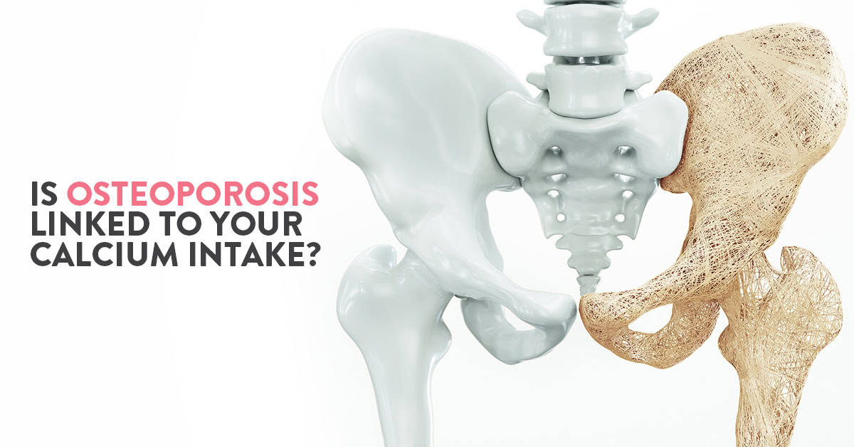
There are many who vouch that increasing calcium intake by having around 3 glasses of milk is the best way to prevent osteoporosis. Osteoporosis is a bone disease characterised by bone loss or weakened bones. People suffering from osteoporosis break their bones easily, sometimes even with a sneeze or cough.
There are also many who believe that just increasing the intake of milk or other dairy products does not have any significant impact on the rate of fractures. In fact, they believe that this intake could be harmful to the body causing problems such as heart disease.
While we may not know the exact answer to this conflict of opinions, let us explore what we actually know about osteoporosis and how our calcium intake affects it.
Table of Contents
The term osteoporosis translates to “porous bones”. Bones affected with osteoporosis look porous or like a honeycomb when viewed under a microscope. It is a condition of the bone which is a result of an imbalance between the rate of bone being built and bone being destroyed.
Osteoporosis is a relatively common condition, becoming more prevalent with age. Ensuring that you have the necessary amount of calcium intake when the rate of bone development is greater (up to the age of 30 years) creates a strong foundation for your bone health in the future. This will not, however, prevent bone loss later in life.
Bone loss due to ageing depends on several factors such as genetic predisposition, physical inactivity and hormone imbalances.
Our body needs a number of minerals and vitamins to function properly. Of these, calcium is important for building and maintaining bones and teeth, blood clotting, transmitting nerve impulses and regulating heartbeats. The bones and the teeth account for 99% of the calcium in the body. The remaining can be found in blood, muscle and tissue.
The body gets calcium either from our daily diet or from sources of calcium in the body, namely bones and teeth. Hence, it is important to have calcium-rich foods such as dairy products with high concentrations per serving of absorbable calcium, leafy vegetables (spinach etc), beans, soy etc.
If our body does not get or is unable to absorb enough calcium from natural sources, it starts “borrowing” calcium from the bones. If this is not replaced, it can have harmful and long term implications.
Read: Your comprehensive guide to joint pains and their treatment
While to the naked eye, we might seem to have stopped growing, our bones are in a state of constant flux. New bone is made while the old one is broken down. Osteoblasts are bone cells responsible for building bones while osteoclasts are cells that break down old bones.
Ideally, till the age of 30 years, the rate of bone being built is greater than that of it being broken down. Hence, you generally reach the peak of your bone mass by the age of 30 years. However, if your body does not have enough calcium, osteoclasts will break down the bone faster to make up for the deficiency, making your bones weak and brittle.
The likelihood of developing osteoporosis increases if you have lower bone mass (difference between bone being built and broken down).
In most of us, bone is rapidly deposited by osteoblasts up to the age of 30 years. During this time we must make sure we have an adequate supply of calcium and vitamin D so that we have more reserves in the future.
However, this will not prevent bone loss due to other factors such as ageing, hormone imbalance and other conditions.
Also Read: Ivy Gourd: Benefits & Side Effects
There are two key things to keep in mind to slow down the onset of osteoporosis.
The first and foremost is to build the healthiest, strongest and densest bone possible in the first thirty years of your life.
You can also prevent or minimise bone loss as you age by:
Apart from the above, you can also limit your caffeine intake, get enough vitamin A and protein in your daily diet to keep your bones healthy and strong.
Also, read: Tips on how to cure Osteoporosis by the orthopaedic expert

Your blood type is either positive or negative. This indicates the presence or absence of a protein called Rh (rhesus) protein in your blood. Checking for blood type is the possibly the most preliminary test done when you are pregnant.
The Rh factor is passed through genes. Which means the foetus will either have a positive or negative blood type depending on the parents’ blood type. If the expectant mother is Rh negative and her foetus is Rh negative, it results in a complication called Rh incompatibility.
Table of Contents
If you are Rh-negative and any amount of Rh-positive blood mixes with yours, your body treats the presence of the Rh protein as an intruder. It then creates antibodies (anti-Rh antibodies) to destroy the Rh-positive protein. These antibodies can then cross the placenta and attack the foetus’s blood cells causing serious health risks for the foetus or new-born which can also prove to be fatal.
Don’t be too alarmed if you discover that your pregnancy has Rh incompatibility. As long as it is detected early in the pregnancy, your doctor would monitor the condition and provide treatment to minimise risk.
In fact, in most cases, your blood doesn’t mix with your baby’s blood during pregnancy. Risk of contact increases during labour and delivery as well as during amniocentesis, bleeding during pregnancy, move the foetus during breech presentation or abdominal trauma during pregnancy.
If you are Rh negative, you would have to undergo an antibody screen during your first trimester. This test detects the presence of anti-Rh antibodies. If it isn’t present, you would be given a dose of Rh immune globulin, so as to prevent your body from producing anti-Rh antibodies during your pregnancy. If your baby is born Rh positive, you might be given another dose of the same medication after delivery, which isn’t required if your baby is Rh negative.
If Rh antibodies crosses the placenta and enters the foetal blood stream, it can result in Rh diseases such as life-threatening anaemia; a condition in which the foetal red blood cells are destroyed faster than they are produced. If the antibody screen detects the presence of Rh antibodies then you are said to be Rh sensitised. In this case, the only course forward is to monitor foetal health and manage the condition. This is done by giving the baby blood transfusions through the umbilical cord during the pregnancy or immediately after delivery as required. For some cases, the doctor might also decide to deliver the baby before full term.
Rh sensitisation can occur in the event of miscarriage, ectopic pregnancy and induced abortion. This can case complications in subsequent pregnancies in case of Rh incompatibility. If you have undergone any of the above events and are not Rh sensitised, your doctor might advise you to take a dose of Rh immunoglobulin to prevent complications in subsequent pregnancies.
First things first, do not panic. Understand the condition clearly by discussing it at length with your doctor. Maintain regular prenatal visits to monitor the condition. Today, with improvements in testing and treatment has significantly improved the prognosis of Rh incompatibility. This is evident as now, most babies with Rh disease survive and lead healthy normal lives. Relax and focus on maintaining a healthy pregnancy by making good lifestyle choices.
Ques: Is Rh incompatibility a problem if I am Rh positive?
If you are Rh positive, it does not cause Rh sensitisation. This is because the absence of Rh protein in the foetal blood stream won’t stimulate the production of Rh antibodies.
Ques: Can being Rh negative result in miscarriage?
Being Rh negative can result in Rh incompatibility if the foetus has Rh positive blood type. With early detection and proper treatment, the condition can be managed. Regular prenatal visits can help in managing this condition and preventing it from complicating.
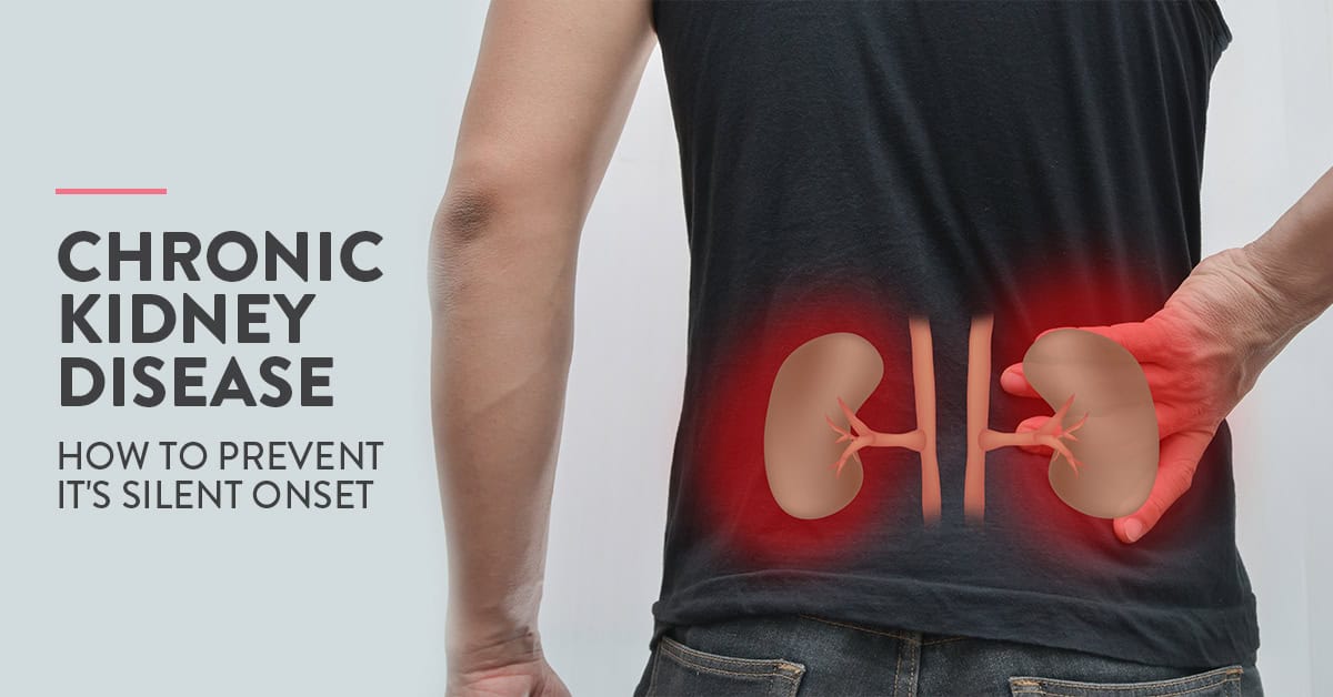
Chronic kidney disease occurs when your kidneys are too damaged to filter the blood properly. It is termed chronic as the damage to the kidneys occur over a period of time. Malfunctioning kidneys result in a build-up of toxins in the body which can in turn have serious repercussions.
CKD is often associated with ageing. However, it can happen to anyone and at any age. In this article let us take a closer look at this silent disease and how we can prevent it from interfering with our daily life.
Table of Contents
Kidneys are two fist-sized organs which are situated at either side of the spine at the lowest level of the rib cage. Kidneys contain millions of nephrons where the blood is filtered.
Most of us know that the basic function of the kidneys is to remove toxins, waste and excess fluid from the blood and push it out of the body in the form of urine. Kidneys also play a major role in regulating the body’s salt, potassium and acid content. Apart from this, kidneys produce hormones that affect other bodily functions such as blood production.
CKD is often considered to be a silent disease as it does not show any symptoms in its early stages. This condition worsens over time and can eventually result in complete renal (kidney) failure. In many cases, CKD is diagnosed during routine check-ups or during investigations for other concerns.
In the early stages, your kidneys may still function well enough that they do not cause any obvious symptoms. People can even lead normal lives with just one kidney. Detecting CKD in early stages is the best way to prevent further damage to your kidneys and avoid kidney failure.
In advanced stages, people with CKD can experience
If left unmonitored, CKD can lead to other health conditions such as heart disease, high blood pressure etc. It also increases your risk of having strokes and heart attacks. There is also a greater risk of developing acute kidney injury (AKI) which is a sudden change in kidney function due to medication, illness or injury.
When we think of any kidney disease, the first word that comes to our mind is dialysis. This is a common misconception. Many also think that CKD means kidney failure. This is far from true. In fact, most people with kidney disease do not even need dialysis as long as they make certain lifestyle and dietary changes on time to protect their kidneys.
The most important thing to remember is that CKD is a progressive disorder. Basically, it worsens with time eventually resulting in kidney failure. Kidney failure occurs when the kidneys are so damaged that they work at less than 15% of capacity. So, if you start your treatment on time and make lifestyle changes, you can avoid long term effects of CKD.
CKD is generally the result of excessive strain on the kidneys. In many cases, a combination of a number of factors causes kidney damage. Some of the most common causes of CKD include diabetes, hypertension, high cholesterol, kidney infections, polycystic kidney disease (genetic disease) and prolonged use of certain medications.
Chronic kidney disease or CKD is diagnosed with the help of blood and urine tests. The presence of increased levels of certain proteins in urine and/or blood generally indicate kidney damage. These tests are also used to define the stage of kidney disease or the degree of damage to the kidneys.
If you are identified as a high-risk candidate for CKD (having a family history of kidney disease or diabetes, high blood pressure etc) then you are advised to go for routine check-ups so as to monitor your renal (kidney) health.
Unfortunately, there is no permanent cure for CKD as the damage to the kidneys is permanent. However, further damage can be avoided with a combination of the following treatments:
The most basic steps to take for better kidney health are:
The trick is to drink the right amount of water. Too little water intake can prevent your kidneys from effectively filtering the blood causing issues such as kidney stones. Too much water can cause an imbalance in the salt levels in your body. According to the National Academies of Sciences, Engineering, and Medicine, the appropriate daily fluid intake is approximately 3.7 litres for men and 2.7 litres for women.
Your fluid intake also depends on the amount of physical activity you do, your environment and your general health.
Diet plays a major role in maintaining renal health. The most important things to keep in mind are:
Many doctors recommend keeping a “food journal” to record and monitor whatever you eat.
Physical activity and exercise are essential for overall health including renal health. Most adults are advised to perform any form of physical activity for at least 30 minutes a day. The amount of exercise as well as the intensity of the physical activity depends on your overall health. So, consult your doctor for a better idea of what would be best for you.
People with obesity or overweight are more prone to a wide range of secondary conditions such as heart disease, high cholesterol etc. Kidney disease is also one such complication that can arise due to overweight or obesity. If you have any of these conditions, you can consult bariatric surgeons for long term solutions in case exercise and diet are not showing the desired results.
Sleep disorders can be associated with a wide range of physiological and psychological issues such as obesity and depression. Medical experts recommend an average of 7-8 hours of sleep every night for adults.
What makes maintaining kidney health so important is that kidney damage occurs over a long period of time with barely any outward signs. So, by the time symptoms start to show, it might be too late to prevent kidney failure. This is especially relevant for people who are more prone to kidney damage such as diabetic patients. In such cases, routine check-ups are advised to monitor renal health.
Also, read: Types of Kidney & Renal stones, and it’s symptoms
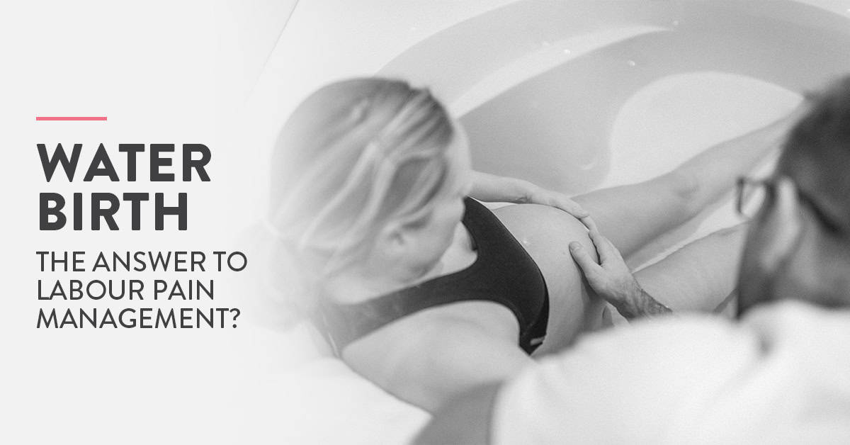
Deciding how you want to deliver your baby is quite an important decision to make during pregnancy. Many expectant women turn pale at the thought of labour pains. They may also find themselves overwhelmed with advice given by well-meaning friends and family. Water birthing to manage labour pains is one such advice that seems to keep popping up.
Let us take a closer look at this birthing technique to see if it is actually enough to effectively manage labour pains.
Table of Contents
To understand if water birthing is the best way to manage labour pains, we need to know more about water birthing. Water birthing is an ancient way of giving birth in water with the help of a deep bath or “birthing pool”. The modern definition states that water birth means having at least a part of your labour and/or delivery in a pool of warm water. It is performed under the guidance of a medical practitioner (doctor or midwife).
The first mention of water birthing dates as far back as 1805, it rose to popularity in the 1980s and 1990s. The idea behind water birthing is to provide a birthing environment as similar to the uterine environment. This is thought to ease the baby’s transition from the womb into the world while also reducing the stress of birthing for the mother.
In the first stage of your labour, when your contractions start but your cervix is not completely dilated, water immersion (in temperature-controlled water) can help ease the pains and make you feel more in control. Studies also suggests that giving birth in water can decrease the risk of severe vaginal tearing however conclusive evidence for this fact is yet to be found.
During stage two of your labour when your cervix is completely dilated and you start pushing to bring your baby into this world, you can choose to step out of the birthing pool or have your baby in the water itself.
If the doctor feels that there is a risk of complication, you might be asked to exit the pool to deliver your baby.
Being immersed in water during labour is a proven way to manage labour pains. Some women also claim to have felt calmer and more controlled during water birthing with an increase sense of privacy.
Warm water is known to encourage relaxation, reduces stress and stimulates the release of endorphins, kicking in the body’s own defence mechanism against pain. This also lowers the mother’s blood pressure which might have risen due to anxiety.
In some cases, water has also been shown to increase a woman’s energy in the latter stages of labour. The water’s buoyancy promotes more efficient uterine contractions and improves the blood circulation to the uterine muscles. This means lesser pain for the mother and more oxygen for the baby.
The water also makes the perineum more elastic and relaxed hence reducing the risk of severe tears while birthing. Perineum is the area between the vaginal opening and the anus. Perineum tears are quite common during childbirth, especially for larger or multiple babies. Based on the extent of the tear, it can be classified into 4 grades. Grade 3 and grade 4 perineum tears require surgical treatment while grade 1 and grade 2 can heal generally heal on their own.
There are several other benefits that are associated with water birth including shorter labours, lower postpartum blood loss, decreased physical contact (hands-off delivery), and greater feeling of satisfaction. However, more research needs to be done for conclusive results.
Many women do report that they felt significantly lesser labour pains when they were immersed in water. The temperature of the water helps relax the muscles, making labour pains more bearable for the mother.
However, pregnancy and birthing experience varies greatly from woman to woman. And water birthing does not take away labour pains entirely. While some women may feel almost zero pain while giving birth in the water, others may need to exit the pool for other pain management alternatives.
Unfortunately, water birth is not suitable for everyone. The following factors can make water birthing an unsuitable birthing option for you:
If your obstetrician or midwife feels that your labour needs to be monitored closely, or if any complications arise, water birth might not be advised. In case this occurs in the middle of water birthing, you may also be asked to exit the birthing pool.
Read: Home remedies to combat Gestational Hypertension
There is no conclusive proof indicating that giving birth in water is any riskier than giving birth out of water. Further research is required to understand the benefits and risks associated with water birthing.
However, before deciding to opt for a water birth, an important factor to note is that the water makes continuous electronic fetal monitoring tough. Hence this birthing technique is possible only for low risk pregnancies.
Hospitals are extremely meticulous about maintaining the hygiene of their birthing pools following strict protocols set by medical authorities. While there is no statistical evidence suggesting that water birthing increases the risk of the baby getting an infection, there have a few exceptions.
If you have a smooth, complication-free pregnancy and you are leaning towards natural birth with alternate, conservative pain management; water birthing is definitely an option you should take a closer look into.
Ques: What do I wear during water birthing?
You should wear something that makes you feel comfortable. Just remember you are essentially in a large tub of water. Many women may choose to wear comfortable robes while birthing, others might wear a bra, vest or t-shirt if they desire more privacy. You can also decide to forgo clothing if that is comfortable to you.
Ques: What is hypnobirthing?
Hypnobirthing using self-hypnosis, deep breathing and other relaxation techniques to manage labour pains. It can also be used in combination with other forms of pain relief. In some cases, it is shown to even shorten the labour. The research in the field of hypnobirthing is limited. Hence the true extent of benefits and risks of hypnobirthing is yet to be explored.
Ques: What are some of the benefits of water birthing?
Benefits of giving birth in water include:
In some cases, water birthing is also shown to have significantly lowered labour time.
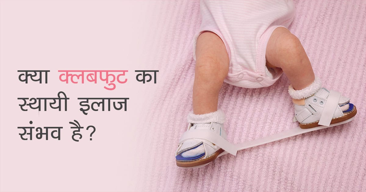
एक रिसर्च के अनुसार, प्रत्येक 1000 जन्मों में से 1 बच्चा क्लबफुट से अवश्य प्रभावित होता है। यह संख्या अलग अलग देशों में अलग-अलग हो सकती है। यदि क्लबफुट का इलाज (Clubfoot treatment in Hindi) सही समय पर और सही तरीके से न किया जाये तो इसका परिणाम आजीवन विकलांगता और असहनीय दर्द हो सकता है। इसके विपरीत, यदि इस बीमारी का जन्म के ठीक बाद इलाज कर लिया जाये, तो इस स्थिति में सुधार के लिए सर्जरी की भी आवश्यकता नहीं पड़ेगी।
दरअसल यह बीमारी इतनी सामन्य नहीं है जिस कारण अधिकतर लोग इससे अभिज्ञ हैं। जागरूकता के आभाव के कारण यह आसानी से सुधारात्मक विकृति बहुत सारे बच्चों में स्थायी विकलांगता का कारण बन जाती है। अतः आवश्यक है कि आपको पूरी जानकारी हो कि क्लबफुट (Clubfoot in Hindi) क्या होता है? इस लेख में, हम आपको विस्तार से बतायेंगे कि क्लबफुट क्या होता है (What is clubfoot in Hindi) और किस प्रकार सही समय पर इसका पता लगाकर इसका इलाज (Clubfoot treatment in Hindi) किया जा सकता है।
Table of Contents
क्लबफुट एक प्रकार की पैर से सम्बन्धित जन्मजात विकृति (जन्म के समय उपस्थित) होती है जिसमें जन्म के समय से ही बच्चे का पैर उसके सामान्य आकार का नहीं होता है। यह या तो बाहर की ओर या अंदर की ओर मुड़ा होता है। यह नवजात शिशुओं में पायी जाने वाली सबसे आम विकृति है जो हड्डियों और जोड़ों से सम्बन्धित होती है।
यह स्थिति सामान्य भी हो सकती है और कुछ परिस्थितियों में यह स्थिति गंभीर भी हो सकती है और शिशु के एक पैर या दोनों पैरों में भी दिखाई दे सकती है। पैर की मांसपेशियों को पैर की हड्डियों से जोड़ने वाले टेंडन्स के छोटे और तंग होने के कारण यह विकृति होती है जिस कारण शिशु का पैर अंदर की तरफ मुड़ जाता है।
यधपि क्लबफुट क्यों होता है इसका सटीक कारण अभी तक स्पष्ट नहीं है, किन्तु कुछ प्रतिष्ठित अनुसंधानों के अनुसार यह स्थिति किसी एक जीन संचरण के कारण नहीं अपितु कुछ आनुवंशिक और पर्यावरणीय कारकों की परस्पर जटिल क्रिया के कारण होती है।
जन्म के साथ क्लबफुट स्थिति की संभावना को बढ़ाने वाले जोखिम कारकों में निम्न शामिल हैं:
आनुवंशिक या न्यूरोलॉजिकल समस्याओं के साथ पैदा होने वाले बच्चों में कभी कभी क्लबफुट का एक अन्य रूप भी देखने मिलता है जो मांसपेशियों के असंतुलन का कारण बनता है। क्लबफुट के इस रूप को “द्वितीयक क्लबफुट” कहा जाता है।
प्रेगनेंसी के दौरान, 20वें सप्ताह के अल्ट्रासाउंड स्कैन में भी क्लबफुट का पता लगाया जा सकता है। हालांकि, अधिकांश मामलों में इस स्थिति का पता जन्म के बाद बाल रोग विशेषज्ञ या बाल रोग आर्थोपेडिक सर्जन द्वारा मेडिकल जांच के बाद ही लगाया जा सकता है।
सौभाग्यवश, क्लबफुट का इलाज पूरी तरह से किया जा सकता है, बशर्ते इसका इलाज सही समय पर किया जाए। इस स्थिति के इलाज की प्रक्रिया में शिशु के पैर के प्रभावित हिस्से पर क्रमिक प्लास्टर (अनुक्रमिक मलहम) किया जाता है जो इसके दीर्घकालिक प्रभाव को रोकने में भी सक्षम होता है। वास्तव में, सही इलाज के बाद इस विकृति के साथ पैदा हुए बच्चों को भविष्य में किसी भी कार्यात्मक कठिनाई का सामना नहीं करना पड़ता।
अच्छे परिणाम के लिए आवश्यक है, विकृति का पता चलते ही जितना जल्दी हो सके इलाज शुरू करा दें। डॉ हर्षवर्धन हेगड़े के अनुसार – जन्म के 5-7 दिनों के बाद कास्टिंग शुरू करा देनी चाहिए। उचित समय पर इलाज से लगभग 95-98% प्रभावित बच्चे पूरी तरह से बिना किसी सर्जिकल सुधार के ठीक हो सकते हैं। यदि गर्भावस्था के दौरान या जन्म के तुरन्त बाद क्लबफुट का निदान कर लिया जाता है, तो इस स्थिति में माता-पिता को चिंतित होने की आवश्यकता नहीं है।
यहाँ यह याद रखना अत्यन्त महत्वपूर्ण है कि यदि समय पर इलाज नहीं कराया जाये तो यह स्थिति उम्र के साथ और बिगड़ सकती है, इसलिए बच्चे के स्वस्थ और सामान्य जीवन जीने के लिए शुरुआती चिकित्सा हस्तक्षेप आवश्यक है।
बिना सर्जरी किये भी क्लबफुट का इलाज (Clubfoot treatment in Hindi) किया जा सकता है। इसके इलाज के प्रोटोकॉल में प्रारंभिक स्ट्रेचिंग, साप्ताहिक कास्टिंग और ब्रेसिंग का समायोजन होता है।
5-7 दिन बाद, बच्चे के पूरी तरह स्वस्थ होने के बाद ही इसका इलाज शुरू किया जा सकता है। यदि शिशु का समय से पहले जन्म हुआ है या उसका वजन जन्म के समय बहुत कम (2.5 किलोग्रा से कम) है, उस स्थिति में शिशु के स्वस्थ हो जाने के बाद ही स्ट्रेचिंग प्रारम्भ करनी चाहिए।
साप्ताहिक कास्टिंग का उपयोग करते हुए पैर के क्रमिक सुधार की इस प्रक्रिया को “पोंसेटि तकनीक” कहा जाता है। स्थिति की गंभीरता के आधार पर, यह प्रक्रिया कुछ महीने लम्बी हो सकती है और इसमें विशेष बूट और बार के उपयोग की भी आवश्यकता हो सकती है।
कार्यात्मक, दर्द मुक्त, सीधे पैर और भविष्य में चलने में किसी प्रकार की परेशानी न होना ही इस इलाज प्रक्रिया को करने का मुख्य उद्देश्य है। सही समय पर सही इलाज शीघ्र रिकवरी में सहायक होता है।
कुछ मामलो में पोंसेटि तकनीक वांछित परिणाम नहीं दे पाती, ऐसा उन परिस्थितियों में होता है जब स्थिति अत्यंत जटिल हो या अंतर्निहित स्थिति (द्वितीयक क्लबफुट) इसका कारण हो। ऐसे मामलों के इलाज (Treatment of clubfoot in Hindi) के लिए सर्जिकल सुधार का उपयोग किया जा सकता है।
इग्नेसियो वी. पोन्सेटि (चिकित्सक जिन्हे आर्थोपेडिक्स के क्षेत्र में अपने योगदान के लिए पहचाना जाता है) के नाम पर रखी गयी पोंसेटि तकनीक दुनिया भर में क्लबफुट के इलाज (Treatment of clubfoot in Hindi) के लिए सबसे व्यापक रूप से इस्तेमाल की जाने वाली तकनीक है। इस तकनीक में एक समयावधि में इस विकृति को ठीक करने के लिए सौम्य स्ट्रेचिंग और अनुक्रमिक कास्टिंग के संयोजन का उपयोग किया जाता है।
इस तकनीक में, बच्चे के पैर को धीरे से स्ट्रेच किया जाता है और उसे सही स्थिति में जोड़ दिया जाता है और इसे एक कास्ट (आमतौर पर पैर की उंगलियों से जांघ तक) की मदद से सही करने का प्रयास किया जाता है। यह प्रक्रिया हर सप्ताह दोहराई जाती है जब तक वांछित परिणाम प्राप्त नहीं हो जाते हैं। इसमें 6-8 सप्ताह या उससे अधिक समय भी लग सकता है।
एक बार जब स्ट्रेचिंग और अनुक्रमिक कास्टिंग पूरी हो जाती है, तो सर्जन एक छोटे से प्रोसीजर की मदद से एच्लीस टेंडन (एड़ी की हड्डी) में टाइटनेस को कम कर देता है। अगले चरण में एक और छोटा प्रोसीजर किया जाता है जिसे टेनोटॉमी कहा जाता है। इस प्रोसीजर में टेंडन को काटने के लिए बहुत पतले उपकरण का उपयोग किया जाता है। इस प्रक्रिया में टाँके लगाने की आवश्यकता नहीं होती है।
इसके बाद 3 सप्ताह के लिए सर्जन टेंडन को सपोर्ट और रक्षा देने के लिए एक कास्ट का प्रयोग कर सकता है। एक बार जब टेंडन वापस से एक निश्चित लम्बाई तक बढ़ जाता है तब क्लबफुट (Clubfoot in Hindi) को पूरी तरह सही माना जाता है।
पोन्सेटि तकनीक में, पैर को धीरे से उसकी स्थिति सही करते हुए कास्ट को हर हफ्ते बदल दिया जाता है। हर बार जब प्लास्टर लगाया जाता है, तो पैर को थोड़ा और सही किया जाता है। इसलिए, आवश्यक प्लास्टर की संख्या विकृति की गंभीरता पर निर्भर करती है। हालांकि, आम तौर पर, औसतन 4-10 कास्ट का उपयोग किया जाता है। बच्चे की उम्र अधिक होने पर यह बढ़ सकता है, यदि विकृति अधिक जटिल है, या लंबे समय तक अनुपचारित छोड़ दिया गया हो। ऐसे मामलों में, सप्ताह में दो बार कास्टिंग की भी आवश्यकता हो सकती है।
एक बार अंतिम कास्टिंग पूरी हो जाने और कास्टिंग हटाने के बाद, बच्चे को मेटल की पट्टी से जुड़े विशेष जूते पहनने की आवश्यकता होगी। इसे डेनिस ब्राउन / मिशेल पोंसेटि स्प्लिंट कहा जाता है। ये जूते आमतौर पर 3 महीने तक एक दिन में 23 घंटे के लिए पहनाये जाते हैं। 3 वर्ष की आयु के बाद इन जूतों को बच्चे के सोने के समय रात में पहनने की सलाह दी जाती है।
माता-पिता को भी बच्चे की स्ट्रेचिंग जारी रखने की आवश्यकता होती है। क्लबफुट विकृति को ठीक करने में जूतों का उचित उपयोग अत्यंत आवश्यक है।
निम्नलिखित मामलों में, क्लबफुट के इलाज के लिए सर्जरी (Surgery for clubfoot treatment) की आवश्यकता हो सकती है:
ऐसे सभी मामलों में, यदि बच्चे में चलने फिरने के दौरान हाइपरएक्टिव टेंडन के लक्षण दिखते हैं तब आमतौर पर टेंडन स्थानांतरण की आवश्यकता होती है।
क्लबफुट के अन्य सर्जिकल उपचारों में सॉफ्ट टिस्सु रिलीज, बाहरी फिक्सर के साथ क्रमिक सुधार, बोन कटिंग सर्जरी या पैर के जोड़ों का संलयन शामिल है।
जब तक सही उपचार प्रोटोकॉल का पालन किया जाता है, इस उपचार तकनीक में कोई महत्वपूर्ण जोखिम शामिल नहीं हैं। कुछ मामलों में, यदि बच्चे की त्वचा अधिक संवेदनशील है या यदि कास्ट का उपयोग उचित तरीके से नहीं किया गया है, तो प्लास्टर त्वचा पर घावों का कारण बन सकता है। इनका आसानी से एंटीबायोटिक्स द्वारा इलाज किया जाता है और लगभग एक सप्ताह तक कास्टिंग की जाती है।
यदि पोन्सेटि तकनीक को सही तरीके से नहीं किया जाता है, तो इसके परिणामस्वरूप रॉकर बॉटम फुट (मिडफुट ब्रेक) या आईट्रोजेनिक कॉम्प्लेक्स क्लबफुट जैसी जटिलताएं हो सकती हैं। कास्टिंग के बाद एड़ी की हड्डी को काटने के लिए इस्तेमाल की जाने वाली प्रक्रिया से रक्तस्राव भी हो सकता है, हालांकि यह आसानी से मैनेज हो सकता है।
सौभाग्यवश, सही उपचार के साथ, इस विकृति के साथ पैदा हुए बच्चे लगभग सामान्य जीवन व्यतीत कर सकते हैं, वो अच्छे से चल सकते हैं, भाग सकते हैं, खेल सकते हैं और सामान्य जूते पहन सकते हैं। कुछ मामलों में प्रभावित पैर, दूसरे पैर से 1 से 1.5 इंच छोटा हो सकता है।
कॉल्फ मसल्स भी छोटी हो सकती हैं, जिससे बच्चे को दर्द या थकान महसूस हो सकता है। हालांकि, यह शायद ही कभी किसी महत्वपूर्ण समस्या का कारण बनता है।
इस स्थिति का इलाज एक प्रशिक्षित बाल चिकित्सा आर्थोपेडिक सर्जन द्वारा किया जाता है। लंबी अवधि की जटिलताओं से बचने और बच्चे के चलने के समय तक पैर की पूर्ण कार्यक्षमता प्राप्त करने के लिए विस्तार पर ध्यान देना महत्वपूर्ण है।
भले ही यह प्रक्रिया अपेक्षाकृत सरल और गैर-सर्जिकल हो, लेकिन पैर को सही ढंग से संरेखित करने के लिए सही इलाज की आवश्यकता होती है। अपने शिशु की स्थिति के इलाज के लिए एक अनुभवी बाल चिकित्सा आर्थोपेडिक सर्जन चुनें, जो क्लबफुट का इलाज (Treatment of clubfoot in Hindi) करने में पारंगत है।
यदि आपके शिशु में गर्भावस्था के दौरान या जन्म के तुरंत बाद क्लबफुट का पता लगता है, तो सबसे महत्वपूर्ण बात यह है कि तुरंत उपचार शुरू किया जाए। बेहतर परिणाम के लिए होम केयर निर्देशों का पालन करें जैसे कि डॉक्टर द्वारा दिए गए बच्चे के लिए व्यायाम कराना इत्यादि ।
Also, read this in English: Finding the permanent solution for clubfoot!

PCOS or polycystic ovary syndrome is an extremely common health problem amongst women of childbearing age. In fact, it is so common that in an effort to raise awareness about this condition, the month of September has been declared “PCOS awareness month”. Awareness is the first step in the fight against the spread of this condition. Let us educate ourselves about PCOS and take a closer look into this syndrome, its impact and how diet & exercise can help in stopping this condition from taking over our lives.
Table of Contents
Polycystic ovary syndrome, commonly known simply as “PCOS” is a condition characterised by an increased production of male sex hormones (androgens). The three main characteristics of PCOS are:
Every month, the ovaries release a mature egg for fertilisation. This process is called ovulation. If the egg is not fertilised, it is released from the body (periods). In some cases, women may experience irregular periods, indicating problems in ovulation.
Small quantities of androgens or “male” hormones are produced by the adrenal glands, ovaries and fat cells. They are vital to normal reproductive function, emotional stability, cognitive function, muscle function and bone strength. High levels of androgens can result in abnormal hair growth (especially facial hair), acne and male patterned baldness.
Polycystic ovaries are characterised by enlarged ovaries containing large number of cysts (fluid-filled sacs). These cysts surround the eggs making it difficult for the ovaries to release the eggs. This results in anovulation.
Women with PCOS experience at least two of the above characteristics.
Read: First period post pregnancy: what is normal and what is not?
Earlier, PCOS was considered to be a simple endocrine disorder, causing hormonal imbalances. However, today with advances in research, we have a better understanding of this syndrome. It is now treated as a complex condition with metabolic, hormonal and psychological factors. Patients too are treated holistically by addressing the phycological impact of the disorder as well as the physiological one.
PCOS can be devastating for women in their childbearing years. It is one of the leading causes of female infertility. For many women, giving birth is an important aspect of their identity, and they face judgment by their partners, family and society if they are unable to do so. It is important to educate the family as well so that they can give the required emotional support to the patient.
Studies also indicate that women with PCOS have higher levels of psychological distress. This can be in part due to symptoms of PCOS such as obesity, hirsutism (excessive hair growth) and infertility.
PCOS is also known to increase the risk of several complications such as developing metabolic syndrome and cardiovascular disease. Early intervention, lifestyle changes and long-term monitoring is key to relieving the symptoms of PCOS.
Read: Pregnancy and Polycystic ovary syndrome
Many women mistakenly feel that something they did cause PCOS. This adds on to the psychological impact of this condition. The exact cause of PCOS is still not known, it is believed that it is caused by a combination of several factors including genetics and having insulin resistance. Obesity is also known to increase the likelihood of developing PCOS.
The symptoms of PCOS depends on the severity of the condition. In a large number of PCOS cases, diagnosis is made when the woman experiences difficulty in conceiving. This is because the symptoms may not be severe enough to cause the patient to seek medical help. Some of the most common symptoms of PCOS are:
As the symptoms need not be evident all the time, especially in the early stages, routine gynaecological check-ups are advised as soon as you start menstruating, especially if you are at a higher risk of developing PCOS.
Read: What to do when you suspect infertility
Home remedies for PCOS are usually the first line of defense against this condition. This can further be classified into two categories i.e.: Dietary changes & Lifestyle changes. Let us take a closer look into home remedies for PCOS management.
Diet tends to influence our health to a great extent. Women affected with PCOS are likely to have higher levels of insulin than the ones without PCOS. This is because insulin resistance is one of the symptoms of PCOS. Insulin resistance indicates that the insulin produced by the pancreas is not utilized for digestion of the sugar in the body, resulting in excess blood sugar.
In addition to this, due to insulin resistance many affected women find it difficult to lose weight, especially around their abdomen. Excess insulin is also linked to increased production of androgens (male hormones) causing male patterned baldness and hirsutism.
Keep the following guidelines in mind while coming up with your own unique PCOS diet plan.
Regular physical activity and weight management are important aspects of PCOS management. The following exercises will help you relieve PCOS symptoms.
In more severe cases, medication can be used to manage PCOS symptoms. Following a healthy lifestyle and managing your weight with regular exercise is the best way to minimise the impact of PCOS.
Over the past few years, recovery time, scarring and pain associated with a wide range of general surgeries have reduced significantly. This is all thanks to the rising popularity of “laparoscopic” procedures or keyhole surgeries. With the help of laparoscopic surgery, surgeons can now treat patients in outpatient procedures itself avoiding unnecessary risks, scarring, pain and expenditure, enabling the patient to recover comfortably at home. In this article we will be exploring the laparoscopic risks and benefits that need to be factored in while deciding to opt for the procedure.
While laparoscopic procedures can be traced as far back as 1805. However, it was Dr Heinz Kalk, a German gastroenterologist who popularised it by inventing a superior laparoscope. This earned him the title of “Father of Modern Laparoscopy”. In the 1930s, Laparoscopy gained popularity in the United States through the work of internist John Ruddock. In the last few decades, laparoscopy has become the first choice of surgical treatment for both doctors and patients.
Table of Contents
According to the National Health Service-UK, laparoscopic surgeries or keyhole surgeries are defined as “a type of surgical procedure that allows a surgeon to access the inside of the abdomen or pelvis without having to make large incisions in the skin”
In this procedure, the surgeon makes one or more small incisions. A small tube is used to pump gas into the body cavity through one of these incisions. This is to help the surgeon get a clearer view inside the body. Using a laparoscope (a small tube with a light source and camera), the surgeon can either see inside the body to make a diagnosis or use it to guide other surgical instruments. Over the years, laparoscopy has become a common procedure to diagnose and treat gynaecological, GI, minimal access and urological conditions.
Laparoscopy is used to treat a wide range of GI, gynaecological and urological conditions. It can be used to perform tubal sterilization, hysterectomy, gallbladder removal, appendectomy, kidney stone removal etc.
Laparoscopy is also a common way to conduct a number of diagnostic tests such to diagnose and treat conditions such as endometriosis, fibroids, ovarian cyst, ectopic pregnancy, pelvic floor disorders and some forms of cancer.
Read: What you need to know about an Ectopic pregnancy
The most obvious and basic benefit of laparoscopic surgery is that large open wounds and incisions can be avoided. This in turn significantly decreases blood loss, pain, discomfort and scarring. Even the side effects caused by anaesthesia can be reduced or avoided. The instruments used in laparoscopy also cause less trauma to the tissue.
Owing to the nature and size of incisions in laparoscopic procedures, the risk of developing postoperative complications associated with the wound is much lower. This includes complications such as dehiscence, infection and incisional hernia amongst others.
Laparoscopic procedures also help avoid cooling, drying, excessive handling and retraction of internal organs that can occur in traditional open surgeries.
These benefits together aid in minimising recovery time and post op pain hence reducing the risk of developing conditions associated with prolonged bed rest and inactivity (such as bone loss, muscle atrophy and urinary retention). Most of the time, laparoscopic surgeries can also be performed as outpatient procedures.
In a more practical sense, laparoscopic procedures minimise direct contact between the surgeon and patient. Consequently, there is a much lower risk of transmission of any type of infection between the two, making it a preferred surgical option for a wide range of treatment and diagnostic needs.
The benefits of laparoscopic surgery are quite obvious and widely accepted. However, like all surgical procedures, it does not come without its own risks. One of the main risks is that laparoscopy takes longer time to perform as compared to open surgery. This increases the exposure of the patient to general anaesthesia which can result in complications.
Other serious complications that can occur (rarely) in laparoscopic procedures are:
Just like any traditional surgical procedure, the complications may not be evident right after surgery. Rather they can start developing over a period of time following the surgery. So, it is generally advised that the patient is not left alone for the first 24 hours after surgery. Even if they have been discharged from the hospital. If you observe any of the following symptoms after a laparoscopic procedure, seek immediate medical attention:
In some cases, due to complications arising during the procedure, the surgeon might also have to switch to open surgery instead of laparoscopic surgeries. Further surgery may also be required to treat some of the serious complications that may arise after a laparoscopic procedure.
Now, let us come back to the main question. Is laparoscopic surgery safe? Considering all of the above facts, it is clear why laparoscopic surgeries are rising in popularity. As compared to traditional open surgeries, they are considered safer as they minimise the time spent by the patient in the hospital or on bed rest. In fact, in many cases, patients are discharged a few hours after the procedure itself.
It is important to remember that laparoscopic surgery is by no means a magic cure to all surgeries. Not all patients are suitable for this procedure. Based on the severity of the condition and the patient’s general health, the doctor can decide between open or laparoscopic procedures.
Laparoscopy during procedure is not considered unsuitable or dangerous to the health of the fetus and the mother during any trimester. However, precautions are to be taken to ensure the safety of the baby and mother such as carefully deciding the type and dosage of anaesthesia as well as ensuring the facility is equipped to handle any complications should they arise.
Patients are generally advised not to eat or drink anything for at least 8 hours before the surgery. This is because laparoscopic procedures are usually performed under general anaesthesia.
The recovery time after a laparoscopic procedure depends on several factors such as the type of treatment and your general health. Surgeries such as appendectomy can take around 3 weeks for recovery.
Robotic-assisted laparoscopy is the latest innovation in the field of laparoscopy. In this procedure, the surgeon uses a console located in the operating theatre to perform the procedure using robotic arms attached with a special camera and surgical equipment. The greatest advantage of this technique is that the system offers a magnified 3D vision and increased range of movement for the instruments inside the body cavity. This helps the surgeon be more precise and accurate. There is also evidence suggesting that robotic assisted laparoscopy can have lower risk of complications as compared to traditional laparoscopy or open surgeries.
Read: Treatment of fibroadenoma without surgery – here’s how you can dissolve it naturally