Filter :
Gaining weight is as difficult as losing weight, and therefore maintaining a balance is essential. People who are underweight find it challenging to slow down their metabolism. This article, suggests ways how one can slow down metabolism in order to gain weight.
Table of Contents
Metabolism allows the body to grow and function. In simple terms, metabolism means the conversion of food into energy. Metabolism is the set of life-sustaining chemical reactions in organisms. The three main purposes of metabolism are: (1) the conversion of the energy in food to energy available to run cellular processes; (2) the conversion of food to building blocks for proteins, lipids, nucleic acids, and some carbohydrates, and (3) the elimination of metabolic wastes.
People who are thin, slow metabolism will help them to gain weight naturally. In a world where everyone is trying to do things faster and faster. For example- people who wish to lose weight want to do that at speed, and people who wish to gain weight want to gain it very fast. But in the real world, this is not possible, or we can say not that easy. It takes time and patience to gain or lose weight.
In the diet world, speed of metabolism is something commonly talked about. It is a known fact that a faster metabolism will surely lead to losing weight. It means the faster your body will digest food, the fewer calories intake your body will need to convert into energy. What we need to talk and understand about is how and what are the ways one can slow down metabolism.
It is beneficial for people who need to gain weight to actually be able to find ways to naturally slow down their metabolism.
Taking calories is not enough but in fact, consuming the right amount of calories is what matters. Eat food rich in carbohydrates like parathas, a plate full of brown rice, whole milk, eggs, potatoes, full-fat curd, etc. These give your body the energy it needs to gain weight. Foods that are high in fat are difficult to digest and thus can slow down your metabolism, so consume food rich in carbohydrates only.
Calculate or track the number of calories you eat during the day. There might be instances where you feel full but still do not consume enough calories needed to gain weight. Therefore, it is important to count the calories you consume.
Indulging in a low-intensity workout for a shorter duration ensures that your body does not become calorie deficient and also helps in slowing down the metabolism. Lower-intensity strength exercise will help you maintain a healthy metabolism while simultaneously increasing muscle mass, and on the other hand, performing weight resistance training or walking and high-intensity workouts can lessen your metabolic rate.
Taking sufficient sleep, i.e. sleeping for long hours, is the easiest way to slow down the metabolic rate. It is because your body does not burn calories while your body remains steady for long hours. Therefore, if you improve your sleep cycle, you will automatically be able to slow down the metabolic rate, thereby helping you to increase weight.
If your appetite is low, then the chances are high that the already existing food will break down faster in the body. Therefore, it is essential to boost the appetite. Eat food rich in nutrients, add more carbs to your meals, try tricking the brain with different size portions and avoid skipping breakfast. Other than these, the doctors might also recommend appetite stimulants which may help you to boost your hunger and, thereby, your appetite. In addition, it is okay to add extra cheat meals to your diet plan as they may help in adding calories to the diet.
Caffeine has been shown to boost metabolism, and therefore it is suggested that people who want to reduce their metabolism should avoid consuming caffeine in any form. Caffeine makes a person more hyper and super active. Sleep more to refrain yourself from consuming caffeine.
There might be some underlying health issues that may be stopping you from gaining weight. And to know what may be the reason behind no weight gain, book an appointment with the internal medicine doctor at CK Birla Hospital.
Water is the driving force of nature. Pure water is the first and foremost medicine, and therefore there is no life without water. Water has x number of benefits, and that is why keeping water clean and hygienic for all should be every individual’s priority. Water is essential and precious for survival, and if not taken care of or contaminated can cause and spread illnesses like water-borne diseases.
In this article, Dr. Rajeev Gupta shares what are and how one can prevent water borne diseases.
Table of Contents
Consumption of contaminated water or food after coming in contact with the person who has it or with the bacteria itself can lead to infection, thereby having water borne diseases. Ingesting contaminated water or coming into contact with excrement can cause illnesses caused by tiny organisms such as viruses and bacteria.
These diseases would not exist if everyone surrounding us was able to practice proper sanitation and hygiene and had access to clean water.
Typhoid fever is an infection that spreads through food that is contaminated and unhygienic water. People living in poor sanitation areas are more prone to be infected with typhoid fever. It can partly be treated by vaccine if proper precautions should be taken to restrict the spread of the infection.
The treatment recommended and prescribed by the doctor will be antibiotics and fluids.
To prevent the infection, timely vaccines are recommended for people who live or travel in places where there is poor sanitation and unhygienic drinking water. In such places, avoid drinking tap water and always prefer bottled water.
Cholera is a bacterial disease that is commonly found in rural villages where there is poor sanitation. Cholera is a disease that spreads through water and can be fatal if it is not treated on time.
The treatment plan for cholera is rehydrating the body with IVF fluids and antibiotics.
Washing your hands properly and often can help prevent cholera or any water borne disease. Eat completely cooked food and wash vegetables with fresh water only.
Giardia, also called beaver fever, is an intestinal infection caused by a parasite named giardia. It is another type of water borne disease that spreads through contaminated water and food. Giardia can also spread person-to-person. However, the infection is cleared from the system in a few weeks but can affect the intestines for years.
Like other water-borne diseases, giardia is also treated by antibiotics and anti-parasite.
Though there is no vaccine available for giardia until now, there are simple changes one can make to avoid the infection. For example, washing hands with soap every now and then, avoiding swallowing swimming pool water, and drinking bottled water only.
Dysentery is an intestinal infection characterised by inflammation and bloody diarrhoea. The bacteria which causes dysentery is shigella bacteria (shigellosis) or an amoeba. It is spread through contaminated water and food.
Increased fluid intake, rehydration solutions, IV fluids, and antibiotics are all possible treatments for dysentery. With rest and drinks, mild dysentery usually goes away, and stomach cramps are relieved with over-the-counter medication.
To prevent dysentery or any water-borne diseases, it is essential to wash hands frequently with soap for at least 60 seconds. In addition, avoid eating food from outside and try to increase your intake of fruits.
Hepatitis A is a contagious liver infection caused by contaminated water or food. Therefore, people travelling to several rural places should be extra cautious of their hygiene to prevent and protect themselves from any water borne diseases.
Though the infection clears away within weeks, it may also vary as per each case and the severity of the infection. Hepatitis A can leave your body feeling tired and weak for days and weeks, so taking extra care and rest is recommended until you fully recover.
The best way to prevent Hepatitis A is a vaccine. Doctors always suggest that every individual get vaccinated against all types of water-borne diseases and others. In some nations, the government has made it mandatory for every individual to get vaccinations. Other than the vaccine, there are few things individuals can do in everyday life to prevent the infection. Maintain hygiene, wash hands before and after eating, use the toilet, sanitise hands every now and then, avoid intake of unwashed fruits and vegetables, and practice safe and protected sexual intimacy.
Waterborne diseases can have adverse effects on the body, which may not be for a few days or a few weeks but for several years. In addition, it can make your intestines and immunity weak making it difficult for one to function normally. So, on doctors’ recommendation, it is advised to get all important vaccinations to lead a healthy life. To know more about water borne diseases, book an appointment with Dr. Rajeev Gupta.
Ques 1. What are the prevention of water borne diseases?
The best way to prevent water borne diseases is to get yourself vaccinated. In addition, other lifestyle changes like washing hands with soap, drinking bottled water only, avoiding street food, and sanitizing hands can help prevent water borne diseases.
Ques 2. What is the best way to achieve primary prevention of water-borne diseases?
Practicing good and healthy personal hygiene and vaccination can help one achieve primary prevention from water-borne diseases.
Reproductive organs are the most sensitive parts of the body. Women are more prone to infections than men. Taking good care of the vagina is something we cannot evade because otherwise, it will lead to vaginal allergies and infections. states that though the vagina is a self-cleaning part of the body, keeping it clean is our duty only. In this article, she gives clarity on what are the causes and treatment of vaginal itching.
Table of Contents
Vulva or Vaginal itching is a common issue in women. But there can be several causes which could have led to itching in the vagina. The vulva is the outer part of the female reproductive organ. Vagina is made of labia, vulva and clitoris. Vaginal irritation can be due to yeast infections, allergies and skin conditions. Vaginal irritation may be inside or outside the vaginal area. Each symptom and cause would have a different treatment plan.
Irritation in and around the vagina can be due to several reasons. The products used around the vaginal area, menstrual pads, underwear fabric, soaps, lubes, creams, or lotions, usage of condoms, or using non-medicated products. The itching and irritation might start to go away after one stops using such products. Unless the medicated product is recommended by a doctor, refrain from using any product around the vagina. When your skin is itchy, avoid scratching or rubbing it, as it will only make the allergy worse.
Bacterial vaginosis is bacterial growth in the vagina. Though the bacteria is treatable is a Short-term infection which resolves within days to weeks but can cause severe itching and bruising inside the vagina. Some common symptoms of bacterial vaginosis are:
Yeast infection takes place when there is an overgrowth of infection or candida in the vagina. Though a yeast infection is a treatable infection which affects the tissues at the opening of the vagina. Yeast infections are treatable and are usually not a serious condition but their signs and symptoms can bother an individual.
There are some common skin infections which can cause severe itching in and around the vagina. Common causes of skin infections are:-
Sexually transmitted infections (STIs) is an infection that can easily be contracted after having sexual contact with a person who has already been diagnosed with STIs. STIs can irritate the vagina and cause pain and discomfort. It is very important to seek medication and treatment for STIs as they can affect the person severely and cause long term issues in the body like pelvic diseases and can also lead to infertility.
There are two ways to treat vaginal itching- medically and through home remedies. Once the cause of vaginal itching is diagnosed the doctor recommends treatment options and the course of treatment will depend on the cause of itching.
Ques 1. Can a UTI cause vaginal itching?
No, there has not been found any relation between vaginal itching and UTIs. Taking antibiotics to treat IUI might trigger the itching in the vagina. But visiting a specialist might be the best option.
Ques 2. Can diabetes cause vaginal itching?
Yes, if your sugar is high then there are chances that your genital area might itch. Therefore, it is suggested to control your sugar levels.
Ques 3. Is vaginal itching normal?
Yes, and No, most vaginal itching is normal but if the itching continues and causes discomfort more than usual then visiting a gynaecologist might be the best.
Ques 4. Does vaginal itching always mean an STD?
No, STD can be one of the causes of vaginal itching but there are several other factors which can lead to vaginal itching.
Ques 5. Does vaginal dryness cause itching?
Yes, vaginal dryness can cause itching, redness, flaky skin around the vagina and pain during sexual intercourse.
Stomach pain in the upper part during early pregnancy can be normal as the baby needs space to grow in your stomach, which is why you might feel discomfort or pain in the stomach. Though the pain you experience might be normal, as an expecting mother, you should be aware of all possible reasons why this pain occurs. It is important to have all the knowledge so that you may be able to recognise symptoms that may cause the pain.
Dr Alka Gupta, Obstetrician, shares insights into why this stomach pain occurs during early pregnancy and all possible symptoms and solutions to the pain.
Table of Contents
Even when one is not pregnant, pain caused due to gas can be really disturbing and painful and so imagine having gas pain during pregnancy. The pain may start from slight discomfort to unbearable pain. Usually, women experience more gas when she is pregnant due to the increased amount of progesterone levels. The pain may either stay or travel from one place to the other. The progesterone relaxes the muscles, due to which the food takes more time to digest, thereby causing gas. Food stays in the colon area for a longer duration, allowing the stomach to create more gas. Also, when the stomach is growing, there is automatically an added pressure on the uterus, which leads to slow digestion of food therefore, an accumulation of gas is there.
Consuming small meals throughout the day and increasing the intake of fluids can help relieve the pain. In addition, light-weight exercises help to aid digestion and reduce abdominal cramps during early pregnancy. Avoid eating food items that may trigger gas pain like greasy foods, beans, cabbage, and carbonated beverages.
Braxton Hicks Contractions are also known as practice contractions. These are more like false alarms and should be differentiated from true contractions. These contractions occur when the uterine muscles contract for a few seconds to minutes. Many pregnant ladies have been reporting that Braxton hacks feel like their stomach muscles are tightening, and their stomach starts to feel hard. Braxton hicks are usually seen during the third trimester of pregnancy. They may make the women uncomfortable, but these contractions are normal during pregnancy.
Drinking loads of water and relaxing can help relieve pain caused due to Braxton hicks. Change your position as it may help to stop the false contractions. Relax and try to calm yourself by taking a warm bath or listening to music.
When the baby is trying to adjust herself/himself in the womb, the uterus and the ligaments stretch, this stretch may cause sharp or dull pain in the abdomen, groin, and hips. In addition, walking, sneezing, or coughing may trigger the round ligament pain. The round ligament pain is usually experienced by the women in their last trimester of pregnancy.
Carefully make movements when you are sitting or lying down, as it may decrease the pain caused due to ligaments. Try bending your hips whenever you get a sudden urge to sneeze or cough. Stretching on a daily basis might also help to relieve round ligament pain.
Feeling constipated? Don’t worry this is the most common concern of every pregnant woman you must have met. But have we ever wondered what causes constipation in pregnant women? Constipation can be caused by a variety of factors, including disturbance in hormones, fewer fluids or fibre intake, lack of exercise, and consumption of iron tablets. Constipation can be extremely painful and discomforting for the pregnant woman. They start to feel weak, uncomfortable, bloated, and irritated.
Include more fibre in your daily routine, increase the intake of fluids, and drink 8-10 glasses of water every day. These are home remedies to treat constipation in pregnant women. If constipation still persists, the doctor might recommend stool softeners that are only meant for pregnancy.
Pregnant women might have pain in the left side of the stomach during early pregnancy due to the changes going on in their bodies. This pain usually occurs because the uterus and the body are trying to make space for the baby. But there could be a more serious issue of the pain in the left side for which immediate assistance may be required.
Treatment for the lower abdominal pain in early pregnancy 2 weeks or pain on the left side of the stomach will depend on the cause of the pain. So, the treatment may be a combination of a doctor’s recommendation and home remedies. Avoid heavy lifting, do Kegel exercises to strengthen your pelvic area, sleep with a pillow between your legs or knees, Use a heating pad, but never for more than 10 minutes at a time, and can also use a maternity belt for extra support.
Ques 1. Are stomach aches normal during early pregnancy?
Yes, the stomach aches are normal during early pregnancy, but if the pain continues, the pregnant woman should immediately seek medical assistance.
Ques 2. Does a miscarriage feel like a stomach ache?
Slight pain in the stomach during early pregnancy may usually mean that the womb, the muscles, and ligaments are expanding and stretching. However, if the woman feels pain and discomfort in the lower abdomen, it may be a sign of miscarriage, and immediate assistance is required.

Let’s plan your Sunday – you start your morning with a plate of chole bhature from the popular food stall in your neighbourhood. At brunch, you have some samosas followed by your favourite tea. For lunch, there is a deluxe thali with four of the spiciest, flavourful dishes. By evening, you plan to go for some cheesy Italian pizza with your friends. And at dinner, you are gorging on some rich biryani. What do you have at the end of the day? Probably, a gas problem.
Gas problems are very common in India, a country that is known for its rich flavours. People across all age groups from toddlers to the elderly are prone to be affected by gastric symptoms. Despite its high and very frequent incidence, a large section of people are unaware of management options and how to reduce gastric problems.
In this article, with insights from Gastroenterologist we will explore what we need to know about a gas problem, gastric problem symptoms and gastric problem remedies.
Table of Contents
A gas problem is a condition that occurs when excess gas in your stomach causes pain. Having gas in your stomach, also known as intestinal gas, is very normal and is considered part of digestion. Intestinal gas is a mix of odourless vapours and usually includes oxygen, carbon dioxide, nitrogen, methane, and hydrogen.
It is also normal for your body to relieve excess gas through burping/belching or in the form of flatus (flatulence). Passing gas is a natural process. According to studies, most people pass gas upto 12 times a day. When excess gas gets trapped inside your stomach, it may cause pain, bloating and other gastric symptoms.
Gastric symptoms are very obvious. However, they are often misinterpreted due to their overlap with other gastrointestinal problems.
Common gastric symptoms are:
These symptoms are usually indicative of other gastrointestinal conditions. If you have any of the following signs on top of gas, you may consult your healthcare provider.
Other symptoms include:
Intestinal gas is a natural part of the digestive process. Excess gas, however, results from a range of issues.
Common factors that cause a gas problem are:
Common foods that may increase your chances of developing gas are:
You are prone to develop excess gas in your stomach if you lack certain enzymes to help aid the digestion process. Due to these enzymes, the small intestine is not able to absorb all the nutrients in the food and the undigested food gets passed on to the large intestine.
We all naturally swallow air while eating food. However, sometimes, due to our eating habits, we may swallow more than enough air. The said air includes nitrogen, oxygen, and carbon dioxide. Most people release this air through belching. But sometimes, your intestines may partially absorb some air and cause gas.
The gas problem can be easily identified because of its signs and symptoms. However, most gastric problems have a similar set of symptoms. This may cause overlap and cause diagnostic dilemmas.
Your healthcare provider is the right person to identify if you have a gas problem. He/she may order a range of tests and procedures to confirm the diagnosis. Your doctor may additionally do some tests to find out the cause of the gas problem in order to provide comprehensive, complete treatment.
Common methods to identify the gas problem are:
The first line of treatment for intestinal gas is to offer pain relief. Your healthcare provider will analyse your health and examine your overall condition before offering any treatment.
Based on the findings of your diagnosis, your doctor will devise a personalised treatment plan to relieve symptoms and treat the root cause of the gas problem.
The following treatment protocols are used for a gas problem:
If left untreated, intestinal gas can cause a range of complications. These include chest pain, elevated risk of cardiovascular problems, and increased risk of indigestion issues.
Yes, it is entirely possible to prevent a gas problem. However, prevention measures are unique for every individual. For instance, your unique food triggers and lifestyle factors will be important to help understand what may be contributing to developing gas and what all can be avoided.
You can practice the following measures to treat gas problems:
Gas problems or the pain caused by intestinal fas can be well managed at home. You may always need immediate clinical help to cope with the pain and discomfort. And applying the above-given measures can also help decrease your chances of developing gas problems every once and a while.
However, if your gas problem is accompanied by the following symptoms, call or visit your doctor as soon as possible.
Gastric issues or more precisely gas problems have become a common piece of information in almost every household with one or even two members experiencing gastric symptoms regularly. However, it is important to manage intestinal gas at the very beginning to avoid its complications.
If you are experiencing the above-given signs and symptoms, you can consult Dr. Amit Javed at the CK Birla Hospital.
Ques 1. Can gastric problems cause shortness of breath?
Some types of gastric problems such as GERD can cause shortness of breath also known as dyspnea.
Ques 2. Which yoga is best for gastric problems?
There is a range of yoga asanas and sets that are considered great for gastric problems. These include paschimottasana, balasana, pavanamuktasana, trikonasana and ardha matsyendrasana.
Ques 3. Can turmeric cause gastric problems?
While turmeric is not known to cause gastric problems, some people might experience bloating, nausea, upset stomach and dizziness because of it.
Ques 4. Can hormonal imbalance cause gastric problems?
Yes, your hormones have a considerable impact on your gastric system. A hormonal imbalance can help affect the functioning of your gastric organs.
Ques 5. Can gastric problems cause headaches?
Yes, some gastric problems or gut disorders can cause headaches.

In some parts of India, especially among the rural and low socio-economic population, it is wrongly believed that having a squint eye is a sign of good luck. Unfortunately, it is due to such misconceptions that there is considerable hesitation in accessing good quality treatment. Squint eyes are a real health condition that requires as much seriousness, attention and awareness. It has been estimated that close to 4% to 6% are affected by this condition and out of these nearly 93% can be treated with effective treatment.
This article is an attempt to escalate awareness about squint eye treatment. With insights from Dr. Vijay Verma, a top ENT specialist in Gurgaon, we will explore the subject of squint.
Table of Contents
Squint eye, also medically known as strabismus, is a condition in which a person has misaligned eyes. It means that each eye points in different directions. Squint can cause one of your eyes to point downward, upward, or sideways while the other eye may point ahead.
Squint eye is an eye disorder that may remain persistently or come and go time and again. This condition most commonly affects children but may also be present in adults.
Squint eyes indicate a misalignment in the direction of the eyes with each one looking in different directions. Based on the direction they are looking in, squint eyes can be categorised into four types. These are:
What are the signs of squint eyes?
Squint eyes symptoms appear during early years and are usually obvious. Even a small squint, in its initial stages as well, can be noticed very easily.
However, sometimes, a cross eye happens only temporarily. Some newborns and infants may experience being cross eyed when they are tired.
Only an ENT specialist near you or your paediatrician would be able to identify and highlight the signs of squint eyes.
Squint eyes causes can be varied. Some common ones include:
While squint eyes are usually congenital, they may also develop later in life resulting from the above-given factors.
It is suggested that children and babies have routine eye check ups to identify any changes or possible risk of eye disorders.
Your ENT specialist or paediatrician will be able to check any signs of squint eyes and may order further testing to confirm the diagnosis for a well-rounded treatment.
Strabismus is usually detected with the help of the Hirschberg test. For this test, your doctor first uses eye drops to dilate the pupils. Followed by this, he/she shines a light in the eye and observes where the light reflects from the corneas in order to assess the condition.
If the light goes directly at the centre of the eyes, then the patient does not have any misalignment. However, if the light reflects away from the centre, then the patient is diagnosed with squint eyes.
Squint eye treatment is based on the findings of the investigations. Your ENT doctor will utilize one or more of the following methods to help treat the condition.
Squint eye treatment protocols include:
ENT specialists all across the globe recommend doing the home-based pencil pushups for squint eye treatment. It is a standard exercise that is an easy, cost-free and effective therapy done using a pencil, as the name suggests.
Steps to do home-based pencil push ups are:
If the squint is appearing after 3 months of age or happens all the time, you should get it treated at the earliest.
If left untreated, squint eye can lead to various other problems, such as:
If your child has developed a lazy eye as a result of squint eye, your ENT doctor will focus on treating the lazy eye first.
Squint eye is a common condition which can be effectively treated with timely clinical intervention. Surgery for squint eye is considered a top line treatment in which the ENT surgeon detaches the eye muscles and connects them to a new position.
If you are seeking squint eye treatment, you can consult Dr. Vijay Verma at the CK Birla Hospital, Gurgaon.
Ques 1. How can I fix squint eyes naturally?
You can fix squint eyes naturally with the help of eye exercises. The best and most common exercise is home-based pencil pushups.
Ques 2. Does squint eye increase with age?
Usually squint eyes develop during the early years of a child’s life. Sometimes, it may increase and develop in adults.
Ques 3. Is squint surgery painful?
No, squint surgery is done under the effects of anaesthesia and does not hurt. However, you may feel some level of discomfort.
Ques 4. Can squint be treated without surgery?
Yes, there are other methods to treat squint eyes which include injections, eye drops, eye patch and exercises.
Ques 5. What are the risks of squint surgery?
The common risks of squint surgery are another surgery, permanent double vision, infection, and loss of vision.
How often do you feel stressed about your work that your levels of worrying exceed? Or have a passive aggressive response to somebody’s polite query? Or feel tired in the morning even after a full night’s sleep? Your answer to these questions are determiners of your current mental health status. In the recent past, mental health awareness and the discourse associated with it was received with frowns and whispers. Thanks to the growing dialogue about mental health on internet platforms, the concept is gaining relevance and prominence. The present attention given to mental health issues is well deserved considering they have the highest prevalence in India.
In India alone, according to a survey done in 2016, nearly 56 million people out of the total population were suffering from core mental health issues. Moreover, it was reported that about 2,00,000 individuals ended their lives owing to these issues. The statistics are even more steep for the worldwide population.
Understandably, mental health disorders and issues require appropriate limelight. It is important to address not these problems in order to spread awareness about their management. In this article, with expert insights, we aim to contribute to this awareness by drawing on the fundamentals of mental health. Take a deep breath and read on.
Table of Contents
Mental health, similar to your physical health, is recognised as the wellness of your mind. It is the status of your psychological and behavioral state in correspondence to the body.
Medically, medical health is defined as the combination of your emotional, social and psychological wellbeing. Your mental health helps determine how you interact, how you live your life, how you do your duties, how you manage stress and how you socialise.
Mental health is not an illness. It is the state of your mind. It can be good or bad and is important for everyone at every stage of life, be it your childhood or your adulthood.
Having a good state of mental health will have you
Imagine you are suffering from the flu. You sit by your office all day sneezing and coughing with a runny nose and headache. It is obvious you are unwell and at this point in time, nobody is expected to ask you to participate and run in a marathon. Simply put because you are ill.
The tables turn when the subject is mental health. Like the flu, it also impacts the functioning of your body. It impacts how you concentrate and relate. The only difference is that the signs often go unnoticed.
This is purely why mental health is important. Poor mental health can go unacknowledged, unseen. The lack of recognition of a poor mental health is the reason why it aggravates in most people and translates into a disorder worsening each day.
The reason why mental health is important is because it defines your cognitive, emotional and social behaviour. Your thoughts, feelings and emotions all face the burnt if you have poor mental health.
In return, this state of mind can negatively impact your life by affecting your productivity, self-esteem, confidence, efficiency and even relationships.
Mental health is a very subjective aspect. It is different for everyone. All our minds work in different ways. We all have unique lives where we feel different feelings based on our experiences.
Therefore, the state of our mental health is also unique. Everyone experiences a different type of change when their mental health gets bad.
Researchers have compiled a list of the most common early signs of mental health. The following signs indicate that your mental health is not its best state.
Your mental health changes each day based on how you live life or the kind of events you experience. Based on your routine, the places you go, people you meet, work you do, and the decisions you make among other things, you are likely to experience good or bad mental health signs.
Your life experiences, choices, biological factors, and family history among other factors affect your mental health.
Biological factors that can make your mental health poor include:
Psychological factors impacting your mental health are:
Environmental factors that impact your mental health are:
Poor mental health, if not taken care of, can soon turn into a mental health disorder. A mental health disorder or illness is an umbrella term to define a range of various conditions that affect your mood, thinking, and behaviour.
There are several different types of mental health disorders, these include:
It is unsaid that if you feel healthy with both your mind and body, you will feel happy. Having good mental health can help you lead a healthier and happier life. If you achieve good mental health, you will not be able to relieve the above-given signs but also have a rather joyful and pleasing life.
Common benefits of good mental health include:
The above-mentioned signs can help you understand your emotional, psychological and behavioural needs better.
Here is a list of steps you can take to improve your mental health:
You can consult our mental health experts for more clarity and information.
Ques. What is the importance of community mental health?
Community mental health is defined by the facility of crisis management, social support, and access to healthcare services. It provides an early detection and management of critical mental health problems.
Ques. What is the importance of healthy self-esteem to mental and emotional health?
Your mental wellness is also dependent on your emotional health. Your self-esteem and emotional wellbeing are critical to having good mental health as it allows you to gain confidence, handle adversity, and have positivity.
Ques. What is the importance of mental health among youth?
Young adults experience a range of changes in their hormones, emotions and environment that puts their mental health under critical purview. It is thus important for them to lead a healthy lifestyle and have good mental health.
Ques. How to Get Help For Mental Health?
If these suggestions deem ineffective, you are advised to seek mental health support from a verified expert, counsellor or therapist.

Table of Contents
पीरियड्स मिस होना (इर्रेगुलर पीरियड्स) कोई बड़ी समस्या नहीं है, लेकिन कुछ मामलों में यह किसी गंभीर बीमारी या समस्या की ओर एक इशारा हो सकता है। आमतौर पर पीरियड मिसिंग प्रॉब्लम का सबसे बड़ा कारण गर्भावस्था यानी प्रेगनेंसी होती है। अगर मासिकधर्म की अनियमितता आपको परेशान कर रही है तो आपको विशेषज्ञ से परामर्श करना चाहिए।
तनाव भी पीरियड मिस होने का एक मुख्य कारण कारण हो सकता है। आमतौर पर पीरियड साइकिल 28 दिनों का होता है जो हर महीने इतने दिन के अंतर पर चलता रहता है। जब पीरियड का समय एक महीने में लंबा और दूसरे महीने में छोटा होता है तो उसे अनियमित पीरियड माना जाता है। पीरियड समय पर लाने यानी उसे नियमित करने के उपाय मौजूद हैं। मासिक धर्म अनियमतता के विषय में विस्तार से समझने के लिए इस ब्लॉग को अवश्य पूरा पढ़ें।
विशेषज्ञ का मानना है की कुछ समस्याओं का उपचार भी पीरियड्स मिस होने का कारण बन सकते हैं जिसमें मुख्य रूप से हाइपोथायरायडिज्म का इलाज और पॉलीसिस्टिक ओवरी डिसऑर्डर का इलाज शामिल है। अगर आप पीरियड न आने के कारण और उपाय के बारे में विस्तार से जानना चाहते हैं तो यह ब्लॉग आपके लिए ख़ास है। इस ब्लॉग में हम पीरियड लाने के ख़ास घरेलू उपाय यानी नुस्खों के बारे में बात करेंगे।
ये भी पढ़े: पीरियड में कम ब्लीडिंग होने के कारण और उपचार
पीरियड मिस होने या लेट आने के अनेक कारण होते हैं। अगर आप गर्भधारण का प्लान नहीं बना रही हैं और उसके बाद भी आपके पीरियड्स में अनियमितता है तो इसके निम्न कारण हो सकते हैं:-
तनाव एक महिला के शरीर को कई तरह से प्रभावित करता है जिससे पीरियड्स में अनियमितता भी शामिल है। तनाव के कारण महिला के शरीर में GnRH हार्मोन की मात्रा कम होती है जिसके कारण ओवुलेशन नहीं होता है या पीरियड्स नहीं आते हैं।
डेली रूटीन में बदलाव जैसे कि नाइट शिफ्ट में काम करना, शहर से बाहर जाना, या घर में किसी की शादी या कोई फंक्शन के दौरान सोने, जागने, खाने-पीने की रूटीन में बदलाव आने के कारण भी पीरियड्स देर से आ सकते हैं।
कुछ मामलों में ब्रेस्टफीडिंग के दौरान महिलाओं को समय पर पीरियड्स नहीं आते हैं। लेकिन जैसे ही वह ब्रेअस्फीडिंग बंद करती हैं उनके पीरियड्स दोबारा नियमित हो जाते हैं।
किसी लंबी बीमारी या अचानक से हुई सर्दी, खांसी या बुखार के कारण पीरियड्स में देरी हो सकती है। हालांकि, यह कुछ समय के लिए होता है। जैसे ही यह समस्या दूर होती है पीरियड फिर से नियमित हो जाते हैं।
बर्थ कंट्रोल पिल्स का सेवन करने से पीरियड साइकिल में बदलाव आता है जिसके परिणामस्वरूप पीरियड्स देर से आने लगते हैं। इन सबके अलावा भी पीरियड लेट आने के दूसरे कारण हो सकते हैं जैसे कि:-
ऊपर दिए गए कारणों के अलावा अनियमित माहवारी के अन्य कारण भी हो सकते हैं जैसे कि:
इसे आम बोलचाल की भाषा में पीसीओएस कहते हैं जो हार्मोन में असंतुलन होने के कारण पैदा होता है। विशेषज्ञ का कहना है कि पीसीओएस के कारण पीरियड्स अनियमित हो सकते हैं।
गर्भाशय में फाइब्रॉइड होने एक गंभीर स्थिति है जिसका समय पर उपचार होना आवश्यक है। फाइब्रॉइड से ग्रसित महिला में पीरियड्स अनियमित होने का खतरा अधिक होता है।
ओवेरियन सिस्ट को पीरियड्स में अनियमितता का अन्य मुख्य कारण माना जाता है। अगर एक महिला को ओवेरियन सिस्ट है तो उसके पीरियड्स अनियमित हो सकते हैं।
हाइपोथायराइडिज्म होने पर महिला के शरीर के रक्तप्रवाह में पर्याप्त थायराइड हार्मोन नहीं होता है और उसका चयापचय धीमा हो जाता है। इस विकार के कारण भी महिला को इर्रेगुलर पीरियड्स अनुभव हो सकता है।
शरीर में शुगर की मात्रा बढ़ने को मेडिकल भाषा में डायबिटीज कहते हैं। यह एक गंभीर समस्या है जिससे पीड़ित लोगों की संख्या लगातार बढ़ रही है। महिला में शुगर बढ़ने पर उसके पीरियड्स अनियमित हो सकते हैं।
अंडाशय नष्ट होना अनियमित पीरियड्स अन्य कारणों में से एक है। जब किसी महिला का अंडाशय नष्ट हो जाता है तो उसके पीरियड्स में अनियमितता आ सकती है।
मासिक धर्म अनियमितता के अनेक कारण हो सकते हैं। आपके अनियमित मासिक धर्म का सटीक कारण क्या है यह जांच के बाद विशेषज्ञ ही बता सकते हैं।
इसे भी पढ़ें: जानिए प्रेगनेंसी में संबंध बनाना चाहिए या नहीं ?
अनियमित माहवारी यानी अनियमित पीरियड्स के लक्षण के लक्षणों में असामान्य गर्भाशय रक्तस्राव के साथ-साथ निम्न भी शामिल हो सकते हैं, जैसे कि:
अगर अपने अवधि शुरू कर दी है, लेकिन कुछ समय के लिए एक भी पीरियड नहीं हुए हैं (लगभग 3 से 6 महीने) के लिए, आपको अपनी अवधि के बीच, सेक्स के बाद या रजोनिवृत्ति के बाद ब्लीडिंग होती है, आप अपने मासिक धर्म के साथ अनियमितताओं का अनुभव करती हैं तोई आपको जल्द से जल्द अपने डॉक्टर मिलना चाहिए।
Book an Appointment with Gynecologist
पीरियड्स न होने के अनेक नुकसान हो सकते हैं। हालांकि, उपचार की मदद से इनसे बचा जा सकता है। पीरियड्स न आने पर निम्न नुकसान हो सकते हैं:-
अगर आपके पीरियड्स लेट आते हैं तो आपको तुरंत एक अनुभवी स्त्री रोग विशेषज्ञ से परामर्श करने की आवश्यकता है।
पीरियड लेट आना या अनियमित पीरियड को नॉर्मल करना आसान है। कुछ खास घरेलू उपाय मौजूद हैं जिनकी मदद से पीरियड मिस होने या पीरियड लेट आने की समस्या को दूर किया जा सकता है। अनियमित पीरियड को नार्मल करने के लिए निम्न घरेलू उपाय अपनाए जा सकते हैं:-
इन सबके अलावा, मासिक धर्म अनियमितता के घरेलू नुस्खे में कच्चा पपीता का सेवन भी शामिल है। इसलिए इसका भी सेवन किया जा सकता है। इस बात का खास ध्यान रखें कि पीरियड को नॉर्मल करने की नियत से किसी भी खाद्य पदार्थ का सेवन करने से पहले डॉक्टर की राय लेना आवश्यक है।
पीरियड को लाने के उपाय में जीवनशैली में सकारात्मक बदलाव लाना, डाइट में हरी पत्तेदार सब्जियों और फलों को शामिल करना, सिगरेट, शराब और अन्य नशीली चीजों से दूर रहना और परेशानी बढ़ने पर डॉक्टर से परामर्श करना शामिल है। पीरियड मिस होने पर घरेलू उपाय के साथ-साथ डॉक्टर कुछ ख़ास दवाओं के सेवन का सुझाव दे सकते हैं। आपके लिए क्या बेहतर है इस बारे में डॉक्टर से बात करें।
माहवारी न होने तथा अनियमित माहवारी की समस्या से बचने के उपाय
माहवारी न होने की समस्या से बचने के लिए आप आप निम्न बातों का पालन कर सकती हैं:-
देरी से पीडियाद आने का मतलब है कि यह उस दिन से 5 या उससे अधिक दिन बाद शुरू नहीं हुआ है जिस दिन आपने इसके शुरू होने की उम्मीद की थी। मिस्ड पीरियड का मतलब है कि आपकी आखिरी अवधि की शुरुआत के बाद 6 या अधिक हफ्तों तक आपके मासिक धर्म का प्रवाह नहीं हुआ है।
अनियमित पीरियड्स से अनेक रोग होते हैं जिसमें मुख्य रूप से पीसीओएस शामिल है। पॉलीसिस्टिक ओवरी सिंड्रोम में, आपके अंडाशय बड़ी मात्रा में एण्ड्रोजन बनाते हैं, जो एक प्रकार का हार्मोन है। यह हार्मोन ओव्यूलेशन को रोकता या देरी करता है, जिससे अनियमित पीरियड्स होते हैं। पीसीओएस से पीड़ित महिलाओं का मासिक धर्म पूरी तरह से बंद हो सकता है।
मासिक धर्म आमतौर पर चार से सात दिनों तक रहता है और लगभग हर 28 दिनों में होता है। अनियमित मासिक धर्म के उदाहरणों में ऐसी अवधियाँ शामिल हैं जो 21 दिनों से कम या 35 दिनों से अधिक होती हैं, लगातार तीन या अधिक मासिक धर्म नहीं होना, और मासिक धर्म प्रवाह जो सामान्य से अधिक भारी या हल्का होता है।
यदि आपके मासिक धर्म चक्र को प्रभावित करने वाली कोई ज्ञात स्थिति नहीं है, तो आपकी अवधि आपके सामान्य चक्र के आधार पर, आपकी अंतिम अवधि के 21 से 35 दिनों के भीतर शुरू हो जानी चाहिए। नियमित अवधि भिन्न हो सकती है। यदि आपका नियमित चक्र 28 दिनों का है और 29वें दिन भी आपकी अवधि नहीं हुई है, तो आपकी अवधि को आधिकारिक तौर पर देर से माना जाता है।
आपका आहार पीरियड्स सहित विभिन्न प्रकार की स्वास्थ्य स्थितियों से निपटने में सहायक हो सकता है। कुछ ऐसे खाद्य पदार्थ हैं जो बिना ज्यादा मेहनत किए या गोलियां खाए आपके पीरियड्स को नियमित यानी रेगुलर करने में मदद कर सकते हैं। इसमें मुख्य रूप से निम्न शामिल हैं:
पीरियड्स अनियमित होने पर मक्खन, क्रीम, बेकन और आलू चिप्स जैसे संतृप्त वसा से बचें; नमक और कैफीन को सीमित करें।

पेट शरीर का सबसे महत्वपूर्ण अंग है जो शरीर की कार्यप्रणाली में एक अमाह भूमिका निभाता है। शरीर में होने वाली अत्यधिक आंतरिक समस्याएं पेट से जुड़ी होती है। पेट खाना ग्रहण कर उससे शरीर को ऊर्जा प्रदान करता है। खान-पान में गड़बड़ी या दूसरी कारणों से पेट में कई तरह की समस्याएं पैदा होती हैं, पेट में दर्द होना भी उन्हीं में से एक है।
Table of Contents
पेट दर्द कई कारणों से हो सकता है, लेकिन अधिकतर मामलों में यह कब्ज, गैस, दस्त, सीने में जलन, अल्सर और गुर्दे की पथरी के कारण होता है। हालांकि, पेट में दर्द के अन्य भी कारण हो सकते हैं।
पेट में दर्द के कई लक्षण होते हैं। हालांकि, इसके मुख्य लक्षणों में निम्न शामिल हो सकते हैं:-
ये भी पढ़े: एसिडिटी से तुरंत राहत पाने के घरेलू नुस्खे
पेट में दर्द (Stomach Pain) कई कारणों से हो सकता है। पेट दर्द के मुख्य कारणों में निम्न शामिल हो सकते हैं:-
इन सबके अलावा, पेट में दर्द के दूसरे भी अनेक कारण हो सकते हैं जैसे कि दिल का दौरा और निमोनिया आदि।
ये भी पढ़े: ट्यूबरक्लोसिस क्या है – कारण, लक्षण और बचाव (Tuberculosis in Hindi)
पेट में दर्द (Stomach Pain) से राहत पाने के लिए अनेक तरीके मौजूद हैं। पेट के दर्द से राहत पाने के लिए घरेलू नुस्खों, अंग्रेजी दवाओं, आयुर्वेदिक दवाओं, होम्योपैथिक दवाओं आदि का उपयोग किया जा सकता है।
अगर पेट में दर्द का कारण बदहजमी, गैस, फूड पॉइजनिंग या कोई दूसरी सामान्य समस्या है तो दवाओं से इसका उपचार किया जा सकता है। अगर पेट दर्द का कारण कोई गंभीर बीमारी या समस्या है तो इस स्थिति में डॉक्टर सर्जरी का उपयोग कर सकते हैं।
पेट में दर्द के सटीक कारण की पुष्टि करने के लिए डॉक्टर जांच का सहारा लेते हैं। जांच का परिणाम आने के बाद, डॉक्टर पेट दर्द के कारण और मरीज की उम्र एवं समग्र स्वास्थ्य को ध्यान में रखते हुए उपचार के माध्यम का चुनाव करते हैं।
पेट में दर्द होने पर नीचे दिए गर खाद्य पदार्थों का सेवन किया जा सकता है:-
पेट में दर्द होने पर आपका डाइट कैसे होना चाहिए इस बारे में अपने डॉक्टर से बात करनी चाहिए। अपने मन मुताबिक पेट दर्द के दौरान किसी भी खाद्य पदार्थ का सेवन आपके लिए नुकसानदायक साबित हो सकता है।
जहां एक तरफ, पेट दर्द होने पर आपको कुछ चीजों का सेवन करने का सुझाव दिया जाता है, वहीं दूसरी तरह आपको कुछ खाद्य पदार्थों से परहेज करने को कहा जाता है। पेट दर्द होने पर आपको निम्न चीजों से परहेज करना चाहिए:-
पेट में दर्द होने पर ऊपर दिए गए खाद्य पदार्थों के सेवन से बचना चाहिए, क्योंकि ये पेट दर्द को बढ़ाकर दूसरी जटिलताएं पैदा कर सकते हैं।
पेट दर्द का घरेलू उपचार किया जा सकता है। अगर पेट में दर्द का कारण कब्ज, बदहजमी या कोई आम समस्या है तो कुछ खास घरेलु नुस्खों से इसका उपचार किया जा सकता है। पेट में दर्द का उपचार निम्न घरेलू नुस्खों से किया जा सकता है:-
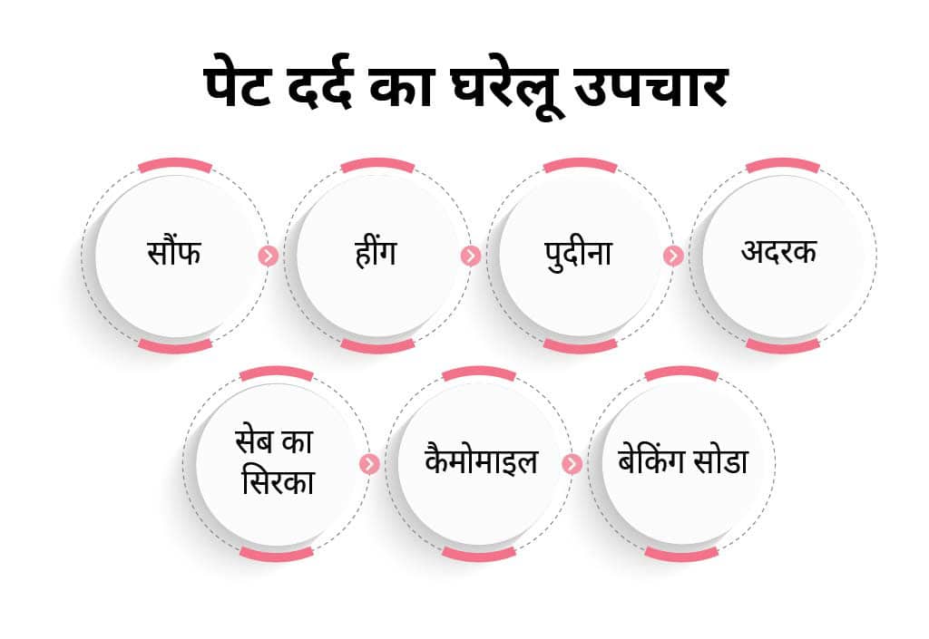
इन सबके अलावा, आप दही और चावल के पानी का भी उपयोग कर सकते हैं। लेकिन ध्यान रहे कि पेट दर्द का उपचार करने की नियत से किसी भी प्रकार के घरेलू नुस्खे का इस्तेमाल या सेवन करने से पहले डॉक्टर से उनकी राय अवश्य लें।
पेट दर्द होने पर आपको अपने जीवन में अनेक बदलाव लाना चाहिए, क्योंकि इससे दर्द को कम करने में काफी मदद मिलती है। पेट में दर्द होने पर आप अपनी जीवनशैली में निम्न बदलाव ला सकते हैं:-
साथ ही, घरेलू नुस्खों या दवाओं से पेट दर्द ठीक नहीं होने पर डॉक्टर से मिलकर अपने पेट दर्द का उचित जांच और इलाज कराएं। लंबे समय तक पेट दर्द को नजरअंदाज करना खतरनाक साबित हो सकता है।