Filter :

Saffron, also known as Kesar, is famous in various cuisines for its rich flavor and color. An exotic spice derived painstakingly from the flowers of Crocus sativus, saffron is also known for its medicinal properties and forms a part of several Ayurvedic recipes.
Many women believe that they must consume saffron during pregnancy for a fair baby. However, it must be noted that the skin color of a child is genetically determined, and it hasn’t been scientifically proven whether saffron can impact the complexion of a newborn baby or not.
Yet, Kesar is highly recommended in India for pregnant women, and they stand to reap the benefits of drinking saffron milk during pregnancy when taken in prescribed quantities.
If you are pregnant and confused about introducing saffron in your diet, here’s all you need to know about this exotic spice and how it can help you.
Table of Contents
Let’s start with the first question that most pregnant women have regarding saffron. Drinking Kesar milk during pregnancy or introducing saffron in your diet in any form is generally safe and beneficial. However, excessive intake of Kesar during pregnancy can lead to some undesirable issues, which is why it is important to regulate your intake.
According to Ayurveda and ancient wisdom, pregnant women may benefit from consuming 125 mg of saffron, twice daily. The best way to consume saffron is by preparing saffron milk in 2 easy steps:
In general, clinical studies have reported dosages up to 1.5 gm per day of pure saffron to be safe. Consuming more than 5 gm of pure saffron may have toxic effects on individuals.
Saffron, the most expensive spice in the world, is traditionally used for treating mild to moderate depression and also recommended to pregnant women with milk for its medicinal properties. Below, we have shared how you can benefit from saffron during pregnancy when taken in a controlled manner:
High blood pressure is a common problem faced by several expecting mothers and can lead to complications if left unchecked. Saffron is proven to reduce blood pressure, owing to the presence of crocin and safranal – which are effective in combating hypertension in humans. However, it is recommended to have Kesar in limited quantities as excessive consumption may lead to a dangerous drop in your BP.
Also, Read: Lifestyle Changes and Blood Pressure Control
Many women complain of constipation, bloating, and heartburn during pregnancy, which is a result of improper digestion. Regular intake of saffron is known to alleviate these symptoms by reducing acidity, improving blood flow, and boosting the metabolism in general.
Morning sickness or nausea can take away the joy of your pregnancy. Unbecoming of its name, morning sickness does not occur only in the early hours of the day. It can happen at any time and leads to extreme discomfort in many cases. Saffron-infused tea, which soothes the digestive system, may relieve morning sickness when taken in limited quantities.
Drinking saffron milk in pregnancy can treat mild to moderate depression thanks to its mood-altering properties. For the same reason, its anti-depressant properties work to reduce mood swings in pregnant women, reducing anxiety, depression, and irritation in the gestational period.
Hormonal fluctuations during pregnancy lead to hair fall in many pregnant women. According to ancient wisdom, applying a paste of saffron, milk, and liquorice can prevent hair fall during pregnancy and also stimulate hair growth.
Many women complain of muscular cramps in legs and abdomen, and joint pain in the third trimester. Saffron, which has anti-spasmodic and antinociceptive properties, can help in relieving excessive contractions in the muscles and relax the joints, reducing muscular pains during pregnancy.
Will saffron make the baby fair? This is a common question asked by pregnant women in India. First and foremost, many Indians have a naturally dusky complexion, which is suitable for the Indian climate. Second, having fair or dark skin does not reflect on the health of the baby, and is determined by the genes of the parents rather than the consumption of any food product or spice by the mother.
Till date, there has been no proven scientific relation between drinking Kesar milk in pregnancy and the complexion of a child. However, saffron is believed to improve skin texture and may be consumed in a controlled manner for healthy skin and other benefits, as mentioned above.
Pregnant women can start taking the recommended dosage of saffron at any time during their pregnancy. However, it is safer to avoid saffron in the first trimester. Ayurveda suggests the consumption of saffron only after the fourth month of pregnancy when you start feeling the movement of the baby in your womb.
Saffron, though beneficial in limited amounts, can lead to toxicity when consumed in larger quantities. Research on rats has indicated that oral consumption of saffron can induce premature labour and abortion, and a possible link to human abortion is suspected when consumed in higher quantities.
Some pregnant women may be sensitive to saffron and experience symptoms like:
Drinking saffron milk during pregnancy may also lead to vomiting in some pregnant women.
Clearly, there are many benefits of saffron milk during pregnancy. However, pregnant women must stick to the recommended dosage to avoid any complications and health issues.
If you face any of the above symptoms, stop consuming saffron immediately and book an appointment with the best gynaecologist in Gurgaon. In general, it is best to discuss any diet changes with your doctor when expecting to avoid any complications or unnecessary discomfort.
Book an online appointment at CK Birla Hospital for instant expert care to ensure a healthy and comfortable journey to motherhood.
Also, Read: How to Prepare Your Body for Pregnancy: A Complete How-to Guide

Picky-eating toddlers are common in every family. Generally, the peak age for fussy eating behaviours in a child is 3 years. A survey on the topic carried out on 4,000 children concluded that the majority of the kids are picky eaters at one time or the other. But, just the condolence that your child is not the only picky eater will not work for you.
You might feel the stress once your kid gets into the habit of throwing tantrums at the dinner table. Before looking at the tips for your fussy-eating toddler, it is necessary for you to know the root cause of the problem.
Table of Contents
In the very first place, you should know that there are different environmental influences and biological factors that can make kids move away from healthy eating habits. We will have a look at them below:
The home eating environment has an important role to play in influencing selective eating behaviours in a child. Simply put, children get accustomed to eating foods that are frequently on their dinner table. They become familiar with and even start accepting general food groups and specific dishes that the family eats together.
For example, if your child gets into the habit of having salty pasta with a bit of variation in spices, veggies, and proteins, he or she might not like having anything other than that. In the same way, if you offer dessert at the end of a meal, your child will always expect sugary treats after meals. So, the kid will ultimately develop an irresistible craving for sugar.
Several studies over the years show that people have this natural bend towards salty and sweet snacks – and children are no exception. Given a choice, a child of almost any age will reach out for innately satisfying food. This can be something sweet or salty over something bitter, bland or sour.
Regardless of the causes behind your picky-eating toddler, the question remains, what do you need to do? Some tips that can help you out are as follows:
Rather than having lunch with the entire family at 1 pm only to switch to 2 pm the next day, meals and snacks should be offered at a set time regularly. The best way to go about this is to allot a particular time for meals and at least two snack-times during the entire day.
In between meal times, your child should just have water and nothing else. This way, he or she will remain hydrated and not be full before mealtime. So, you will have a hungry child at the table.
Your kid’s willingness to taste a certain food will completely depend on the environment. Low-stress and pleasant mealtimes can be of good help here. Work on making mealtimes social, regular, and happy occasions. Do not worry about spilled food or drinks on the floor.
When preparing healthy food for picky eaters, starting small will help to a considerable extent. Guide your child on trying out new food and praise him for making an attempt. Do not force your kid into trying any food he doesn’t want to. Ignoring your fussy eater will encourage him or her to keep away from this habit.
Snacks and meals are essential for children. Therefore, they should have them properly and in adequate proportions. Distractions like computers, mobile phones, and television might not help them develop healthy eating habits. Mealtime should typically involve having food and interacting with the entire family.
Books, toys, music and TV are for playtime and not for mealtime. Make sure your child is away from these distractions if you want to change their fussy eating habit.
Of course, you must look for variety in the food you serve. But this does not mean you must make compromises on choosing healthy food for picky eaters. Try offering different types of fruits and vegetables along with foods high in their protein content such as deboned fish and meat. Serve these to your child at least twice during the week.
An important tip for fussy-eating toddlers is to guide your child in exploring new food textures and flavours. Also, work your way out in minimising waste. If you are introducing them to any new food, start with small quantities. You also need to wait for at least one week before offering the same food all over again.
Ans: Always keep in mind that kids love choosing what they want to have for a meal. So, it will work for you to ask your child about his preferences.
Of course, you must plan a balanced meal for your kid while taking his choices into account. If you factor in your child’s food preferences every day, with time, he will stop being a picky eater.
Ans: Picky eaters will change their habits only if they get small portions of healthy food regularly. Parents have to acknowledge that improving food habits in children is a gradual process and requires a fair amount of time.
To make the process easier, you can play into your child’s tendency to imitate you. As a parent, you can be a role model for their healthy eating habits. Your toddler will learn to eat things you eat and soon give up the habit of being a fussy eater.
Picky eaters are not just children who do not like to eat fruits or vegetables. They may also have a habit of eating the same food repeatedly. So, you need to follow these tips for picky eaters and toddlers and plan accordingly to change their practices.
As a parent, it is quite natural to be concerned about the health and diet of your child. Understand that being selective around food is normal during the developmental stages of a toddler. Patiently guide your kid on the path towards healthy and nutritious eating.
If nothing seems to be working, consult a pediatrician. They can help in troubleshooting problems and ensure that your child has a balanced and nutritious diet regularly.
Related Read: Taking Care of Your Fussy Eating Toddlers

The prostate is a part of the male reproductive system. It is a small gland situated below the bladder and ahead of the rectum. The prostate gland is responsible for making some of the ejaculatory fluid in your semen.
If you are anxious about your prostate health or experiencing any discomfort or pain during urination, then this article may help shed light on some or all of your issues.
Table of Contents
Prostate enlargement is a common occurrence for the prostate to grow in size as men age. As the prostate becomes larger, it firmly presses against the urethra. As a consequence of this, problems like the delayed release of urine, frequent urination, and pain during urination arise.
The condition mentioned above, commonly known as ‘Benign Prostatic Hyperplasia (BPH)’, is noticeable in approximately one-third of men older than 50 years and prevalent in up to 90% of the men by the age of 85.
Benign prostatic hyperplasia is not unusual just as it is not serious, typically. However, a range of treatments and procedures exist to treat prostate enlargement, just in case it gets serious. Contrary to popular belief, an enlarged prostate is not cancerous and does not cause prostate cancer, but severe BPH can result in other complications.
The exact causes of prostate enlargement are unclear. Popular speculation and large evidence suggest four main reasons for BPH:
An individual may have an enlarged prostate if he is showing one or more of the following BPH signs and symptoms.
Despite a frequent urge to urinate, there may be a delay before the urine is released. You might have to put in more effort for the urine to release and sometimes it could even be a false alarm. These are highly ubiquitous BPH symptoms.
If your prostate is enlarged, you might notice that it is increasingly difficult to control your urine for longer periods. You may be unable to sit for longer periods without having to urinate soon.
One of the signs of BPH is ‘nocturia.’ It refers to the condition where you are unable to sleep for continuously long hours without having to wake up and urinate. Generally, you might have to urinate every 6 to 8 hours.
As the enlarged prostate presses against the urethra, the tube that releases the urine and squeezes it, you may experience mild or severe pain in the bladder. The intensity of the pain depends on the severity of your BPH condition.
Even after you have just urinated, you may feel that it was incomplete and your bladder has not been emptied yet.
Urinary Tract Infection (UTI) and the presence of blood in the urine are among the prostatic hyperplasia symptoms but are rarely seen. These symptoms are more common in the extreme cases of BPH.
The treatments for BPH can range from simple dietary changes to minimally invasive surgery, and in some cases, even the removal of the prostate gland.
If the BPH symptoms that you are observing do not seem to bother you much, then it is likely not very severe and can be cured over time. Simple lifestyle and dietary changes can do wonders, especially if you are young, and your condition is not severe.
Consult a doctor and have regular checkups to monitor your health and condition.
A common practice while treating BPH with allopathic medication is the usage of ‘alpha-blockers.’ Alpha-blockers help relax the prostate and bladder that make urination easier and relieve you of some or all of the symptoms.
Since the exact type of medication also depends on other factors such as your overall health and age, kindly consult a doctor before proceeding further.
There are several types of surgical operations to treat BPH symptoms. Below is a list:
Laser therapy is among the preferred surgical methods as it is minimally invasive (apart from TUMT and PUL) and has relatively lower side effects. But it may not be necessarily the best treatment for you.
Kindly consult a doctor to help you figure out the most suitable surgical method to treat your BPH condition.
You can incorporate a multitude of practices in your routine to prevent prostate enlargement. Although these methods are not 100% effective, they certainly reduce the likelihood of developing BPH.
Cutting down alcohol consumption is essential to treat the symptoms of benign prostatic hyperplasia. Alcohol increases the frequency of urination. Additionally, it is important to stay active by exercising and also to follow a healthy diet.
Be sure to incorporate anti-inflammatory foods in your diet, such as:
Zinc supplements are useful in preventing BPH. You can also lean towards dietary sources of zinc like:
Drinking green tea is also a good practice as it helps to limit excess urination and is rich in antioxidants.
Any male above 40 or 50 years of age is naturally susceptible to prostate enlargement than those younger to them.
You are likely to be at greater risk if you have the following:
Turning a blind eye to benign prostatic hyperplasia symptoms can aggravate the condition and call the need for surgical intervention. It is strongly advised that you don’t jump to conclusions through self-diagnosis. If you experience any symptoms, consult a doctor to acquire more clarity.
There is no standardised treatment procedure for all patients. The treatment varies from one person to another and depends on various factors like allergies, overall health, fitness, height and weight. Consulting with an expert will help you make appropriate decisions for what is best suitable for you.
Also, Read: The Growing Problem Of An Enlarged Prostate Gland

Muscles, bones, tendons, and ligaments build up the feet that aid in all kinds of physical activity. All of these together are responsible for making one walk, run, or even stand. Individuals must take care that all the elements are working correctly for maximum movement.
Although minor issues in the foot can be checked over by general medical doctors, in case of specific ache or problem, the foot needs to be diagnosed by a podiatrist.
Table of Contents
Although there are many types associated with foot pain, there are four aches that need special attention.
Metatarsalgia affects the ball of the foot. Although it is a common cause of foot pain, the pain is usually ignored and not treated medically. The condition is common to see in people who are overweight or spend a lot of time running or standing.
Even shoes with improper soles could cause pain in the ball of the foot. Many other aches are similar to metatarsalgia; hence it is vital to get the condition checked out by a podiatrist to get a proper diagnosis.
The standard treatment options for metatarsalgia include:
Surgery is a last resort to treat the issue, depending on what is causing the foot issue.
Ingrown toenails are a physical condition where the edges of your nails grow into the skin surrounding the nail. Although it can affect any toe, it is mostly seen in the big toe of the foot. The toe becomes inflamed where the nail pieces the skin, followed by swelling and pus discharge.
Ingrown toenails are primarily seen in middle-aged and older people, and also in those people who have sweaty feet. No research indicates that ingrown toenails are genetic; it is usually caused by wearing tight footwear and following incorrect foot hygiene.
You can treat ingrown toenails at home with simple remedies which include:
if these are ineffective, the wound requires minor surgery to remove the accumulated pus or part of the toenail.
A bunion is a bump that forms on the joint that connects the big toe to the foot. A bunion mainly affects the big toe and causes it to lean in towards the second toe. In this way, bunions can cause pain and discomfort in the second and third toe.
Other toes may develop physical deformities like a hammertoe, claw toe, or mallet toe. Any pain in the toe or swelling around the joints should be communicated to a podiatrist as soon as possible.
You can get bunions due to the following conditions:
Necessary measures to treat bunion pain and stop further progression of the condition include:
In case these measures are insufficient, the next step is surgery, where the big toe is manually aligned back to its place.
Heel pain is one of the most common foot problems. Heel pain usually manifests in the back of the ankle and is often caused by plantar fasciitis (heel spur movement). Conditions like arthritis, tendonitis, stress fractures, nerve irritation and in rare cases, cysts can also cause heel pain. The pain is generally mild, which goes away on its own without any treatment. However, if the pain escalates and becomes chronic, a podiatrist is needed to provide accurate diagnosis and treatment.
Heel pain is typically seen in:
The treatment methods for heel pain entail:
Also, Read: Foot and ankle pain: Everything you need to know
Usually, a simple discussion, along with physical examination, is enough in case of minor foot problems. In case of severity or lack of detailed information, the doctor would recommend some of the following options:
Foot ache caused by heel pain, bunions, metatarsalgia, and ingrown toenails can first be treated with home remedies. Soaking feet in warm water is one of the most common ways to help relieve pain.
Podiatrists can properly diagnose severe foot issues. Depending on the severity and complexity of the problem, a podiatrist would prescribe drugs or recommend surgery.
Any significant foot problems must be taken care of by a specialist, i.e., a podiatrist rather than a general doctor. A podiatrist would have a more practical and effective approach in comparison to a general medical practitioner.
While treating foot pain is easy and manageable, you can take some measures to prevent foot pain altogether.
Although foot pain is quite common, it is easily preventable if you learn to take proper care. If you want to avoid these conditions, the first indicator that you should look out for is foot pain. In such cases, the worst thing to do is ignore the discomfort.
Immediate medical attention is necessary if basic remedies are not enough to reduce the pain. You can book an appointment with Dr Praveen Tittal, a Gurgaon-based podiatrist.

The third trimester of pregnancy is when the fetus gains most of its weight. During this period, its lungs fully develop, hair and nails grow, movement increases, and more. This is also the period when fat starts to build up to help keep its body temperature as well as energy.
To support all this growth, your doctor may advise you to increase your food intake. According to the American Pregnancy Association, your baby gains around 1/2 pound (266.6 gms) each week during the 9th month of pregnancy. This requires adequate nourishment and monitoring to ensure that your baby’s growth is not hampered.
Table of Contents
Knowing the estimated foetal weight is crucial as it can help monitor the baby’s growth and ensure proper development. It aids in identifying potential issues and guiding appropriate prenatal care. It is advisable to consult your healthcare provider for accurate assessments.
Each person is different and subject to multiple factors that affect weight and response to food intake. Some common reasons why you might not be hitting the weight marks could be:
Make sure to keep a note of your weight gain and food habits as this impacts your baby’s growth.
You Can Also Read: Breech Baby – everything you need to know
Foetal weight is typically estimated using ultrasound. The measurement involves assessing the baby’s head circumference, abdominal circumference, and femur length. These values are used in a formula to estimate weight.
The last few weeks of pregnancy are when the baby’s weight and development rapidly increase. Below is a chart of the normal weight of a baby during the 9th month of pregnancy.
Deficiencies in certain vitamins and minerals can stunt your baby’s growth. To increase the weight of your baby in the 9th month of pregnancy, you can include the following foods in your diet:
Balance is key when it comes to foods that increase fetal weight in the 9th month of pregnancy. Monitor your diet and do not go overboard with any particular food.
Ensure that whatever you eat is unprocessed and avoid canned foods. Take the time to read food labels and understand the nutritional value of what you purchase and consume.
Whatever you consume directly impacts your baby’s health. Try eating around 5 to 6 times a day in controlled quantities in case you aren’t comfortable with large meals or are experiencing a loss of appetite.
You Can Also Read: Hypertensive Disorders in Pregnancy
In addition to ensuring the safe and healthy growth of your baby, you also benefit from the following by maintaining a healthy baby weight during the 9th month of pregnancy:
This chart is an average estimate; your doctor’s recommendations to your pregnancy may vary.
Caring for yourself during pregnancy involves three essentials: physical well-being, mental well-being and fetal well-being. While most of your concentration will be on fetal well-being, you should maintain a positive balance among all three. Below are a few pointers:
Various factors play a role when it comes to maintaining an ideal baby weight in the 9th month of pregnancy. Some methods may work, and others might not, depending on your body type and medical history. Be sure to take your doctor’s advice and reach out immediately if you see signs out of the ordinary like:
Also, Read | How to Prepare Your Body for Pregnancy: A Complete How-to Guide
To support healthy foetal growth and increase baby weight, you have to focus on your well-being by eating a balanced diet, taking prenatal vitamins, attending regular check-ups, staying hydrated, getting adequate rest and managing stress.
Fruits rich in essential nutrients for foetal development include bananas (potassium, folate), avocados (healthy fats, folate), oranges (vitamin C, folate), berries (antioxidants, fibre), papayas (vitamin C, folate) and apples (fibre, vitamins).
Foetal weight is typically estimated using ultrasound. The measurement involves assessing the baby’s head circumference, abdominal circumference, and femur length. These values are used in a formula to estimate weight.
Yes, knowing the estimated foetal weight can help monitor the baby’s growth and ensure proper development. It aids in identifying potential issues and guiding appropriate prenatal care. Consult your healthcare provider for accurate assessments.

Diabetes is a health problem that can affect several body parts, one by one. One in six people with diabetes resides in India with an estimated number of 77 million. It has been observed that 10-15% of people with diabetes will develop a foot ulcer at some point of time. A survey done in 2014 revealed that only 54% of diabetics in India were aware that diabetes could lead to foot problems and only 22% had their feet examined by a health worker or doctor.
Do you know that it can affect your feet too? If you have a minor cut on your feet, it can turn into a significant wound because diabetes impairs the wound healing in your body. Also, you may not be able to feel anything in your feet because of nerve damage. Because of diabetes, blood flow to your feet is obstructed as well. This will cause a small wound to amplify into a stubborn infection, due to which you face the risk of amputation.
In case you suffer from diabetes, you need to take precautions to protect your feet and hence avoid any serious foot problems. Guidelines you need to follow to avoid Diabetic foot problem are as follows:
You must keep your foot clean always. Treat your foot like you will take care of a newborn baby. Use lukewarm water to soak in your foot and clean them. Avoid excessively hot water.
You need to check your feet regularly for blisters, cuts, swelling, redness, or nail problems. You can use a magnifying hand mirror to check the bottom of your feet. Do seek help from your doctor if you notice something suspicious.
Use a soft sponge and washcloth to wipe your feet. Dry your feet by gently patting it, especially between the toes.
You need to cut your nails straight across. Also, do not forget to file the edges of your nails. Don’t trim your nails too short as it risks the development of ingrown toenails. If you are worried about your toenails, then you can seek the advice of your foot and ankle specialist.
Always use a moisturizer to prevent itching and cracking of your dry skin. But you must always keep in mind that the place between your toes should not be moisturized as this may risk the development of a fungal infection.
If there are corns and calluses, then you should not try to treat them all by yourself. You must visit your doctor to get rid of them.
This is one of the essential guidelines that you need to follow if you want to avoid the Diabetic foot problems. You must get your feet regularly examined from your foot and ankle expert.
It is always necessary to keep a check on your blood sugar level to prevent Diabetic foot problems.
Smoking will accentuate worsening of blood flow in your feet. Hence if you are a diabetic person and also have a habit of smoking, then it is high time that you quit smoking forever.
You cannot afford to walk barefoot even at your home. You must wear a sandal or boots to avoid any cut or injury at your feet.
Always shake and feel for the inside of the shoe to look for any pebbles or nail inside as you may not feel it when you wear it.
Walking helps in controlling the weight and improves circulation. Wear properly measured and fitted shoes as the size and shape of your foot may change with time.
Related Read: Complications in Diabetes And How to Avoid Them
![Are genetic testing and IVF interlinked? [Complete guide]](https://backend.ckbhospital.com/wp-content/uploads/2020/06/Genetic_testing_and_ivf-e1767611341408.jpg)
The rate of seeking fertility treatments is rapidly escalating due to our current ways of living. Many couples prefer to delay their first pregnancy because of a heightened focus on career development in both the parents’ life or financial instability.
The results of blood tests taken during the mother’s period are used to determine the fertility of her eggs. A similar analysis is conducted on the father’s semen sample to assess the fertile semen. The results of these two tests determine the course of action.
Before making any decisions, it is crucial to understand IUI, IVF, and the differences between them.
The IUI is the procedure suggested to solve the problems of conceiving due to low fertility. A doctor or fertility expert do a transvaginal ultrasound on the mother to check the progress of the eggs before ovulation. Once the eggs have attained the optimal size, the patient is ready for the next stage of the treatment.
The treatment includes an injection that stimulates ovulation in the woman. Then, a fresh semen sample is inserted into the uterus via the cervix with the use of a thin tube. The IUI is quite a simple procedure, taking not more than five minutes from start to finish. The woman can usually resume daily activities after successfully finishing the process.
After many attempts of IUI, if the mother still does not conceive, the next step is IVF. IVF is the last resort, as the time taken to produce a viable embryo is more. Several steps are taken to make both the egg from the mother and sperm from the father ready for fertilisation.
In IVF, many factors contribute towards success – key among them being the age of the mother. A single IVF treatment revolves around the mother’s menstrual cycle.
The IVF process involves many steps. First is extracting a viable egg from the mother, and then inseminating it with a semen sample from the father. The last step is inserting the resulting embryo into the uterus of the mother via the cervix.
Other than these, the mother undergoes weekly shots of hormones and oral medicine to make implantation successful.
Both IUI and IVF have helped families achieve the happiness of a successful pregnancy. IUI is less expensive and less invasive as compared to IVF. Doctors and fertility experts suggest IUI in case of infertility or repeat miscarriages. The IUI treatment is usually successful in cases where the woman is below the age of 40.
IVF, on the other hand, is more invasive, and as a result, quite expensive. But, unlike IUI, the success rate of IVF is more.
According to the National Help Portal of India (NHP), congenital disabilities account for around 6-7% of births, which is about 1.7 million congenital disabilities annually. With most pregnancies happening naturally, it is impossible to detect all anomalies before delivery. Most blood tests are done during the first trimester to rule out common genetic disabilities.
Congenital disabilities can be classified into two categories – structural and functional & developmental. Structural defects that are apparent at birth could be heart defects, cleft lip, spina bifida, or clubfoot. Functional & developmental defects include down syndrome, sickle cell disease, or cystic fibrosis.
Many defects go undetected for months or even years unless they obstruct a baby’s growth.
Before opting for IVF, you must have done tons of research into each aspect of the procedure. You might have come across preimplantation genetic testing. The test takes place before the embryo is implanted to find and select the most viable and healthy embryo.
The biopsy process extracts 3-8 cells from each embryo to send for testing. Problems regarding suspected gene problems (PGD) and an abnormal number of chromosomes (PGS) can be identified with these tests. The chances of selecting a healthy embryo for implantation increase after analysing the test reports.
Preimplantation Genetic Diagnosis is an advantage to those couples who have some family history of severe or deadly diseases. Getting a PGD test done reduces the risk of passing forward those defective genes onto the next generation. Single gene disorders like sickle cell anaemia and cystic fibrosis have specific markers that are identified in the test reports.
Preimplantation Genetic Screening (PGS) is the process of determining that the potential embryo contains the usual number of chromosomes, i.e. 46. An embryo containing an abnormal number of chromosomes has the potential of developing into some disability. The test helps the doctors or fertility experts in choosing the healthiest embryo for implantation.
After trying naturally for years and undergoing multiple unsuccessful rounds of IUI, you have decided on trying an IVF procedure. IVF is costly, and the success rate is between 30-35%.
When the success rate is not so high, it is imperative to get the most out of what could be your only attempt at the IVF procedure.
Genetic testing can help your doctor or fertility expert in implanting you with the healthiest embryo out of the ones formed in the lab. This can help in ruling out a lot of factors that can develop into congenital disabilities and trouble the child at birth or even at an advanced age.
However, you must be entirely sure about your decision to go for genetic testing. There might be instances where the embryo could be damaged at the time of cell retrieval, making no embryo viable for implantation. It is also essential to understand the risk that even with successful implantation, there might be chances of miscarriage due to some unknown factors.
The mental stress the decision causes on the potential parents is also not negligible. The patients must be informed and given proper guidance in taking the decision that is best for them.
The key takeaway from here is to understand all the risks and rewards associated with genetic testing. Only with complete information on the IUI and IVF procedures will you be able to make the right choice.
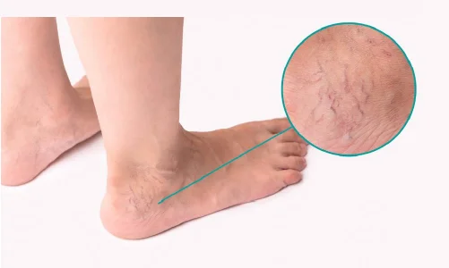
Varicose veins are a relatively common and painful condition, with over 10 million cases in India each year. Scientifically referred to as varicose or varicosities, this condition is caused by your veins becoming swollen, dilated, and overloaded with blood.
To the naked eye, varicose veins may look bluish-purple, and sometimes red. The most common cases of varicose veins are in the lower parts of your legs. This is because they are the furthest from your heart, and gravity prevents the blood from automatically flowing upwards.
Table of Contents
The primary cause of varicose veins is a problem with the way your veins function. The blood in your veins flows in one direction, controlled by valves. The function of this process is to ensure your blood is pumped effectively through the body. But in some cases, the valves stop working. This causes the blood to enlarge the appearance of your veins by collecting in them.
This condition could be brought up by other changes in your body that put a lot of pressure on your abdomen or legs. It could also be a result of your lifestyle or certain habits that you practice. Some of these have been mentioned below:
An increase in age can also cause varicose veins, especially in individuals who are over 50 years old, as veins can weaken with age. You are likely to get varicose veins if there is a family history of the condition.
The most common symptoms of varicosity are the appearance of the veins. The enlarged veins are almost always superficial veins that lie just below the surface of your skin. In addition to the veins looking bluish, enlarged capillaries in the vicinity give the appearance of a spider web. This condition is known as spider veins.
In some cases, although quite rare, the affected vein could be one of the interior ones of the leg, causing more pain than usual.
Since this condition is most common in the legs, it may also be accompanied by swollen ankles and itchy skin.
In the case of mild varicose veins, you need not visit a doctor. You can try the following tips for the prevention and treatment of varicose veins:
If you see that these methods are not working for you, it may be time for you to consult a vein doctor.
The vein specialist will provide several procedures to treat varicose veins. The three most common varicose veins treatment methods are as follows:
Varicose vein surgery is one of the main ways to address the condition if the previously suggested remedies don’t work and if varicose veins lower your overall functioning.
Endoscopic vein surgery involves blocking off the vein through an invasive process. A scope is inserted via an incision, which then blocks off the vein.
Since surgery is an invasive way to treat the condition, over time, this has been replaced by less invasive methods.
This method involves injecting a solution into the vein to cause the blood vessels therein to collapse and the blood to clot. Over time, this fades into scar tissue and is no longer painful or a cause of varicose veins.
Sclerotherapy is considered a more useful way to deal with the problem of varicose or spider veins. A small Indian study found that ultrasound-guided foam sclerotherapy proved to be more effective in treating primary varicose veins than surgery.
However, it is vital to inquire about the sclerotherapy cost before choosing to go with it.
Endovenous ablation therapy can help you deal with the irritation and pain that comes with varicose veins, in a less invasive manner than surgery. In this process, the doctor burns and closes each varicose vein. This is done using radiofrequency or light energy and leaves almost no scars.
Although this process is invasive, it is not as invasive as surgery. A special light known as an endoscopic transilluminator is placed under the skin using a small incision. This light is then used to cut off the problematic vein, which is removed using suction. You may have some bruising for a few days following this procedure; however, this will fade with time.
In the case that you feel or see any of the following symptoms, you should make an appointment with a trained medical professional who can advise you on the best course of treatment.
Keep in mind that not treating venous diseases in the initial stages can cause serious problems in the long run.
It is in the interest of the patient to keep an eye out for any varicose veins beginning to stand out. If you just see varicose veins form, the home remedies suggested may help you control it at the outset. However, if the varicose veins interfere with your daily activities, book an appointment at the vein centre closest to you for varicose veins pain relief.

Breast pain, also known as mastalgia or mastodynia, is a common condition. About 60-70% of women experience breast pain at some point in their life. However, only 10-20% of these cases require medical attention.
The severity of breast pain may vary, ranging from mild breast tenderness to burning sensation or stabbing pain in the breast. Similarly, you could experience pain in both breasts or pain in one breast alone, such as pain under the left or right breast.
There are several breast pain causes; some of these include:
One of the most typical breast pain reasons is a poorly-fitting bra. About 80% of women wear the wrong bra size.
Bras that are either too big or old and stretched out will do a poor job at supporting your breasts. Consequently, it can cause discomfort and soreness not only to your breasts but also to your neck, shoulders, and back. Conversely, tight bras can compress your chest and result in breast sensitivity.
Breast cysts are yet another common reason for breast pain. The filling up of fluid in the lobules or ducts can result in cyst formation, which, although asymptomatic, can be rather painful during your periods.
Cysts that are closer to the surface are softer, whereas the deeper cysts are hard lumps in the breast that hurts relatively more. Hereditary could put some women at higher risk of developing breast cysts.
However, if you notice the formation of new lumps or worsening in pain, consider consulting your doctor.
Breast tenderness and pain is a side effect of medication, hormone supplements, and replacements. Several oral contraceptives contain reproductive hormones like estrogen and progesterone, which could cause breast soreness. Thus, if you are on birth control pills or are undergoing infertility treatment or hormone replacement therapy, the medication could be the reason behind breast pain.
Have you noticed a sharp pain in your right breast that comes and goes as you carry out household chores or lift heavy things? The chances are that you are experiencing pain in your breast bone.
Muscular strains, characterised by sharp, shooting pain in breast, leads to sternum pain. In this case, the source of the pain is your pectoral muscles. The pecs are located beneath and around your breasts. Hence, any strain to these muscles could make it appear that you have breast pain.
Typically, this kind of pain is limited to pain in one breast.
Costosternal syndrome, also known as Costochondritis, is an inflammatory disease of the cartilage that connects the breastbone to the rib. Women aged 40 and above are more susceptible to this syndrome.
Those suffering from Costochondritis may also experience a burning sensation in their breast, in addition to pain in the breastbone. This condition often co-exists with autoimmune diseases like arthritis. However, it can also occur due to an injury or physical strain.
Fibrocystic breast changes are also known as cyclic mastalgia due to its periodic nature. It results in lumpy and swollen breasts due to the buildup of fibrous tissues and fluid-filled cysts. These cysts may enlarge before the onset of your period. The breast pain or tenderness before periods appears on the upper and outer areas of the breasts, and you may also notice nipple discharge.
Fortunately, this condition is harmless. More than 50% of women have experienced fibrocystic breast changes at some point in their lives. Further, it is more common for women between the ages of 20 and 50.
An infection of the breast tissues can lead to mastitis. This painful condition is most common among breastfeeding women and is a result of clogged milk ducts. However, it can happen to anyone. Mastitis typically affects only one breast.
Symptoms of mastitis include swollen breasts, redness, breast tenderness, fever, fatigue, burning sensation in the breast, and chills. When left untreated, mastitis can develop an abscess. Hence, consult your physician immediately.
Interestingly, breast pain is rarely an indicator of cancer, as most breast cancers do not cause any pain. However, in the case of inflammatory breast cancer, the tumours can cause mastodynia and general discomfort.
If you notice painful lumps in the breast that do not subside even after menstruation, then you must immediately contact your doctor. Also, talk to your physician if you notice persistent breast pain without any known cause.
Some women experience breast pain before the start of their period. On the other hand, some develop sore breasts due to hormonal changes during the menstrual cycle. The release of hormones like estrogen and progesterone increase the size and number of milk ducts, which causes breast tenderness. In some cases, breast pain may radiate towards the armpit, arm, and back. The pain, swelling, and tenderness go away once your period ends.
If you have undergone breast surgery, especially for cancer removal, you may experience breast pain of varying intensity. The pain could be due to the formation of scar tissues, inflammation, or nerve damage.
Patients report sharp pain in the breast for the initial days, followed by moderate pain after some passage of time. Some individuals may continue experiencing mild, persistent pain even six months after surgery.
Breast pain could be a common occurrence and not a cause of grave concern. Start by getting fitted for an appropriately-sized bra for extra support.
At the same time, several women experience cyclical breast pain that starts with their period and ends once their period is over. Similarly, women of reproductive age may also experience breast tenderness at some point before they reach menopause. Pregnancy and breastfeeding also lead to swollen breasts, which can cause breast pain.
Clearly, most of the breast pain causes are natural and unavoidable.
However, if you have been experiencing mastalgia for prolonged durations, new lump formations, blood discharge from nipples, and other symptoms, it is time to see a doctor.
Schedule a breast examination appointment with expert Breast cancer doctors as soon as possible.

