Filter :

जब भी आपके सिर में तेज दर्द होता है और आपकी माँ बस यही कहती है, “यह सब इसलिए है क्योंकि आप अपने मोबाइल फोन का बहुत अधिक उपयोग करते हैं.. लेकिन यह सच नहीं है, कम से कम हर बार तो नहीं। ऐसे कई कारक हैं जो सिरदर्द का कारण बन सकते हैं, और कोई आश्चर्य नहीं कि केवल एक नहीं बल्कि विभिन्न प्रकार के सिरदर्द हैं।
ये भी पढ़े: जानिए काला पीलिया क्यों होता है ? इसके कारण, लक्षण, उपचार
Table of Contents
सरल शब्दों में, सिरदर्द सिर में एक धड़कता हुआ दर्द पैदा करने वाली असुविधाजनक बेचैनी है जो दैनिक और छोटे-मोटे कामों को भी मुश्किल बना देती है। सबसे प्रचलित शिकायतों में से एक और किसी भी स्थिति या बीमारी के सबसे विशिष्ट संकेतकों में से एक सिरदर्द है। इसलिए, यदि आप एक कष्टदायी और धड़कते सिरदर्द से पीड़ित हैं, तो आप अकेले नहीं हैं।
सिरदर्द सिर के किसी भी हिस्से में गंभीर परेशानी पैदा कर सकता है और इसके साथ अतिरिक्त लक्षण भी हो सकते हैं।
ये भी पढ़े: गले में सूजन का कारण और उपचार | Swelling in Throat in Hindi
यह लेख सबसे सामान्य प्रकार के सिरदर्दों पर चर्चा करेगा, उनके कारण क्या हैं, और उनका प्रबंधन या उपचार कैसे करें।
अपनी जीवनशैली में कुछ बदलाव करने से बार-बार होने वाले तनाव सिरदर्द का इलाज करने और उससे बचने में मदद मिल सकती है। इसमे शामिल हैं:
ये भी पढ़े: हर्पीस बीमारी क्या है? इसके कारण, लक्षण और उपाय
जाहिर है, मासिक धर्म का दर्द शरीर को बहुत पीड़ादायक और थका देने वाला बना देता है। लेकिन ऐसा नहीं है कि कई बार मासिक धर्म चक्र भी सिरदर्द का कारण बनता है, इसलिए इसे हार्मोन सिरदर्द या मासिक धर्म माइग्रेन कहा जाता है।
मासिक धर्म के दौरान एस्ट्रोजेन के स्तर में परिवर्तन के कारण माइग्रेन हो सकता है। पीरियड्स से संबंधित सिरदर्द अक्सर चक्र के 2-3 दिन बाद या ओव्यूलेशन चरण के दौरान होता है।
ये भी पढ़े: सर्वाइकल पेन के लक्षण, कारण और घरेलू इलाज
ये भी पढ़े: टॉन्सिल (टॉन्सिलाइटिस) के कारण, लक्षण और इलाज
ये भी पढ़े: सिर में भारीपन के कारण और घरेलु इलाज
सिर में चोट लगने के तुरंत बाद सिरदर्द होता है। सिर में चोट लगने के बाद सिर में दर्द होना और उल्टी जैसा महसूस होना सामान्य बात है। यह ध्यान केंद्रित करने या ध्यान केंद्रित करने या अपने आस-पास की बुनियादी चीजों को याद रखने की व्यक्तिगत क्षमता को भी प्रभावित करता है।
एक व्यक्ति को दृष्टि में समस्या, गर्दन में दर्द और कान में दर्द, चक्कर आना और भ्रमित होना जैसे अन्य लक्षण भी हो सकते हैं, जो सभी सिर की चोट वाले सिरदर्द का कारण बनते हैं।
ये भी पढ़े: लो बीपी (लो ब्लड प्रेशर) : लक्षण, कारण और उपचार
ये भी पढ़े: बीपी (ब्लड प्रेशर) कम करने के 5 उपाय
यदि सिर में अचानक तेज दर्द हो, जैसे बिजली की गड़गड़ाहट हो, तो यह वज्रपात सिरदर्द का संकेत है। थंडरक्लैप सिरदर्द, हालांकि, सिर में असामान्य दर्द है जो जानलेवा बीमारी भी बन सकता है। थंडरक्लैप सिरदर्द के लक्षण बहुत नाटकीय और अचानक होते हैं। सिर में अचानक दर्द हो सकता है जो 60 सेकंड के भीतर चरम पर पहुंच जाता है और इसके साथ मतली या लगातार उल्टी होती है, साथ ही दौरे भी पड़ सकते हैं।
ये भी पढ़े: ट्यूबरक्लोसिस क्या है – कारण, लक्षण और बचाव (Tuberculosis in Hindi)
आज के समय में सिर दर्द एक आम समस्या है, लेकिन ज्यादातर सिरदर्द को घरेलू उपचार या ओटीसी दवाओं से नियंत्रित किया जा सकता है। हालांकि, अगर किसी को अचानक असहनीय दर्द का अनुभव होता है जिससे खुद को संतुलित करना या बोलना भी मुश्किल हो जाता है, तो उन्हें चिकित्सा सहायता लेनी चाहिए।
प्रमुख ईएनटी विशेषज्ञ डॉ अनीश गुप्ता सिरदर्द के संकेतों और लक्षणों के आधार पर सही निदान और सही उपचार योजना प्रदान करने में सक्षम होंगे। सही नुस्खे से माइग्रेन, स्पाइन और क्लस्टर्स जैसे सिरदर्द का इलाज किया जा सकता है।
सिरदर्द से कुछ हद तक बचा जा सकता है। उदाहरण के लिए, एक स्वस्थ आहार खाना, नियमित रूप से व्यायाम करना, भोजन छोड़ने से बचना या भोजन में अनावश्यक अंतराल बनाना, कैफीन का सेवन कम करना, अपने तनाव के स्तर को प्रबंधित करना और पर्याप्त नींद लेना।
ये भी पढ़े: गले में इन्फेक्शन का कारण, लक्षण, बचाव और घरेलू उपचार
जब सिर में दर्द लगातार हो और 24 घंटे से अधिक समय तक रहे, तभी चिंता शुरू होनी चाहिए। दैनिक सिरदर्द के संभावित कारण तनाव, चिंता, ज्यादा सोचना, मस्तिष्क में और उसके आसपास सूजन, मेनिनजाइटिस जैसे संक्रमण और सिर में उतार-चढ़ाव हो सकते हैं।
सिरदर्द कोविड-19 के पहले संभावित लक्षणों में से एक है। अन्य लक्षणों में बुखार, थकान, गंध या स्वाद की कमी और बुखार शामिल हैं।
जैसा कि सभी व्यक्तियों के शरीर अलग-अलग होते हैं, यहां तक कि थोड़ी सी भी निर्जलीकरण सिरदर्द का कारण बन सकता है। और निर्जलीकरण के अन्य लक्षण चक्कर आना, थकान महसूस करना और मुंह सूखना है। डिहाइड्रेशन के कारण होने वाले सिरदर्द का इलाज पर्याप्त पानी पीने और जरूरत पड़ने पर ओटीसी दवा लेने जैसे घरेलू उपचारों से किया जा सकता है।
गर्भावस्था आपके शरीर पर भारी प्रभाव डाल सकती है। लेकिन गर्भावस्था के दौरान आपको जितनी नींद की जरूरत होती है, वह हमेशा आसानी से नहीं आती। समझें कि गर्भावस्था नींद को कैसे प्रभावित करती है और बिना किसी परेशानी के आराम करने के लिए आप क्या कर सकती हैं।
प्रारंभिक गर्भावस्था के दौरान, हार्मोन प्रोजेस्टेरोन का स्तर बढ़ जाता है और आपका चयापचय उच्च चल रहा होता है। इससे दिन में नींद और थकान हो सकती है। यदि आप गर्भवती हैं और आपको एक बच्चा है देखभाल करने के लिए तो आप और भी अधिक थका हुआ महसूस कर सकती हैं।
ये भी पढ़े: प्रेगनेंसी में कितने समय के बाद सम्बन्ध बनाना बंद कर देना चाहिए
गर्भावस्था के दौरान कई सामान्य लक्षण आपकी नींद को प्रभावित या बाधित कर सकते हैं, जिनमें शामिल हैं:
इन सबके आल्वा, नींद संबंधी विकार, जैसे कि नींद सोने में परेशानी होना आदि भी गर्भावस्था के दौरान गंभीर हो सकते हैं।
इन बातों पर विचार करें:
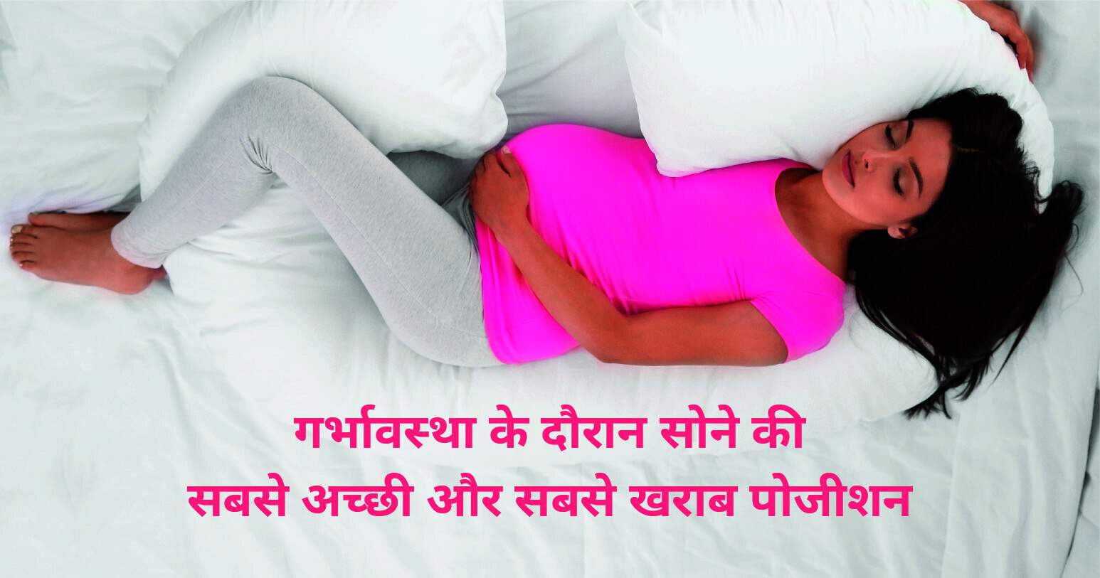
अपना पक्ष खुद तय करें: अपनी पीठ के बल सोने से बचें, जो आपके गर्भाशय का भार आपकी रीढ़ और पीठ की मांसपेशियों पर डाल सकता है। लेकिन अगर आप अपनी पीठ के बल उठती हैं तो चिंता न करें।
तकिए का प्रयोग करें: सावधानी से तकिए का इस्तेमाल करना आपको आराम देने में मदद कर सकता है। अपने मुड़े हुए घुटनों के बीच या अपने पेट के नीचे एक तकिया रखने की कोशिश करें।
आप गर्भावस्था के दौरान नींद की गड़बड़ी को प्रबंधित करने के लिए कुछ कदम उठा सकती हैं। उदाहरण के लिए:
अपने मूड मुताबिक माहौल तैयार बनाएं: एक अंधेरा, शांत और आरामदेह वातावरण और एक आरामदायक तापमान नींद को प्रोत्साहित करने में मदद कर सकता है। हर दिन एक ही समय पर सोने और उठने से आपकी नींद की सेहत में सुधार हो सकता है। अपने शयनकक्ष से इलेक्ट्रॉनिक उपकरणों को हटा दें।
सक्रिय यानी चुस्त रहें: गर्भावस्था के दौरान नियमित शारीरिक गतिविधि करने से आपको आसानी से नींद आने में मदद मिल सकती है। आज शाहम में हल्का-फुल्का व्यायाम, योग और मेडिटेशन कर सकती हैं।
डाइट पर खास ध्यान दें: छोटे-छोटे, बार-बार भोजन करें और सोने से तीन घंटे पहले खाने से बचें। अपने सिर को ऊंचा करके बाईं ओर सोने से भी सीने में जलन के लक्षणों को कम किया जा सकता है। विश्राम तकनीकों का अभ्यास करें। सोने से पहले उन्हें करना मददगार हो सकता है।
यदि आपको गर्भावस्था के दौरान सोने में परेशानी होती है, तो अपने डॉक्टर से बात करें। एक विकल्प अनिद्रा के लिए कॉग्निटिव बिहेवियरल थेरेपी नामक टॉक थेरेपी प्रोग्राम हो सकता है। यह प्रोग्राम आपको उन विचारों और व्यवहारों को पहचानने और बदलने में मदद करता है जो नींद की समस्याओं का कारण बनते हैं।
आप अपनी गर्भावस्था के दौरान बहुत सी बातों को लेकर चिंतित हो सकती हैं। आपकी नींद की स्थिति को सूची में सबसे ऊपर होने की आवश्यकता नहीं है। आपको और आपके बच्चे को इष्टतम रक्त प्रवाह देने के लिए डॉक्टर दाएं या बाएं की तरफ आराम करने की सलाह देते हैं।
इसके अलावा, आप अपने लिए सबसे आरामदायक स्थिति में आने के लिए कुछ तकिए के प्रॉप्स का उपयोग करने की कोशिश कर सकते हैं। अपने बच्चे के जन्म से पहले पूरी नींद लें। और अपने डॉक्टर से परामर्श करें यदि आपके पास अन्य प्रश्न हैं कि आपके लिए कौन सी स्थिति सबसे अच्छी है।
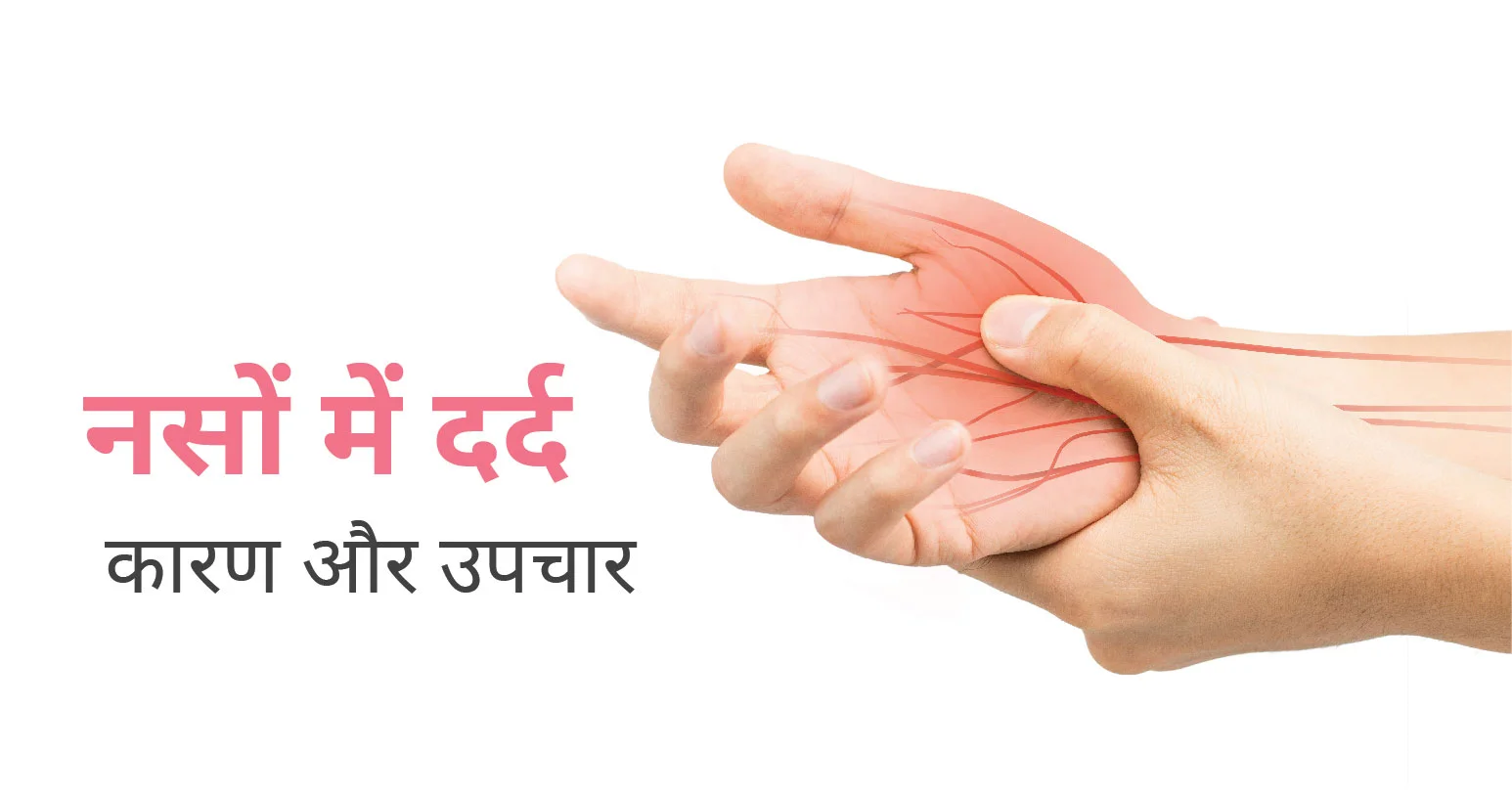
यदि आपकी तंत्रिका तंत्र क्षतिग्रस्त है या ठीक से काम नहीं कर रही है तो न्यूरोपैथिक दर्द यानी नसों में दर्द हो सकता है। आप तंत्रिका तंत्र के विभिन्न स्तरों-परिधीय तंत्रिकाओं, रीढ़ की हड्डी और मस्तिष्क में से किसी भी स्तर से दर्द महसूस कर सकते हैं। साथ में, रीढ़ की हड्डी और मस्तिष्क को केंद्रीय तंत्रिका तंत्र के रूप में जाना जाता है।
परिधीय नसें (Peripheral Nerves) वे हैं जो आपके शरीर के बाकी हिस्सों जैसे कि बाहों, पैरों, उंगलियों और पैर की उंगलियों जैसे स्थानों पर फैली हुई हैं। क्षतिग्रस्त तंत्रिका तंतु दर्द केंद्रों को गलत संकेत भेजते हैं। तंत्रिका क्षति के स्थल पर, साथ ही केंद्रीय तंत्रिका तंत्र (केंद्रीय संवेदीकरण) के क्षेत्रों में तंत्रिका कार्य बदल सकता है।
न्यूरोपैथी कार्य की गड़बड़ी या एक या कई नसों में परिवर्तन है। न्यूरोपैथी के लगभग 30% मामलों के लिए मधुमेह जिम्मेदार है। न्यूरोपैथिक दर्द का स्रोत बताना हमेशा आसान नहीं होता है। ऐसी सैकड़ों बीमारियाँ हैं जो इस तरह के दर्द से जुड़ी हैं।
ये भी पढ़े: यूरिक एसिड बढ़ने का कारण, लक्षण, इलाज और आसान घरेलू नुस्खे
Table of Contents
यानी नसों में दर्द अनेक रोगों के कारण हो सकता है, जिनमें निम्न शामिल हैं:
अन्य कारणों में शामिल हैं:
ये भी पढ़े: डायबिटिक के मरीज के लिए कम्पलीट डाइट चार्ट
नसों में दर्द के प्रत्येक व्यक्ति के लक्षण थोड़े भिन्न हो सकते हैं, लेकिन ये लक्षण सामान्य हैं:
इन सबके अलावा, पुराने दर्द, नींद की कमी, और आप कैसा महसूस कर रहे हैं इसे व्यक्त करने में कठिनाई के परिणामस्वरूप भावनात्मक समस्याएं
ये भी पढ़े: फैटी लिवर का कारण, लक्षण और इलाज
एंटीकॉन्वल्सेंट और एंटीडिप्रेसेंट दवाएं अक्सर शुरुआत उपचार होती हैं। कुछ न्यूरोपैथिक दर्द के अध्ययनों से पता चलता है कि गैर-स्टेरॉयड एंटी-इंफ्लैमेटरी ड्रग्स (एनएसएआईडी), जैसे कि एलेव या मोट्रिन, दर्द को कम कर सकते हैं। कुछ लोगों को एक मजबूत दर्द निवारक दवा की आवश्यकता हो सकती है।
यदि मधुमेह जैसी कोई अन्य स्थिति शामिल है, तो उस विकार का बेहतर प्रबंधन दर्द को कम कर सकता है। स्थिति का प्रभावी प्रबंधन आगे तंत्रिका क्षति को रोकने में भी मदद कर सकता है। ऐसे मामलों में जिनका इलाज करना मुश्किल है, एक दर्द विशेषज्ञ दर्द को प्रभावी ढंग से प्रबंधित करने के लिए एक इनवेसिव या इम्प्लांटेबल डिवाइस का उपयोग कर सकता है।
न्यूरोपैथिक दर्द में शामिल नसों की विद्युत उत्तेजना दर्द के लक्षणों को महत्वपूर्ण रूप से नियंत्रित कर सकती है। अन्य प्रकार के उपचार भी न्यूरोपैथिक दर्द में मदद कर सकते हैं। इनमें से कुछ में शामिल हैं:
दुर्भाग्य से, नसों का दर्द अक्सर मानक दर्द उपचार के लिए खराब प्रतिक्रिया करता है और समय के साथ बेहतर होने के बजाय कभी-कभी खराब हो सकता है। कुछ लोगों के लिए, यह गंभीर अक्षमता का कारण बन सकता है।
ये भी पढ़े: सर्वाइकल पेन के लक्षण, कारण और घरेलू इलाज
नसों के दर्द को अक्सर सूथिंग या जलन दर्द के रूप में वर्णित किया जाता है। यह अपने आप दूर जा सकता है, लेकिन अगर ऐसा नहीं होता है तो आपको डॉक्टर से मिलकर उपचार के बारे में बात करनी चाहिए।
न्यूरोट्रोपिक बी विटामिन कोएंजाइम के रूप में तंत्रिका तंत्र की सेहत में महत्वपूर्ण भूमिका निभाते हैं। विशेष रूप से विटामिन बी 1 (थियामिन), बी 6 (पाइरिडोक्सिन), और बी 12 (कोबालिन) एक स्वस्थ तंत्रिका तंत्र के रखरखाव में अनिवार्य रूप से योगदान करते हैं।

बालों का झड़ना (एलोपेसिया) को गंजापन के नाम से बी जानते हैं। यह सिर्फ आपकी खोपड़ी यानी सिर ही नहीं, बल्कि आपके पूरे शरीर को प्रभावित कर सकता है, और यह अस्थायी या स्थायी हो सकता है। यह आनुवंशिकता, हार्मोनल परिवर्तन, चिकित्सा स्थितियों या उम्र बढ़ने के सामान्य भाग का परिणाम हो सकता है।
हर किसी के सिर के बाल झड़ सकते हैं, लेकिन यह पुरुषों में अधिक आम है। गंजापन आमतौर पर आपके खोपड़ी से अत्यधिक बालों के झड़ने को संदर्भित करता है। उम्र के साथ वंशानुगत बालों का झड़ना गंजेपन का सबसे आम कारण है। कुछ लोग अपने बालों के झड़ने को अनुपचारित और अनछुए तरीके से चलने देना पसंद करते हैं। अन्य इसे केशविन्यास, मेकअप, टोपी या स्कार्फ के साथ कवर कर सकते हैं।
बालों के झड़ने का इलाज करने से पहले, अपने बालों के झड़ने के कारण और उपचार के विकल्पों के बारे में अपने डॉक्टर से बात करें।
Table of Contents
आमतौर पर लोगों के एक दिन में 50 से 100 बाल झड़ते हैं। यह आमतौर पर ध्यान देने योग्य नहीं होता है, क्योंकि एक ही समय में नए बाल बढ़ रहे होते हैं। बालों का झड़ना तब होता है जब नए बाल गिरे हुए बालों की जगह नहीं लेते हैं। बालों का झड़ना आमतौर पर निम्नलिखित कारकों में से एक या अधिक से संबंधित होता है:
एलोपेसिया का मुख्य लक्षण सामान्य से अधिक बाल झड़ना है, लेकिन इसे पहचानना आपके विचार से अधिक कठिन हो सकता है। इसके निम्नलिखित लक्षण हो सकते हैं:
चौड़ा करने वाला भाग: यदि आप अपने बालों को विभाजित करते हैं, तो आप देख सकते हैं कि आपका हिस्सा चौड़ा हो रहा है, जो बालों के पतले होने का संकेत हो सकता है।
सिर के मध्य में से बालों का घटना: इसी तरह, अगर आपको लगता है कि आपकी हेयरलाइन सामान्य से अधिक ऊंची दिख रही है, तो यह बालों के पतले होने का संकेत हो सकता है।
कंघी में बालों का जमा होना: अपने ब्रश या कंघी को इस्तेमाल करने के बाद चेक करें। क्या यह सामान्य से अधिक बाल जमा कर रहा है? अगर ऐसा है तो यह बालों के झड़ने का संकेत हो सकता है।
दर्द या खुजली: यदि आपके बालों के झड़ने के कारण आपकी अंतर्निहित त्वचा की स्थिति है, तो आपको दर्द भी महसूस हो सकता है या आपकी सिर में खुजली का अनुभव हो सकती है।
बालों के झड़ने के लिए उपचार के कई विकल्प हैं, लेकिन आपके लिए सबसे अच्छा विकल्प इस बात पर निर्भर करेगा कि आपके बालों के झड़ने का कारण क्या है। आमतौर पर, सबसे आम प्रकार के बालों के झड़ने का उपचार सामयिक या मौखिक दवाओं के साथ किया जाता है, जो संभवतः उपचार का पहला कोर्स होगा।
ओवर-द-काउंटर (ओटीसी) दवाओं में आमतौर पर सामयिक क्रीम, जैल, समाधान या फोम शामिल होते हैं जिन्हें आप सीधे खोपड़ी पर लगाते हैं। सबसे आम उत्पादों में मिनोक्सिडिल नामक एक घटक होता है। प्रिस्क्रिप्शन दवाएं, जैसे फायनास्टराइड (प्रोपेसिया), एंड्रोजेनिक बालों के झड़ने को रोकने में मदद कर सकती हैं, विशेष रूप से पुरुष पैटर्न गंजापन के लिए।
बालों के झड़ने को धीमा करने के लिए आप रोजाना इस दवा का सेवन करते हैं, हालांकि कुछ लोग फायनास्टराइड लेने पर नए बालों के विकास का अनुभव करते हैं। यदि बालों का झड़ना एक ऑटोइम्यून स्थिति से संबंधित लगता है, तो आपका चिकित्सक कॉर्टिकोस्टेरॉइड्स जैसी सूजन-रोधी दवाएं लिख सकता है।
जिन नए उपचारों की खोज की जा रही है, उनमें लेजर थेरेपी के कुछ रूप, पीआरपी के साथ माइक्रोनीडलिंग, साथ ही अन्य मौखिक दवाएं शामिल हैं। इनमें से कई उपचार अभी भी शुरुआती परीक्षण चरणों में हैं, और अधिक शोध आवश्यक होगा।
हेयर ट्रांसप्लांट सर्जरी में आपके सिर के गंजे हिस्सों में त्वचा के छोटे-छोटे प्लग, प्रत्येक में कुछ बाल होते हैं। यह वंशानुगत गंजापन वाले लोगों के लिए अच्छा काम करता है, क्योंकि वे आमतौर पर सिर के शीर्ष पर बाल खो देते हैं। क्योंकि कुछ बालों का झड़ना प्रगतिशील हो सकता है, आपको समय के साथ कई प्रक्रियाओं की आवश्यकता हो सकती है। यह ध्यान देने योग्य है कि इस विधि से एलोपेसिया के निशान वाले लोगों को लाभ या सहायता की संभावना नहीं है।
बालों के झड़ने के साथ केवल राइबोफ्लेविन, बायोटिन, फोलेट और विटामिन बी 12 की कमी को जोड़ा गया है।
जी हां, बालों को झड़ने से रोकने में नारियल का तेल कारगर और सुरक्षित साबित हुआ है। दरअसल पुराने समय में लोग अपने बालों को स्वस्थ, मजबूत और घना रखने के लिए इसका इस्तेमाल करते आ रहे हैं।
अगर आपके बाल झड़ रहे हैं तो डॉक्टर से परामर्श करने के बाद आप निम्न चीजों को सेवन कर सकते हैं:
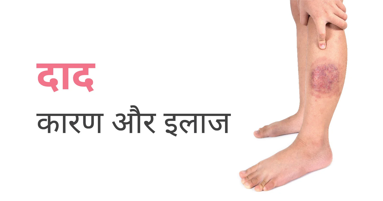
दाद एक फंगल संक्रमण है जो त्वचा पर अंगूठी के आकार का संक्रमण पैदा करता है। एक्जिमा और सोरायसिस जैसी स्थितियां कभी-कभी दाद के समान हो सकती हैं, लेकिन उपस्थिति और उपचार में महत्वपूर्ण अंतर होते हैं। दाद को डर्माटोफाइटिस, डर्माटोफाइट संक्रमण या टिनिया के रूप में भी जाना जाता है
फंगस की लगभग 40 अलग-अलग प्रजातियां दाद का कारण बन सकती हैं। वे आमतौर पर ट्राइकोफाइटन, माइक्रोस्पोरम और एपिडर्मोफाइटन प्रकार के होते हैं। ये कवक आपकी त्वचा और अन्य सतहों, विशेष रूप से नम क्षेत्रों पर रह सकते हैं। वे मिट्टी में बीजाणुओं के रूप में भी लंबे समय तक जीवित रह सकते हैं। कवक मनुष्यों में चार तरीकों से फैल सकता है:
इन सबके आल्वा, कवक ले जाने वाली मिट्टी के सीधे संपर्क के बाद मनुष्य और जानवरों को दाद हो सकता है।
ये भी पढ़े: हर्पीस बीमारी क्या है? इसके कारण, लक्षण और उपाय
डॉक्टर आपकी त्वचा की जांच करके और संभवतः प्रभावित क्षेत्र को देखने के लिए एक काली रोशनी का उपयोग करके दाद का निदान करेंगे। कवक के प्रकार के आधार पर, यह कभी-कभी काले प्रकाश के नीचे फ्लोरोसेंट (चमक) हो सकता है। डॉक्टर कुछ परीक्षणों का अनुरोध करके दाद के निदान की पुष्टि कर सकते हैं जैसे कि:
यदि आपको या तो त्वचा की बायोप्सी या फंगल कल्चर मिल रहा है, तो डॉक्टर आपकी त्वचा का एक नमूना लेकर या ब्लिस्टर से निकालेंगे और इसे फंगस की उपस्थिति के परीक्षण के लिए लैब में भेजेंगे।
यदि आप केओएच परीक्षा कर रहे हैं, तो डॉक्टर प्रभावित त्वचा के एक छोटे से क्षेत्र को एक स्लाइड पर खुरच कर हटा देंगे और उस पर पोटेशियम हाइड्रॉक्साइड (केओएच) नामक तरल की बूंदों को रख देगा।
केओएच विशिष्ट त्वचा कोशिकाओं को अलग कर देता है, जिससे सूक्ष्मदर्शी के नीचे कवक तत्वों को देखना आसान हो जाता है।
दाद के लिए आपके लिए आवश्यक उपचार इस बात पर निर्भर करता है कि आपके शरीर में संक्रमण कहाँ है और यह कितना गंभीर है। कई मामलों में, डॉक्टर ऐसी दवा की सिफारिश कर सकते हैं जिसे आप अपने स्थानीय दवा की दुकान पर काउंटर (ओटीसी) से खरीद सकते हैं। दूसरों को नुस्खे की जरूरत होती है।
यदि संक्रमण आपकी त्वचा पर है – जैसे एथलीट फुट या जॉक खुजली के मामले में, तो डॉक्टर एक ओटीसी ऐंटिफंगल क्रीम, लोशन या पाउडर का सुझाव दे सकते हैं।
ज्यादातर मामलों में, आपको यह सुनिश्चित करने के लिए 2 से 4 सप्ताह तक अपनी त्वचा पर दवाओं का उपयोग करना होगा कि आप दाद पैदा करने वाले फंगस को खत्म कर रहे हैं। ऐसा करने से इसके वापस आने की संभावना भी कम होगी।
यदि आपके सिर पर या आपके शरीर पर कई अलग-अलग जगहों पर दाद है, तो ओटीसी उपचार पर्याप्त नहीं हो सकते हैं। ऐसे में डॉक्टर एक एंटिफंगल दवा के लिए एक ओरल प्रिस्क्रिप्शन लिखेंगे जिसे आपको 1 से 3 महीने तक मुंह से लेना होता है।
चीजों को साफ रखने से संक्रमण आपके शरीर के अन्य भागों के साथ-साथ आपके घर के अन्य लोगों और जानवरों में भी फैलने से रोकता है। एक बार आपका ठीक हो जाने के बाद यह पुन: संक्रमण को भी रोकेगा। दाद का संक्रमण होने पर चीजों को साफ रखने के नीचे कुछ तरीके दिए गए हैं:
इन सबके अलावा, प्रभावित क्षेत्र को साबुन से साफ करें, और अपने शरीर के बाकी हिस्सों से अलग तौलिये से सुखाएं। हर दिन ताजे कपड़े पहनें, खासकर अंडरगारमेंट्स पहनें। अपने कपड़ों को नियमित रूप से धोएं और उपयोग में न होने पर उन्हें सुखा कर रखें।
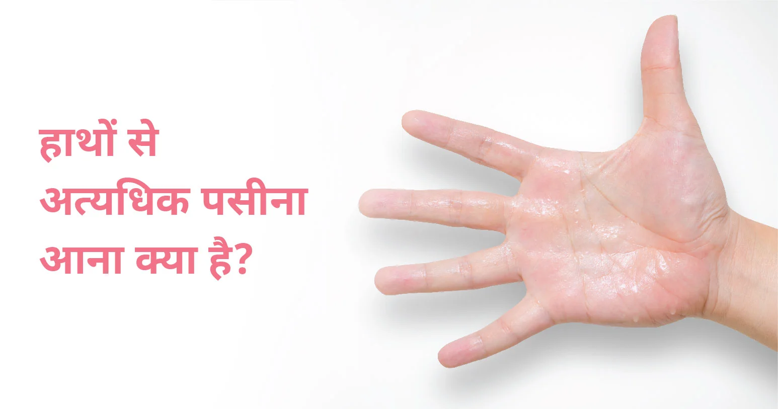
पसीने से हथेलियाँ तर होने की स्थिति को मेडिकल भाषा में पाल्मर हाइपरहाइड्रोसिस के रूप में भी जाना जाता है। हालाँकि, आम बोलचाल की भाषा में इसे पसीने से हथेलियाँ भीगना या तर होना कहा जाता है। यह पैरों के तलवों में पसीने से जुड़ा हो सकता है (पैरों में पसीना आने को प्लांटर हाइपरहाइड्रोसिस कहा जाता है)।
पसीने से तर हथेलियाँ प्राथमिक हाइपरहाइड्रोसिस का एक उपसमूह है – एक ऐसी स्थिति जिसके कारण हाथ पैरों, अंडरआर्म्स और चेहरे में अत्यधिक पसीना आता है। पाल्मर हाइपरहाइड्रोसिस सहित हाइपरहाइड्रोसिस, 2 से 3% आबादी को प्रभावित करता है, लेकिन प्रभावित लोगों में से 40% से कम चिकित्सा उपचार चाहते हैं।
पसीने से तर हथेलियों का मुख्य लक्षण खुद अत्यधिक मात्रा में अनियंत्रित पसीना आना है। आपकी हथेलियां चिपचिपी या गीली महसूस हो सकती हैं, जिससे आप किसी से हाथ मिलाने, मीटिंग में पेपर देने या कीबोर्ड पर टाइप करने में असहज महसूस कर सकते हैं। पसीना बिना ट्रिगर के आएगा, न कि किसी बाहरी कारक जैसे व्यायाम या शरीर के तापमान में वृद्धि के कारण। यह किसी भी तापमान या किसी भी मौसम में हो सकता है। तनाव या चिंता के समय लक्षण बढ़ सकते हैं।
आप एक बच्चे के रूप में पसीने से तर हथेलियों के लक्षणों को देख सकते हैं, जब आप यौवन में प्रवेश करते हैं तो लक्षणों में वृद्धि होती है। जैसे-जैसे आप अपने 40 और 50 के दशक में पहुंचते हैं, पसीने से तर हथेलियों के लक्षण अक्सर कम हो जाते हैं, जब तक कि यह किसी अन्य चिकित्सीय स्थिति के कारण न हो।
हाथों में पसीने की ग्रंथियां अत्यधिक सक्रिय ग्रंथियों के कारण होती हैं और ऐसा होने के कई कारण होते हैं। यह परिवारों में चल सकता है, और यह हाइपरहाइड्रोसिस के अन्य रूपों या कुछ चिकित्सीय स्थितियों से जुड़ा हो सकता है। इसके संभावित कारणों में निम्न शामिल हो सकते हैं:
यह स्थिति दोनों लिंगों को समान रूप से प्रभावित करती है, लेकिन हाथों से अत्यधिक पसीना आने का पर इसके इलाज के लिए महिलाओं की अधिक संभावना हो सकती है।
हाथों से अत्यधिक पसीना आना आपके शारीरिक स्वास्थ्य को नुकसान नहीं पहुंचाती हैं, लेकिन यह निश्चित रूप से आपके जीवन की गुणवत्ता और भावनात्मक स्वास्थ्य को प्रभावित कर सकती हैं। उपचार के कई विकल्प हैं। आपके लक्षणों की गंभीरता के आधार डॉक्टर इस बात पर चर्चा कर सकते हैं कि कौन सा उपचार आपके लिए बेहतर विकल्प है। इस समस्या के उपचार में शामिल हो सकते हैं:
पसीने की ग्रंथियों को अवरुद्ध करने में मदद करने के लिए हथेलियों पर प्रतिस्वेदक का उपयोग करना
एंटीकोलिनर्जिक दवाएं, जो पसीने के उत्पादन के लिए जिम्मेदार न्यूरोट्रांसमीटर को ब्लॉक करने में मदद करती हैं
चिकित्सीय प्रक्रियाएं जो पसीने से तर हथेलियों के इलाज में मदद कर सकती हैं:
बोटुलिनम टॉक्सिन (बोटॉक्स): यह एक इंजेक्शन है जिसका उपयोग पसीने वाली हथेलियों के इलाज के लिए एसिटाइलकोलाइन, एक न्यूरोट्रांसमीटर जारी करके किया जाता है, जिससे आपकी हथेलियों में पसीने की ग्रंथियों का उत्पादन कम हो जाता है।
योणोगिनेसिस: हथेलियों को पसीने से रोकने के लिए एक चिकित्सा उपकरण त्वचा के माध्यम से एक आयनित पदार्थ पारित करने के लिए पानी और एक विद्युत प्रवाह का उपयोग किया जाता है।
एंडोस्कोपिक थोरैसिक सिम्पैथेक्टोमी (ईटीएस): यह एक न्यूनतम इनवेसिव प्रक्रिया है जो तंत्रिका तंत्र से हाथों की हथेलियों तक के रास्ते से छुटकारा दिलाती है, जिससे हथेलियों से पसीने की क्षमता समाप्त हो जाती है।
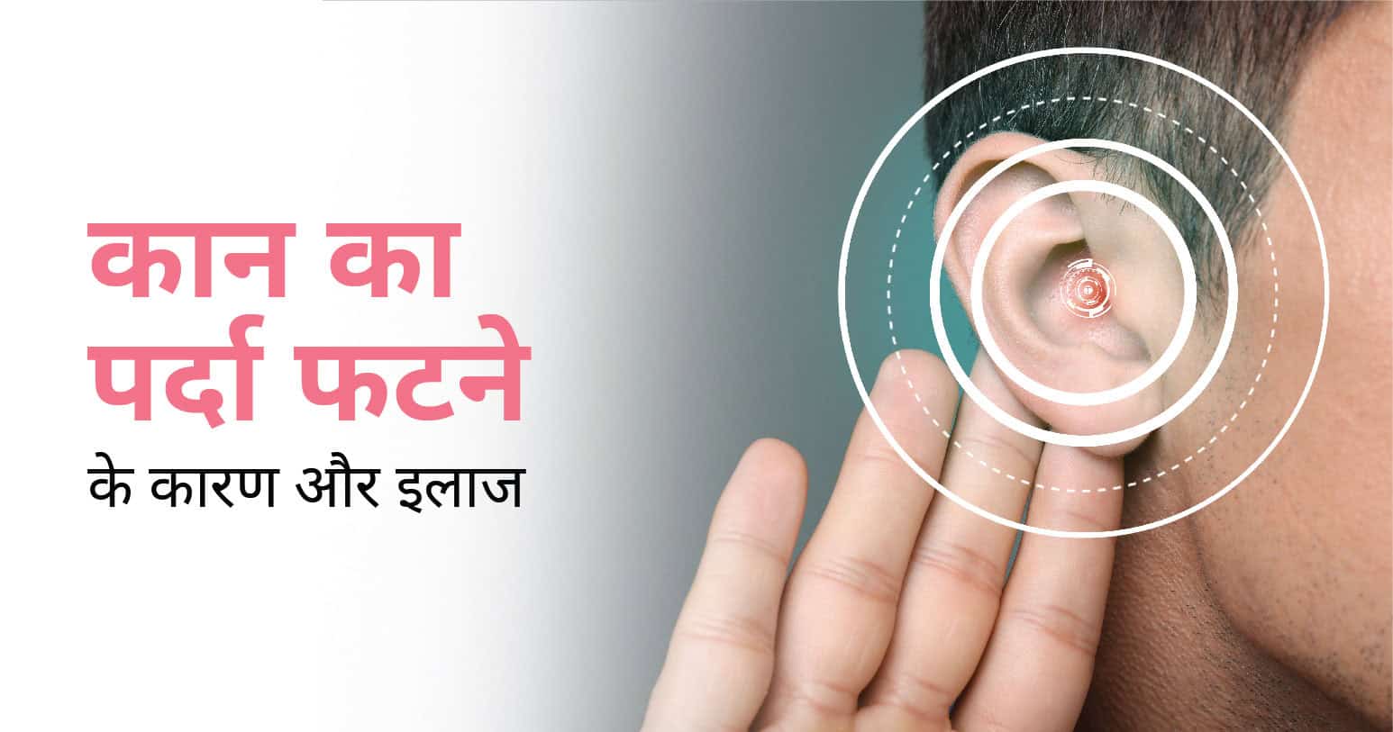
कान के परदे को अंग्रेजी में ईयरड्रम कहते हैं जो ध्वनियों के कंपन को प्राप्त करके ध्वनि सुनने में मदद करता है। यह एक ऊतक है जो ईयर कैनाल को मध्य कान से अलग करता है। पर्दा फटना मतलब कान के पर्दे में छेद होना है जिससे सुनने की क्रिया बाधित हो सकती है।
यह बच्चों में सबसे आम है। हालांकि, यह बिना किसी हस्तक्षेप के कुछ दिनों में ठीक हो जाता है, लेकिन कभी-कभी इसके लिए सर्जिकल उपचार की आवश्यकता हो सकती है। कान के संक्रमण से अक्सर कान का पर्दा फट सकता है। संक्रमण से द्रव का निर्माण हो सकता है, जिससे कान के ड्रम में टियर हो सकता है। इस समस्या से पीड़ित मरीजों को दर्द और परेशानी महसूस हो सकती है।
Table of Contents
कुछ लक्षण यह संकेत दे सकते हैं कि कान का पर्दा फट गया है। इन लक्षणों में शामिल हैं:
कान का पर्दा फटने के कई कारण हो सकते हैं, जो इस प्रकार हैं:
ईएनटी विशेषज्ञ कुछ उपकरणों का उपयोग करके कान का पर्दा फटने के लक्षण वाले रोगियों के कानों का निरीक्षण करेंगे। डॉक्टर किसी भी प्रकार की समस्या का पता लगाने के लिए कुछ परीक्षणों की सिफारिश भी कर सकते हैं, जो इस प्रकार हैं:
आमतौर पर, फटा हुआ ईयरड्रम कुछ ही हफ्तों में अपने आप ठीक हो जाता है। यदि कोई संक्रमण है, तो डॉक्टर कुछ दवाएं लिखेंगे, जिनमें ड्रॉप्स, एंटीबायोटिक गोलियां आदि शामिल हैं। यदि ये फटे हुए कान के परदे को ठीक करने में विफल रहते हैं, तो डॉक्टर कुछ अन्य उपचार विधियों का सुझाव दे सकते हैं:
हालांकि, फटा हुआ कान का पर्दा अक्सर उपचार के बिना ठीक हो जाता है, मरीज़ अपने कान के पर्दे में गंभीर चोट के कारण दर्द और परेशानी से जूझ सकते हैं। पहले बताए गए किसी भी लक्षण का सामना करने वालों को जल्द से जल्द अपने स्वास्थ्य सेवा प्रदाता से संपर्क करना चाहिए।
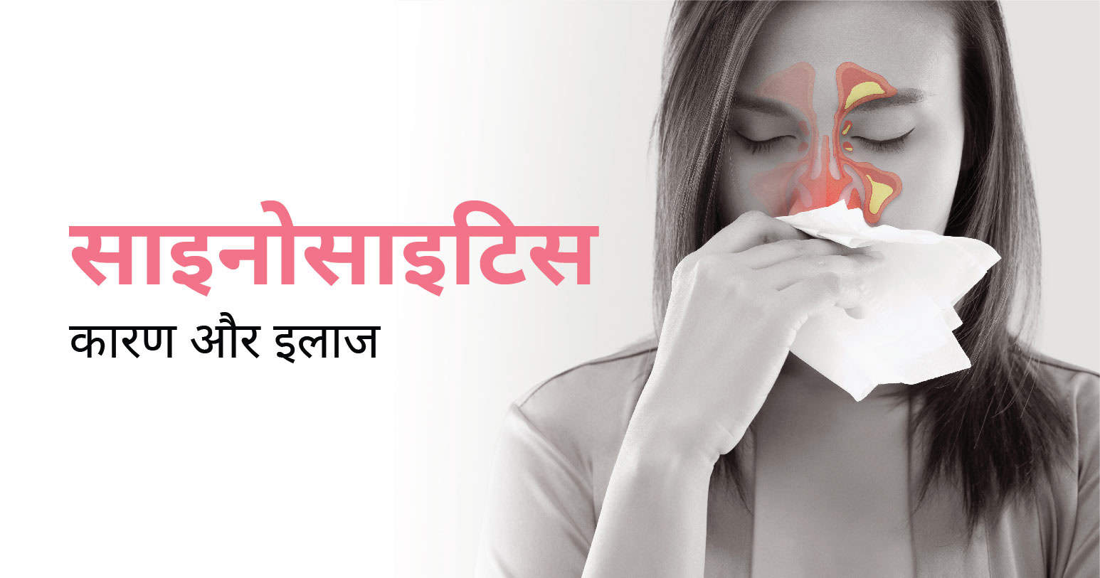
साइनोसाइटिस एक अधिकतर लोगों को प्रभावित करने वाली सामान्य समस्या है जो नाक के साइनस नामक तंत्र की समस्या होती है। यह तंत्र नाक के अंदर फ़्लेम बनाता है जो आपके चेहरे के पीछे और आपके आंतरिक ऑडियो कनेक्शन के पास स्थित होते हैं।
साइनोसाइटिस की सामान्य लक्षणों में नाक से पानी निकलना, नाक बंद होना, सीने में भारीपन, दर्द या सिरदर्द शामिल हो सकते हैं। यह भी हो सकता है कि आपको बुखार, जुकाम, खांसी, गले में खराश या गले के पीछे की खुजली हो।
साइनोसाइटिस अक्सर सामान्य अंगीकृत उपचार से ठीक हो जाता है, जैसे कि एंटीबायोटिक दवाएं और नाक के लिए दवाएं। लेकिन अगर समस्या लंबे समय तक चलती है, तो आपको डॉक्टर से चेकअप कराना चाहिए ताकि वे आपकी समस्या के सटीक कारण की पुष्टि करके उचित इलाज निर्धारित कर सकें।
ये भी पढ़े: सिर में भारीपन के कारण और घरेलु इलाज
Table of Contents
साइनोसाइटिस कई कारणों से हो सकता है। इसके मुख्य कारणों में निम्नलिखित शामिल हो सकते हैं:
इन सबके अलावा, अन्य कारणों से भी साइनोसाइटिस हो सकता है जैसे कि अधिक धूल-प्रदूषण वाले क्षेत्रों में रहना या नाक के अंदर की नसों में संक्रमण होना आदि।
क्रोनिक साइनोसाइटिस के सामान्य लक्षणों में निम्न शामिल सकते हैं:
अन्य संकेतों और लक्षणों में शामिल हो सकते हैं:
क्रोनिक साइनोसाइटिस और तीव्र साइनोसाइटिस के समान संकेत और लक्षण हैं। लेकिन तीव्र साइनोसाइटिस साइनस का एक अस्थायी संक्रमण है जो अक्सर सर्दी से जुड़ा होता है। क्रोनिक साइनोसाइटिस के संकेत और लक्षण कम से कम 12 सप्ताह तक रहते हैं, लेकिन क्रोनिक साइनोसाइटिस विकसित होने से पहले आपको तीव्र साइनोसाइटिस के कई एपिसोड हो सकते हैं। बुखार क्रोनिक साइनोसाइटिस का एक सामान्य संकेत नहीं है, लेकिन तीव्र साइनोसाइटिस में आपको बुखार हो सकता है।
अगर आप निम्न लक्षणों को अनुभव करते हैं तो आपको जल्द से जल्द विशेषज्ञ से परामर्श करना चाहिए:
साइनोसाइटिस के इलाज की विधि इसके कारण और गंभीरता पर निर्भर करती है। कुछ मामलों में, साधारण घरेलू उपचार और धैर्य के साथ साइनोसाइटिस ठीक हो सकता है। लेकिन कुछ मामलों में, दवाओं या चिकित्सा उपचार की जरूरत हो सकती है।
साइनोसाइटिस के कुछ मामलों में दवाओं का इस्तेमाल भी किया जा सकता है। अधिकतर मामलों में, एंटीबायोटिक दवाएं संक्रमण को ठीक करने में मदद कर सकती हैं। एक नाक स्प्रे भी संक्रमण और जलन को कम करने में मदद कर सकता है।
साधारणतया, साइनोसाइटिस के उपचार के लिए सर्जरी की आवश्यकता नहीं होती है। लेकिन, कुछ मामलों में सर्जरी की जरूरत पड़ सकती है जब उपचार और दवाओं से संक्रमण दूर नहीं होता है या अधिक गंभीर मामलों में।
सर्जरी के दौरान, एक सर्जिकल इंस्ट्रुमेंट का इस्तेमाल किया जाता है जो साइनस के अंदर जमा मल को हटाने और साइनस के मुख्य नलिका में फ्रीली फ्लो ऑफ़ ऑयर की अनुमति देने में मदद करता है।
लेकिन, सर्जरी से पहले आपके चिकित्सक को आपके मामले की जांच करनी चाहिए। यह जांच आपके लक्षणों, रूट कारणों और उपचार इत्यादि के आधार पर की जाती है जिससे आपके चिकित्सक सही उपचार या सलाह दे सकें।
ऐसे कई घरेलू उपचार हैं जो साइनस की परेशानी को कम करने में मदद कर सकते हैं। पर्याप्त मात्रा में तरल पदार्थ का सेवन करें और हवा को नम रखने के लिए ह्यूमिडिफायर का उपयोग करें। आप नासल इर्रिगेशन, चिकन सूप या शहद भी इस्तेमाल कर सकते हैं, लेकिन अगर आपके लक्षण 10 दिनों से अधिक समय तक रहते हैं तो डॉक्टर से परामर्श करना सबसे अच्छा है।
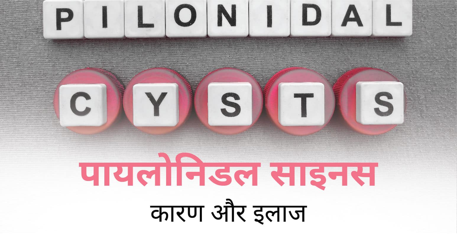
पायलोनिडल को पिलोनाइडल या पायलोनाइडल भी कहा जाता है। पायलोनिडल साइनस को पायलोनिडल सिस्ट या पायलोनिडल फोड़ा कहते हैं। यह एक पुराना त्वचा संक्रमण है जो टेलबोन के ठीक ऊपर, नितंबों के बीच के क्षेत्र को प्रभावित करता है।
Table of Contents
इस समस्या के कारण बाहर से दिखाई नहीं देते हैं। यह त्वचा के अंदर बालों या अन्य कचरे के इकट्ठे हो जाने से होता है। इससे संक्रमण फैलने की सम्भावना बढ़ जाती है। इस समस्या का मुख्य कारणों में निम्न शामिल हो सकते हैं:
अगर आप ऊपर दिए गए कारणों को ध्यान में रखकर कुछ सावधानियां बरतें तो खुद को इस समस्या से बचा सकते हैं।
पायलोनिडल साइनस के कुछ लक्षण हैं जो इस समस्या की पहचान करने में मदद कर सकते हैं। इसके लक्षणों में निम्न शामिल हो सकते हैं:
अगर आप ऊपर दिए गए लक्षणों को खुद में अनुभव करते हैं या इस समस्या से परेशान हैं तो विशेषज्ञ डॉक्टर से परामर्श करने का सुझाव दिया जाता है।
पायलोनिडल साइनस का इलाज कई तरह से किया जा सकता है जिसमें सर्जरी, दवाएं और घरेलू नुस्खे आदि शामिल हैं।
अधिकतर मामलों में, पायलोनिडल साइनस के इलाज के लिए सर्जरी की जाती है। सर्जरी में साइनस के चारों ओर की त्वचा को काट दिया जाता है और साइनस के अंदर जमा मल को निकाला जाता है।
कुछ मामलों में, एंटीबायोटिक्स का उपयोग संक्रमण से निपटने के लिए किया जा सकता है। इसके अलावा, कुछ दवाओं का उपयोग साइनस के मुख को फोड़ने या खोलने से पहले साइनस में मल को निकालने में मदद कर सकता है।
साथ ही, सफाई और हाइजीन भी साइनस के इलाज में मददगार साबित हो सकती है। साइनस क्षेत्र को धोने के लिए अच्छे गुणवत्ता वाले साबुन और गर्म पानी का उपयोग किया जाना चाहिए। इसके अलावा, साइनस क्षेत्र को स्वच्छ और सूखा रखना भी बहुत जरूरी होता है।
पायलोनिडल साइनस के उपचार के लिए सर्जरी सबसे सामान्य और सफल उपाय है। हालांकि, कुछ मरीजों में सर्जरी के बिना भी ठीक होने की संभावना होती है। इसके लिए कुछ उपचार विकल्प होते हैं जो निम्नलिखित हैं:
पायलोनिडल साइनस के लिए घरेलू उपचार कुछ इस प्रकार हैं:
साथ ही, इस बात का भी ध्यान रहे कि आप विशेषज्ञ डॉक्टर से परामर्श किए बिना किसी भी घरेलू नुस्खों का इस्तेमाल नहीं करना चाहिए। अगर आप घरेलू नुस्खों से पायलोनिडल साइनस का दूर करना चाहते हैं तो पहले डॉक्टर से अवश्य परामर्श करें।