Filter :

Table of Contents
प्रेगनेंसी का दूसरा महीना काफी ख़ास होता है। इस दौरान, जैसे-जैसे गर्भावस्था आगे बढ़ती है, कई महिलाओं को ध्यान देने योग्य शारीरिक और भावनात्मक परिवर्तन का अनुभव होने लगता है। यहां कुछ सामान्य लक्षण दिए गए हैं जो इस चरण के दौरान हो सकते हैं:
यह याद रखना महत्वपूर्ण है कि गर्भावस्था के लक्षण हर महिला में अलग-अलग हो सकते हैं, और सभी महिलाओं को समान लक्षण या समान स्तर का अनुभव नहीं होगा। यदि आपको अपनी गर्भावस्था के बारे में कोई चिंता है या आप गंभीर लक्षणों का अनुभव करते हैं, तो व्यक्तिगत सलाह और मार्गदर्शन के लिए विशेषज्ञ से परामर्श करना महत्वपूर्ण है।
इसे भी पढ़ें: प्रेगनेंसी में कितने समय के बाद सम्बन्ध बनाना बंद कर देना चाहिए
गर्भावस्था के दूसरे महीने के दौरान, माँ और विकासशील बच्चे दोनों की बढ़ती ज़रूरतों को पूरा करने के लिए स्वस्थ और संतुलित आहार बनाए रखना महत्वपूर्ण है। यहां कुछ आहार संबंधी विचार दिए गए हैं:
अपनी विशिष्ट आवश्यकताओं और आपके किसी भी आहार प्रतिबंध के आधार पर वैयक्तिकृत सलाह के लिए किसी डॉक्टर या डायटीशियन से परामर्श करें।
गर्भावस्था के दूसरे महीने के दौरान, बढ़ते भ्रूण को सहारा देने के लिए महिला के शरीर में महत्वपूर्ण परिवर्तन होते हैं:
याद रखें कि प्रत्येक महिला का अनुभव अलग-अलग हो सकता है, और यदि आपको कोई चिंता है, तो मार्गदर्शन के लिए विशेषज्ञ से परामर्श लें। आपके मन में यह प्रश्न उठ सकता है कि प्रेगनेंसी के दूसरे महीने में ब्लीडिंग हो तो क्या करें तो इसका सीधा जवाब है कि आपको तुरंत अपने डॉक्टर से मिलना चाहिए।
इसे भी पढ़ें: जानिए प्रेगनेंसी में संबंध बनाना चाहिए या नहीं ?
गर्भावस्था के दूसरे महीने के दौरान, बच्चे का विकास तेजी से जारी रहता है क्योंकि महत्वपूर्ण संरचनाएँ और अंग बनने लगते हैं:
यह ध्यान रखना महत्वपूर्ण है कि ये विकास दूसरे महीने के दौरान तेजी से होते हैं, और बच्चे के विकास की निगरानी करने और स्वस्थ गर्भावस्था सुनिश्चित करने के लिए नियमित प्रसवपूर्व देखभाल महत्वपूर्ण है।
गर्भावस्था के दूसरे महीने के दौरान, स्वयं की देखभाल और स्वस्थ जीवनशैली बनाए रखने पर ध्यान देना महत्वपूर्ण है:
अपनी गर्भावस्था यात्रा के दौरान व्यक्तिगत सलाह और मार्गदर्शन के लिए अपने स्वास्थ्य सेवा प्रदाता से परामर्श करें।
गर्भावस्था के दूसरे महीने के दौरान, हल्के व्यायाम करना महत्वपूर्ण है जो आपके और आपके बच्चे दोनों के लिए सुरक्षित और फायदेमंद हों। यहां दूसरे महीने के लिए उपयुक्त कुछ व्यायाम दिए गए हैं:
कोई भी व्यायाम आहार शुरू करने से पहले अपने डॉक्टर से परामर्श करें। वे आपके स्वास्थ्य, फिटनेस स्तर और आपकी गर्भावस्था से संबंधित किसी भी विशिष्ट विचार के आधार पर वैयक्तिकृत सलाह प्रदान कर सकते हैं।
गर्भावस्था के दूसरे महीने के दौरान, शिशु के स्वास्थ्य और विकास की निगरानी के लिए कई स्कैन और परीक्षण की सिफारिश की जा सकती है। इसमे शामिल है:
आपकी गर्भावस्था के लिए अनुशंसित विशिष्ट स्कैन और परीक्षणों के बारे में अपने डॉक्टर से चर्चा करना महत्वपूर्ण है, क्योंकि वे व्यक्तिगत कारकों और चिकित्सा इतिहास के आधार पर भिन्न हो सकते हैं।
गर्भावस्था के दूसरे महीने में क्या करें?
गर्भावस्था के दूसरे महीने के दौरान अपने स्वास्थ्य और बढ़ते बच्चे की देखभाल पर ध्यान दें। प्रसवपूर्व जांच में भाग लें, पौष्टिक आहार लें, हाइड्रेटेड रहें, हल्का व्यायाम करें, पर्याप्त आराम करें, तनाव का प्रबंधन करें और हानिकारक पदार्थों से बचें। व्यक्तिगत मार्गदर्शन और सहायता के लिए अपने डॉक्टर से परामर्श करें।
गर्भावस्था के दूसरे महीने में क्या ना करें?
गर्भावस्था के दूसरे महीने में, शराब, धूम्रपान, नशीली दवाओं, कुछ दवाओं, अत्यधिक कैफीन से बचें और किसी भी चिंता के बारे में अपने डॉक्टर से चर्चा करें।
गर्भावस्था के दूसरे महीने के दौरान सावधानियां
गर्भावस्था के दूसरे महीने के दौरान कुछ सावधानियां बरतें, प्रसव पूर्व जांच में भाग लें, स्वस्थ आहार बनाए रखें, शराब, धूम्रपान और नशीली दवाओं से बचें, कैफीन का सेवन सीमित करें, सुरक्षित रूप से व्यायाम करें, पर्याप्त आराम करें, तनाव का प्रबंधन करें और अपने डॉक्टर से परामर्श लें।
गर्भावस्था के दूसरे महीने के दौरान होने वाली समस्याएं
गर्भावस्था के दूसरे महीने के दौरान, कुछ सामान्य समस्याएं जो महिलाओं को अनुभव हो सकती हैं उनमें सुबह की मतली, थकान, स्तन कोमलता, मूड में बदलाव, भोजन की लालसा या नापसंद, पेशाब में वृद्धि और कब्ज जैसी पाचन संबंधी समस्याएं शामिल हैं। यदि आप किसी भी संबंधित लक्षण का सामना करते हैं, तो मार्गदर्शन और सहायता के लिए डॉक्टर से परामर्श करना चाहिए।

गर्भावस्था के पहले महीने में महिला के शरीर में कई महत्वपूर्ण बदलाव होते हैं, क्योंकि इस समय निषेचित अंडाणु गर्भाशय की दीवार में जाकर स्थापित होता है। हालांकि, गर्भावस्था की गणना आमतौर पर अंतिम मासिक धर्म के पहले दिन से की जाती है, इसलिए वास्तविक गर्भधारण आम तौर पर दूसरे सप्ताह के अंत तक होता है।
Table of Contents
पहले महीने में, निषेचित अंडा, जिसे जाइगोट कहा जाता है, तेजी से कोशिका विभाजन से गुजरता है, क्योंकि यह फैलोपियन ट्यूब के माध्यम से गर्भाशय की ओर बढ़ता है। तीसरे सप्ताह के आसपास, यह गर्भाशय की परत में प्रत्यारोपित हो जाता है, जहां यह एक भ्रूण के रूप में विकसित होता है।
इस दौरान, हार्मोनल परिवर्तन होते हैं, और शरीर मानव कोरियोनिक गोनाडोट्रोपिन (एचसीजी) का उत्पादन शुरू कर देता है, जो गर्भावस्था परीक्षणों में पाया जाने वाला हार्मोन है। यह हार्मोन गर्भावस्था का समर्थन करता है और गर्भाशय की परत को बनाए रखने में मदद करता है।
पहले महीने के अंत तक भ्रूण की बुनियादी संरचनाएं बननी शुरू हो जाती हैं। तंत्रिका ट्यूब, जो मस्तिष्क और रीढ़ की हड्डी में विकसित होगी, आदिम हृदय और रक्त वाहिकाओं के साथ विकसित होने लगती है। प्लेसेंटा भी बनना शुरू हो जाता है, जिससे मां और विकासशील भ्रूण के बीच पोषक तत्वों और अपशिष्ट का आदान-प्रदान आसान हो जाता है।
जबकि पहला महीना भ्रूण के विकास के लिए महत्वपूर्ण होता है, कई महिलाओं को इस दौरान पता भी नहीं चलता कि वे गर्भवती हैं, क्योंकि गर्भावस्था के शुरुआती लक्षण अभी तक ध्यान देने योग्य नहीं हो सकते हैं। जिन महिलाओं को संदेह है कि वे गर्भवती हो सकती हैं, उनके लिए गर्भावस्था परीक्षण कराना या पुष्टि के लिए डॉक्टर से परामर्श करना और प्रसवपूर्व देखभाल शुरू करना महत्वपूर्ण है।
इसे भी पढ़ें: पीरियड्स मिस होने से पहले प्रेग्नेंसी के क्या लक्षण दिखाई देते हैं?
गर्भावस्था के पहले महीने के दौरान, कई महिलाओं को ध्यान देने योग्य लक्षणों का अनुभव नहीं हो सकता है। हालाँकि, गर्भावस्था के कुछ सामान्य प्रारंभिक लक्षण दिखाई देने लग सकते हैं:
यह ध्यान रखना महत्वपूर्ण है कि ये लक्षण हर महिला में बहुत भिन्न हो सकते हैं, और कुछ महिलाओं को पहले महीने में कोई भी ध्यान देने योग्य संकेत अनुभव नहीं हो सकता है। यदि आपको संदेह है कि आप गर्भवती हो सकती हैं, तो गर्भावस्था परीक्षण करना या स्वास्थ्य सेवा प्रदाता से परामर्श करना गर्भावस्था की पुष्टि करने का सबसे विश्वसनीय तरीका है।
इसे भी पढ़ें: जानिए प्रेगनेंसी में संबंध बनाना चाहिए या नहीं ?
गर्भावस्था के पहले महीने के दौरान, माँ और विकासशील भ्रूण दोनों के स्वास्थ्य को बनाए रखने के लिए संतुलित और पौष्टिक आहार बनाए रखना आवश्यक है। विभिन्न प्रकार के पोषक तत्वों से भरपूर खाद्य पदार्थों के सेवन पर ध्यान दें, इसमें शामिल हैं:
व्यक्तिगत आवश्यकताओं और किसी विशिष्ट आहार प्रतिबंध या विचार के आधार पर व्यक्तिगत आहार संबंधी सलाह के लिए विशेषज्ञ से परामर्श करना महत्वपूर्ण है।
इसे भी पढ़ें: प्रेगनेंसी के शुरूआती लक्षण क्या होते हैं (Pregnancy ke lakshan)?
गर्भावस्था के पहले महीने के दौरान, शिशु का विकास बुनियादी संरचनाओं के निर्माण और अंग विकास की शुरुआत पर केंद्रित होता है। यहां कुछ मुख्य अंश दिए गए हैं:
इस बात का ध्यान रखना महत्वपूर्ण है कि ये विकास तीव्र गति से हो रहे हैं, लेकिन पहले महीने के दौरान अल्ट्रासाउंड में ये दिखाई नहीं दे सकते हैं या पता लगाने योग्य नहीं हो सकते हैं।
इसे भी पढ़ें: प्रेगनेंसी टेस्ट के घरेलू उपाय | Home Remedies For Pregnancy Test
गर्भावस्था के पहले महीने के दौरान, स्वस्थ गर्भावस्था के लिए कुछ सावधानियां बरतना महत्वपूर्ण है:
अपने व्यक्तिगत स्वास्थ्य और गर्भावस्था की जरूरतों के आधार पर वैयक्तिकृत सलाह और दिशानिर्देशों के लिए स्वास्थ्य सेवा प्रदाता से परामर्श लें।
इसे भी पढ़ें: प्रेगनेंसी में कितने समय के बाद सम्बन्ध बनाना बंद कर देना चाहिए
1. 1 महीने से पहले प्रेगनेंसी का पता कैसे लगाएं?
एक महीने से पहले गर्भावस्था का पता लगाना चुनौतीपूर्ण हो सकता है, क्योंकि शुरुआती लक्षण ध्यान देने योग्य नहीं हो सकते हैं। हालाँकि, मासिक धर्म न होने के समय लिया गया एक संवेदनशील गर्भावस्था परीक्षण एक संकेत प्रदान कर सकता है। डॉक्टर के क्लिनिक में रक्त परीक्षण भी गर्भावस्था का पहले ही पता लगा सकता है, आमतौर पर गर्भधारण के एक सप्ताह के भीतर।
2. 1 महीने की प्रेगनेंसी में ब्लड क्यों आता है?
गर्भावस्था के पहले महीने में ब्लड कई कारणों से हो सकता है। यह आरोपण रक्तस्राव से संबंधित हो सकता है जब निषेचित अंडा गर्भाशय की परत से जुड़ जाता है। हार्मोनल परिवर्तन, गर्भाशय ग्रीवा की संवेदनशीलता या गर्भपात या अस्थानिक गर्भावस्था जैसी संभावित जटिलताएँ भी रक्तस्राव का कारण बन सकती हैं।

Table of Contents
गर्भावस्था में रक्तस्राव को सामान्य नहीं माना जाता है और इसका मूल्यांकन विशेषज्ञ द्वारा किया जाना चाहिए। यह हल्के धब्बे या रक्तस्राव आरोपण या हार्मोनल परिवर्तन जैसे कारकों के कारण हो सकता है। लगातार रक्तस्राव होने पर तुरंत मेडिकल सहायता लेने की आवश्यकता होती है। अगर आपके मन में यह प्रश्न उठता है कि प्रेगनेंसी में ब्लीडिंग कितनी होती है तो इसका सरल उत्तर है निम्न या अधिक मात्रा में जो हर महिला में भिन्न हो सकता है।
यह गर्भपात या अस्थानिक गर्भावस्था जैसी गंभीर स्थितियों का संकेत हो सकता है, जिसमें तत्काल चिकित्सा हस्तक्षेप की आवश्यकता होती है। संक्रमण, सूजन या गर्भाशय ग्रीवा में परिवर्तन के कारण भी रक्तस्राव हो सकता है। गर्भावस्था के दौरान किसी भी रक्तस्राव के कारण और उपचार के उचित तरीके को निर्धारित करने के लिए विशेषज्ञ से परामर्श करना महत्वपूर्ण है।
गर्भावस्था के दौरान रक्तस्राव चिंता का कारण हो सकता है और इसका मूल्यांकन एक विशेषज्ञ द्वारा किया जाना चाहिए। हालाँकि, चिंता महसूस करना सामान्य है, लेकिन यह ध्यान रखना ज़रूरी है कि इस चरण के दौरान रक्तस्राव के विभिन्न कारण हो सकते हैं।
अगर आपके मन में यह प्रश्न उठता है कि प्रेगनेंसी में ब्लीडिंग कितने दिन तक होती है तो हम आपको बता दें कि यह ब्लीडिंग कुछ दिनों से लेकर लंबे समय तक हो सकती है। अनियमित ब्लीडिंग होने पर आपको तुरंत विशेषज्ञ से परामर्श करना चाहिए।
प्रत्यारोपण रक्तस्राव एक संभावित कारण है। जब एक निषेचित अंडा गर्भाशय की परत से जुड़ जाता है, तो कुछ महिलाओं को हल्के धब्बे या रक्तस्राव का अनुभव हो सकता है। यह आमतौर पर अपेक्षित मासिक धर्म के समय के आसपास होता है और आमतौर पर नियमित अवधि की तुलना में हल्का और छोटा होता है।
गर्भावस्था के दौरान हार्मोनल परिवर्तन भी रक्तस्राव में योगदान कर सकते हैं। बढ़ते भ्रूण को सहारा देने के लिए शरीर महत्वपूर्ण हार्मोनल बदलावों से गुजरता है, और इन परिवर्तनों के परिणामस्वरूप कभी-कभी हल्का रक्तस्राव या स्पॉटिंग हो सकता है। हालाँकि, लगातार रक्तस्राव का मूल्यांकन डॉक्टर द्वारा किया जाना चाहिए।
दुर्भाग्य से, गर्भावस्था के दौरान रक्तस्राव भी गर्भपात का संकेत हो सकता है। गर्भपात का तात्पर्य 20वें सप्ताह से पहले गर्भावस्था के नष्ट होने से है, और यह भावनात्मक रूप से परेशान करने वाला हो सकता है। गर्भपात के लक्षण में योनि से रक्तस्राव, पेट में ऐंठन और योनि से ऊतक का बाहर निकलना शामिल हो सकते हैं। यदि आप इन लक्षणों का अनुभव करते हैं, तो तत्काल चिकित्सा सहायता लेना महत्वपूर्ण है।
एक और गंभीर स्थिति जो प्रारंभिक गर्भावस्था में रक्तस्राव का कारण बन सकती है वह है अस्थानिक गर्भावस्था। यह तब होता है जब निषेचित अंडा गर्भाशय के बाहर, आमतौर पर फैलोपियन ट्यूब में प्रत्यारोपित होता है। एक्टोपिक गर्भधारण व्यवहार्य नहीं है और अगर तुरंत पता न लगाया जाए और इलाज न किया जाए तो यह जीवन के लिए खतरा हो सकता है। रक्तस्राव के अलावा, लक्षणों में पेट दर्द, कंधे में दर्द और चक्कर आना शामिल हो सकते हैं।
गर्भाशय ग्रीवा, योनि या पेल्विक क्षेत्र में संक्रमण या सूजन के कारण भी गर्भावस्था के दौरान रक्तस्राव हो सकता है। इन स्थितियों में जटिलताओं को रोकने के लिए चिकित्सा उपचार की आवश्यकता हो सकती है। इस स्थिति में पीरियड मिस होने के बाद ब्लीडिंग होना या प्रेगनेंसी में लाल पानी आना गंभीर स्थिति को ओर इशारा भी ही सकता है, इसलिए बिना देरी किए आपको डॉक्टर से परामर्श करना चाहिए।
इसके अलावा, गर्भावस्था के दौरान गर्भाशय ग्रीवा में परिवर्तन इसे अधिक संवेदनशील बना सकता है और रक्तस्राव का खतरा हो सकता है। संभोग या पैल्विक परीक्षण कभी-कभी गर्भाशय ग्रीवा में जलन पैदा कर सकता है और कुछ मामलों में रक्तस्राव का कारण बन सकता है। प्रेगनेंसी के फर्स्ट मंथ में ब्लीडिंग होना भी संभव है।
यह याद रखना महत्वपूर्ण है कि हालांकि यह जानकारी गर्भावस्था के दौरान रक्तस्राव के कुछ संभावित कारण प्रदान करती है, लेकिन यह एक विस्तृत सूची नहीं है। यदि आपको या आपके किसी जानने वाले को गर्भावस्था के दौरान रक्तस्राव का अनुभव हो रहा है, तो उचित मूल्यांकन के लिए स्त्री रोग विशेषज्ञ से संपर्क करना महत्वपूर्ण है। वे विशिष्ट परिस्थितियों के आधार पर व्यक्तिगत सलाह और आवश्यक चिकित्सा देखभाल प्रदान करने में सक्षम होंगे।
यदि आप गर्भावस्था में ‘रक्तस्राव परीक्षण’ की बात कर रहे हैं, तो यह स्पष्ट करना महत्वपूर्ण है कि गर्भावस्था के दौरान रक्तस्राव परीक्षण नामक कोई विशिष्ट परीक्षण नहीं है। हालाँकि, यदि आपको गर्भावस्था के दौरान रक्तस्राव का अनुभव हो रहा है तो विशेषज्ञ विभिन्न परीक्षण कर सकते हैं।
इन परीक्षणों में हार्मोन के स्तर की जांच करने के लिए रक्त परीक्षण, बच्चे के विकास का आकलन करने और किसी भी संभावित समस्या का पता लगाने के लिए अल्ट्रासाउंड स्कैन, और संभवतः गर्भाशय ग्रीवा का मूल्यांकन करने और किसी भी असामान्यता की जांच करने के लिए एक पैल्विक परीक्षा शामिल हो सकती है।
इसे भी पढ़ें: जानिए प्रेगनेंसी में संबंध बनाना चाहिए या नहीं ?
घरेलू उपचार अंतर्निहित कारण का समाधान नहीं कर सकते हैं और संभावित रूप से हानिकारक या अप्रभावी हो सकते हैं। यदि आपको गर्भावस्था के दौरान रक्तस्राव का अनुभव हो रहा है, तो चिकित्सा सहायता लेते समय पालन करने के लिए यहां कुछ सामान्य दिशानिर्देश दिए गए हैं:
याद रखें, ये उपाय पेशेवर चिकित्सा सलाह के विकल्प नहीं हैं। रक्तस्राव का कारण निर्धारित करने और उचित मार्गदर्शन प्राप्त करने के लिए तुरंत स्त्री रोग विशेषज्ञ से परामर्श करना महत्वपूर्ण है। वे आपकी विशिष्ट स्थिति का आकलन करने और आपकी आवश्यकताओं के आधार पर व्यक्तिगत देखभाल प्रदान करने में सक्षम होंगे।
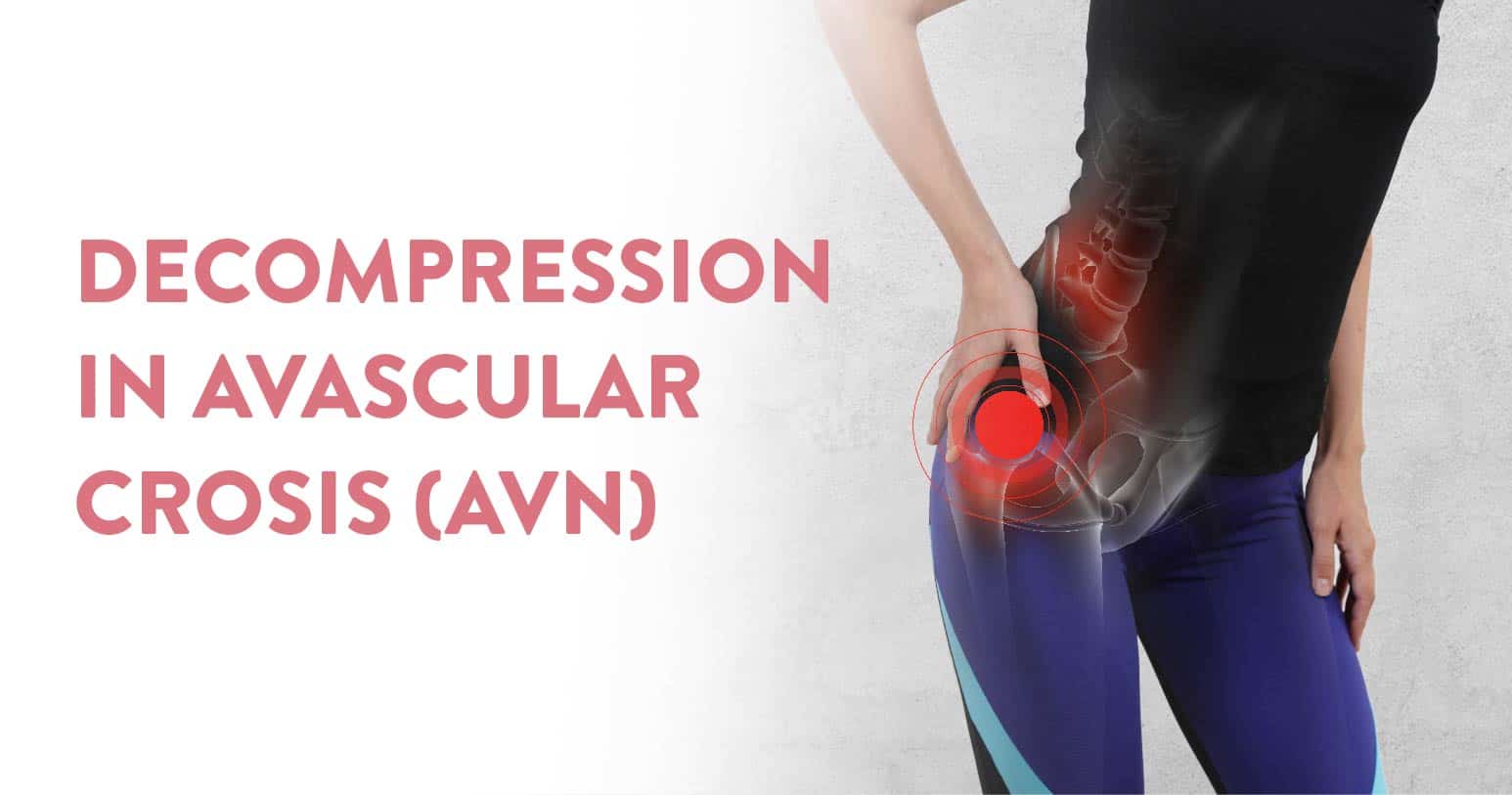
Avascular necrosis, also called osteonecrosis, is a medical condition that arises when there is inadequate blood supplied to the bone tissue. Due to lack of blood and oxygen the tissue starts decaying and eventually achieves the form of necrosis or decay. This fatal condition of avascular necrosis can affect any joint in the body, however, it has been observed that most reported cases are due to pain in the hip joint. Left untreated, the avascular necrosis of the hip can progress towards significant pain and disability. Surgery is the only option for restoring joint function.
Out of these surgical options, the most frequently chosen option is core decompression. In this process the goal is to relieve pressure within the bone tissue of the hip joint. In this blog post, we’ll explore what core decompression is, how it works, and what you can expect if you are considering this procedure.
Table of Contents
The surgical approach of core decompression aims to relieve pressure within the bone tissue of the hip joint. The major goals of this surgical approach are:
Here is a step by step flow of how a core decompression surgery for AVN is performed:
Core decompression is performed under general anesthesia, and usually takes about an hour to complete. Patients are likely to go home in under 24 hours. This procedure can be done successfully in the initial stages of AVN. However, stage 4 level necrosis often poses a much bigger risk, hence timeliness and precision is vital.
Post-operative care is essential as this has an impact on your overall body balance. Patients sometimes may experience some pain and discomfort, for which pain medication is prescribed. Further physiotherapy may be prescribed to help improve the range of motion and strength in the affected hip joint. Some restrictions may be imposed on physical activity or weight-bearing, to ensure a full and speedy recovery.
According to a study published in the Journal of Orthopaedic Surgery and Research, core decompression had a success rate of 76% in patients with early stage avascular necrosis of the hip. Factors affecting the outcome of a core decompression surgery are
The study also found that the success rate decreased to 60% in patients with more advanced stages of the condition. Other studies have reported similar success rates for core decompression in the treatment of avascular necrosis of the hip. However, it is important to note that these success rates can vary depending on the individual patient and the specific circumstances of their condition.
The success rate of core decompression can be significantly improved by choosing a reliable healthcare partner and orthopaedic surgeons for the procedure. When choosing a healthcare provider opt for state-of-the-art facilities supported by experienced medical teams that deliver on the global healthcare standards. A study published in the Journal of Orthopaedic Surgery and Research found that a successful core decompression significantly relies on the skill and judgement of the surgeon.
In conclusion, if you are considering core decompression for avascular necrosis of the hip, it is important to talk to your doctor about the risks and benefits of this procedure, as well as what you can expect during and after the procedure. By working closely with your healthcare team and following their instructions, you can help to ensure the best possible outcome from core decompression. If you are facing any of the symptoms of AVN or need a second opinion, reach out to our award winning orthopaedic department at the CK Birla Hospital, Gurugram. We offer trusted expertise and personalised care across multiple specialties under one roof.
This is an orthopaedic ailment that can be treated by an experienced orthopaedic surgeon. This is a fatal condition and needs prompt attention, diagnosis and treatment to prevent death due to blocked blood circulation in the body.
A cure for avascular necrosis does not exist. However, treatment can sometimes slow down the progress, but there is still no confirmed cure. Most people diagnosed with avascular necrosis eventually end up having surgery, which often includes joint replacement. However, in some cases, people with avascular necrosis can also develop a high risk of severe osteoarthritis.
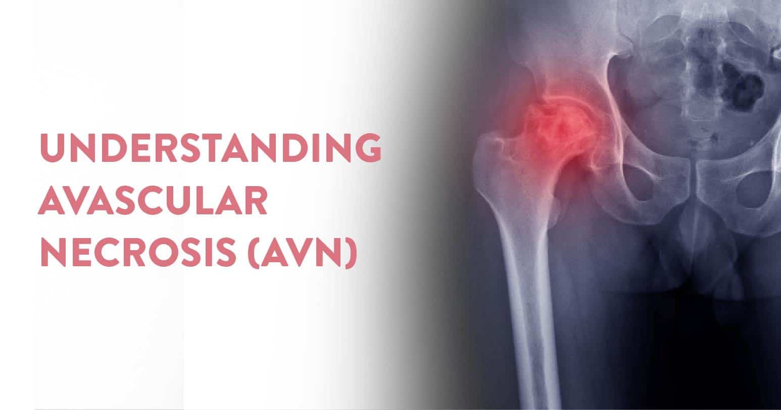
Table of Contents
AVN, also known as osteonecrosis, is a critical medical condition where a part of the bone tissue starts dying due to insufficient blood supply. Avascular Necrosis of the femoral head is when the rounded end of the thigh bone (femur) does not get any blood flow. Eventually it leads to the death and decay of the bone cells, causing the bone to collapse, and the patient ends up with either arthritis and chronic pain. Although avascular necrosis is commonly associated with the hip joint, it can affect other joints as well, including the knee, shoulder, ankle, and wrist
The root causes can be linked to several factors, such as trauma on impact, prolonged use of corticosteroids, heavy alcohol consumption, radiation therapy, and specific medical conditions like sickle cell anaemia and systemic lupus erythematosus. This will surface in the form of pain, specifically in the hip, groin, or thigh regions, limited range of motion, and in severe cases, collapse of the femoral head.
In this blog post, we’ll discuss avascular necrosis of joints, including its causes, symptoms, diagnosis, and treatment options.
The primary cause is the disruption of blood supply to the bone tissue. This can further be traced back to
In some cases, avascular necrosis cannot be explained. These AVNs occur for no apparent reason, which is why they are referred to as idiopathic avascular necrosis. While the exact cause is unknown, it might be due to a combination of genetic and environmental factors.
The symptoms of avascular necrosis vary depending upon the location of pain and severity of the condition. In the majority of reported cases, avascular necrosis is found where there is pain in the affected joint. This tends to worsen when the joint is overused or bears weight. Other symptoms may include stiffness, limited range of motion, and joint instability.
In some cases, the condition can further deteriorate to the point where the affected bone may collapse causing a sudden onset of severe pain and disability. Hence it is vividly clear that AVN needs prompt medical attention and a quick as well as safe diagnosis.
The diagnosis of avascular necrosis requires a certain level of urgency since this is primarily triggered by a compromised circulation of blood. Initially it starts with a physical exam and assessment of prior medical history. Further your doctor may also prescribe more investigation in the form of imaging tests, such as an X-ray, MRI, or CT scan, to help confirm the diagnosis and determine the extent of the damage.
In some cases, your doctor may also recommend a full body bone scan, which involves injecting a small amount of radioactive material into the body bloodstream, which will flow through the body and help identify areas of bone that are low on blood supply in the imaging report.
Impaired circulation will lead to a smaller window for treatment, hence prompt action is essential. Based on the stage of necrosis here are the various diagnostics performed:
The treatment of avascular necrosis also depends on the location and severity of the condition. But it differs based on the underlying cause. The ultimate goal of an AVN treatment, surgical or non-surgical are:
There are no guaranteed ways to prevent avascular necrosis, however, you can reduce your risk, by avoiding excessive alcohol consumption, and managing other medical conditions.
A person suffering from AVN will not be able to detect the problem easily. Circulation impairment is always a fatal condition which needs timely medical intervention. If and when you notice recurring excruciating pain in the joint areas, it is always recommended to speak to an expert. In this case the specialists would be an orthopaedic surgeon, since most AVN conditions quickly escalate towards surgery.
At the CK Birla Hospital, we ensure patients get holistic medical support which includes treatment in a compassionate environment. This patient-centric approach not only helps patients heal better but also ensures they are aware of the preventive measures as well. In case you need to consult an orthopaedic surgeon, do reach out to our award winning Department of Orthopaedics or book a direct appointment with Dr Ashwani Maichand at the CK Birla Hospital. (Booking Link)
This is an orthopaedic ailment that can be treated by an experienced orthopaedic surgeon. This is a fatal condition and needs prompt attention, diagnosis and treatment to prevent death due to blocked blood circulation in the body.
A cure for avascular necrosis does not exist. However, treatment can sometimes slow down the progress, but there is still no confirmed cure. Most people diagnosed with avascular necrosis eventually end up having surgery, which often includes joint replacement. However, in some cases, people with avascular necrosis can also develop a high risk of severe osteoarthritis.
Stage 1: Diagnosis from normal radiographs
Stage 2: Cystic changes and sclerosis visible on imaging
Stage 3: Subchondral collapse or femoral head flattening
Stage 4: Joint space narrowing
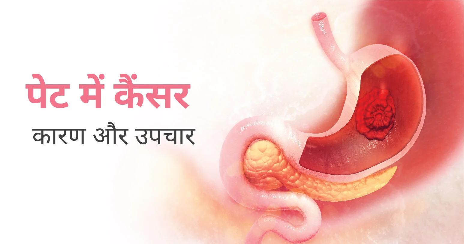
पेट के कैंसर को गैस्ट्रिक कैंसर के नाम से भी जाना जाता है। यह एक ऐसी बीमारी है जिसमें पेट की परत में घातक (कैंसर) कोशिकाएं बन जाती हैं। उम्र, आहार और पेट की बीमारी गैस्ट्रिक कैंसर के विकास के जोखिम को प्रभावित कर सकती है। गैस्ट्रिक कैंसर के लक्षणों में अपच और पेट में बेचैनी या दर्द शामिल है।
Table of Contents
पेट का कैंसर आमतौर पर शुरुआती चरणों में लक्षण पैदा नहीं करता है। यहां तक कि पेट के कैंसर के सबसे आम शुरुआती लक्षण – अक्सर अस्पष्टीकृत वजन घटना और पेट में दर्द – आमतौर पर तब तक दिखाई नहीं देते जब तक कि कैंसर अधिक उन्नत न हो जाए। पेट के कैंसर के लक्षणों में निम्न शामिल हो सकते हैं:
इनमें से कई लक्षण अन्य स्थितियों में भी सामान्य हैं। यह जांचने के लिए अपने डॉक्टर को देखें कि क्या आपके लक्षण पेट के कैंसर या किसी अन्य बीमारी के संकेत हैं।
पेट का कैंसर तब बनता है जब आपके पेट की कोशिकाओं के डीएनए में आनुवंशिक परिवर्तन (परिवर्तन) होता है। डीएनए वह कोड है जो कोशिकाओं को बताता है कि कब बढ़ना है और कब मरना है। उत्परिवर्तन के कारण, कोशिकाएं तेजी से बढ़ती हैं और अंततः मरने के बजाय एक ट्यूमर बन जाती हैं।
कैंसर कोशिकाएं स्वस्थ कोशिकाओं से आगे निकल जाती हैं और आपके शरीर के अन्य भागों में फैल सकती हैं (मेटास्टेसाइज़)। शोधकर्ताओं को पता नहीं है कि म्यूटेशन का क्या कारण है। फिर भी, कुछ कारक पेट के कैंसर के विकास की संभावना को बढ़ाते प्रतीत होते हैं। इसमें शामिल हैं:
पेट के कैंसर के बढ़ते जोखिम के साथ कई आनुवंशिक स्थितियां जुड़ी हुई हैं, जिनमें शामिल हैं:
पेट के कैंसर का आमतौर पर निम्न में से एक या अधिक के साथ इलाज किया जाता है:
पेट में कैंसर कोशिकाओं का इलाज करने के अलावा, उपचार का लक्ष्य कोशिकाओं को फैलने से रोकना है। पेट का कैंसर, जब अनुपचारित छोड़ दिया जाता है, तो यह फैल सकता है:
अकेले पेट के कैंसर को रोका नहीं जा सकता। हालाँकि, आप सभी कैंसर के विकास के अपने जोखिम को कम कर सकते हैं:
कुछ मामलों में, डॉक्टर ऐसी दवाएं लिख सकते हैं जो पेट के कैंसर के जोखिम को कम करने में मदद कर सकती हैं। यह आमतौर पर उन लोगों के लिए किया जाता है जिनके पास कैंसर पूर्व स्थिति है या अन्य बीमारियां हैं जिन्हें दवा सेफायदा होता है।
आप एक अर्ली स्क्रीनिंग टेस्ट कराने पर भी विचार कर सकते हैं। यह टेस्ट पेट के कैंसर की पहचान करने में मददगार हो सकता है। एक डॉक्टर निम्नलिखित पेट कैंसर स्क्रीनिंग परीक्षणों में से एक का उपयोग करके रोग के संकेतों की जाँच कर सकते हैं:
यदि कोई स्वास्थ्य स्थिति समय-समय पर आती और जाती रहती है, तो यह कैंसर होने की संभावना नहीं है। कैंसर लक्षणों का एक निरंतर सेट दिखाता है जो समय के साथ खराब हो जाता है, समय के साथ कुछ नए लक्षण जुड़ जाते हैं।
पेट के कैंसर के लक्षणों में शामिल हो सकते हैं: भूख कम लगना, वजन कम होना और पेट में दर्द होना आदि।

पेट में सूजन तब होती है जब आपका पेट क्षेत्र सामान्य से बड़ा होता है। इसे कभी-कभी एक विकृत पेट या सूजे हुए पेट के रूप में जाना जाता है। एक सूजा हुआ पेट अक्सर असहज या दर्दनाक भी होता है। सूजे हुए पेट के कई संभावित कारण होते हैं और यह एक सामान्य घटना है।
ये भी पढ़े: कब्ज क्या है – कारण, लक्षण, जांच और इलाज (Constipation in Hindi)
पेट में सूजन, या व्याकुलता, अक्सर एक गंभीर बीमारी की तुलना में अधिक खाने के कारण होती है। यह समस्या निम्न कारणों से भी हो सकती है:
आपके पेट में सूजन के कारण के आधार पर, आप अपने लक्षणों का इलाज घर पर आसानी से कर सकते हैं। यदि आपका पेट इसलिए सूज गया है क्योंकि आपने बहुत अधिक खा लिया है, तो बस अपने भोजन के पचने की प्रतीक्षा करना आपकी समस्या का समाधान कर सकता है। छोटे भोजन खाने से भविष्य में इस समस्या को रोकने में मदद मिल सकती है। इसके अलावा, अपने भोजन को संसाधित करने के लिए अपने पेट को समय देने के लिए अधिक धीरे-धीरे खाने पर विचार करें।
यदि आपका पेट गैस के कारण सूज गया है, तो उन खाद्य पदार्थों से बचने की कोशिश करें जो गैस पैदा करने के लिए जाने जाते हैं। इनमें से कुछ खाद्य पदार्थ बीन्स और क्रूसिफेरस सब्जियां हैं जैसे ब्रोकोली और गोभी। कार्बोनेटेड पेय पीने और स्ट्रॉ से पीने से बचें। धीरे-धीरे खाने से भी आपको हवा निगलने से रोका जा सकता है, जिससे गैस बनती है।
डेयरी उत्पादों से बचने से लैक्टोज असहिष्णुता के कारण होने वाली पेट की सूजन को दूर करने में मदद मिल सकती है। आईबीएस के मामले में, आपके तनाव के स्तर को कम करने और अपने फाइबर सेवन को बढ़ाने से लक्षणों से छुटकारा पाने में मदद मिली है। यदि आपको जलोदर है, तो बिस्तर पर आराम करें और अपने सोडियम सेवन को कम करने से आपके शरीर को अतिरिक्त तरल पदार्थ से छुटकारा मिल सकता है।
यदि आराम करना और अपने आहार में सोडियम की मात्रा कम करना लक्षणों से राहत पाने के लिए काम नहीं करता है, तो आपका डॉक्टर मूत्रवर्धक का उपयोग करने का सुझाव दे सकता है। मूत्रवर्धक आपके गुर्दे को सूजन पैदा करने वाले तरल पदार्थ को अधिक निकालने में मदद करेंगे। दुर्लभ मामलों में, आपके जलोदर द्रव में संक्रमण विकसित हो सकता है। यदि ऐसा होता है, तो आपको एंटीबायोटिक दवाओं के साथ कठोर उपचार से गुजरना होगा।
आईबीएस और लैक्टोज असहिष्णुता के कारण सूजन वाले पेट से छुटकारा पाने के लिए बहुत अधिक चिकित्सा उपचार उपलब्ध नहीं है। जलोदर आमतौर पर सिरोसिस जैसे शरीर में एक और गंभीर समस्या का दुष्प्रभाव होता है। आपको देखभाल की योजना के बारे में अपने डॉक्टर से बात करनी चाहिए।
कारण बीमारी का इलाज करने के अलावा, आपको द्रव को हटाने की आवश्यकता हो सकती है। द्रव हटाने की प्रक्रिया, या पैरासेन्टेसिस, अवधि में भिन्न होती है क्योंकि यह इस बात पर निर्भर करती है कि कितना तरल पदार्थ निकालने की आवश्यकता है।

गले का सूजन ऑरोफरीनक्स के श्लेष्म झिल्ली की सूजन है। अधिकतर मामलों में, यह एक संक्रमण के कारण होता है, या तो बैक्टीरिया या वायरल। इसके अन्य कम सामान्य कारणों में एलर्जी, आघात, कैंसर, रिफ्लक्स और कुछ टॉक्सिन्स शामिल हैं।
ये भी पढ़े: गले में दर्द का कारण, लक्षण और उपचार (Gale Mein Dard)
कई वायरल और बैक्टीरियल एजेंट हैं जो गले का सूजन का कारण बन सकते हैं। वे सम्मिलित करते हैं:
गले में खराश का सबसे आम कारण वायरस हैं। गले का सूजन आमतौर पर वायरल संक्रमण जैसे कि सामान्य सर्दी, इन्फ्लूएंजा या मोनोन्यूक्लिओसिस के कारण होता है। वायरल संक्रमण एंटीबायोटिक दवाओं का जवाब नहीं देते हैं, और उपचार केवल लक्षणों से छुटकारा पाने में मदद के लिए आवश्यक है।
बहुत कम मामलों में गले का सूजन एक जीवाणु संक्रमण के कारण होता है। जीवाणु संक्रमण के लिए एंटीबायोटिक दवाओं की आवश्यकता होती है। गले का सबसे आम जीवाणु संक्रमण स्ट्रेप थ्रोट है, जो ग्रुप ए स्ट्रेप्टोकोकस के कारण होता है। बैक्टीरियल गले का सूजन के दुर्लभ कारणों में गोनोरिया, क्लैमाइडिया और कॉरीनेबैक्टीरियम शामिल हैं।
जुकाम और फ्लू के बार-बार संपर्क में आने से गले का सूजन का खतरा बढ़ सकता है। यह उन लोगों के लिए विशेष रूप से सच है जो स्वास्थ्य देखभाल, एलर्जी और अक्सर साइनस संक्रमण में नौकरी करते हैं। सेकेंड हैंड धुएं के संपर्क में आने से भी आपका जोखिम बढ़ सकता है।
ये भी पढ़े: निमोनिया क्या होता है – कारण, लक्षण, जांच और उपचार (Pneumonia Meaning in Hindi)
इन्क्यूबेशन पीरियड यानी ऊष्मायन अवधि आमतौर पर दो से पांच दिन की होती है। गले का सूजन के साथ आने वाले लक्षण अंतर्निहित स्थिति के आधार पर भिन्न होते हैं।
गले में खराश, सूखापन या खरोंच के अलावा, सर्दी या फ्लू के निम्न लक्षण हो सकते हैं:
गले में खराश के अलावा, मोनोन्यूक्लिओसिस के लक्षणों में शामिल हैं:
स्ट्रेप थ्रोट, एक अन्य प्रकार का गले का सूजन भी पैदा कर सकता है:
संक्रामक अवधि की लंबाई आपकी अंतर्निहित स्थिति पर भी निर्भर करेगी। यदि आपको वायरल संक्रमण है, तो आप तब तक संक्रामक रहेंगे जब तक कि आपका बुखार अपना कोर्स नहीं करता। यदि आपके पास स्ट्रेप थ्रोट है, तो आप एंटीबायोटिक दवाओं पर 24 घंटे बिताने तक शुरुआत से ही संक्रामक हो सकते हैं।
सामान्य सर्दी आमतौर पर 10 दिनों से कम समय तक रहती है। बुखार सहित लक्षण लगभग तीन से पांच दिनों में चरम पर हो सकते हैं। यदि गले का सूजन एक ठंडे वायरस से जुड़ा हुआ है, तो आप अपने लक्षणों के इस अवधि तक बने रहने की उम्मीद कर सकते हैं।
ये भी पढ़े: क्या सामान्य ज़ुकाम, खांसी या ठंड निमोनिया में बदल सकती है
यदि कोई वायरस आपके गले का सूजन का कारण बन रहा है, तो घरेलू देखभाल से लक्षणों को दूर करने में मदद मिल सकती है। घरेलू देखभाल में शामिल हैं:
दर्द और बुखार से राहत के लिए, एसिटामिनोफेन या इबुप्रोफेन जैसी ओवर-द-काउंटर दवा लेने पर विचार करें। थ्रोट लोजेंजेस भी एक दर्दनाक, खराश वाले गले को शांत करने में सहायक हो सकता है।
गले का सूजन के इलाज के लिए कभी-कभी वैकल्पिक उपचार का उपयोग किया जाता है। हालांकि, दवाओं के अंतःक्रियाओं या अन्य स्वास्थ्य जटिलताओं से बचने के लिए उनका उपयोग करने से पहले आपको अपने डॉक्टर से संपर्क करना चाहिए।
ये भी पढ़े: गले में इन्फेक्शन का कारण, लक्षण, बचाव और घरेलू उपचार

खांसी एक सामान्य प्रतिक्रिया है जो आपके गले के बलगम या बाहरी जलन को साफ करती है। जबकि हर कोई समय-समय पर अपना गला साफ करने के लिए खांसी करता है, कई स्थितियों में बार-बार खांसी हो सकती है।
खांसी के अधिकांश एपिसोड 2 सप्ताह के भीतर साफ हो जाएंगे या कम से कम काफी सुधार होंगे। अगर आपकी खांसी कुछ हफ्तों में ठीक नहीं होती है तो डॉक्टर या स्वास्थ्य देखभाल पेशेवर से संपर्क करें। यह अधिक गंभीर स्थिति का संकेत दे सकता है।
Table of Contents
तीव्र खांसी के ये सामान्य कारण हैं – दो महीने से कम समय तक रहना:
ये भी पढ़े: ट्यूबरक्लोसिस क्या है – कारण, लक्षण और बचाव (Tuberculosis in Hindi)
यदि आपको ऐसी खांसी है जो 2 सप्ताह में ठीक नहीं हुई है या इसमें सुधार नहीं हुआ है, तो डॉक्टर से संपर्क करें। यह अधिक गंभीर समस्या का लक्षण हो सकता है।
यदि आप अतिरिक्त लक्षण विकसित करते हैं तो तत्काल चिकित्सा प्राप्त करें। इन लक्षणों में शामिल हो सकते हैं:
साथ ही, खून खांसी या सांस लेने में कठिनाई होने पर भी तत्काल विशेषज्ञ से परमर्श करना चाहिए।
ये भी पढ़े: सूखी खांसी: कारण, लक्षण, घरेलू उपाय और निदान
यदि डॉक्टर आपकी खांसी का कारण निर्धारित नहीं कर सकते हैं, तो वे अतिरिक्त परीक्षणों का आदेश दे सकते हैं। इन परीक्षणों में शामिल हो सकते हैं:
खांसी के लिए दिल की समस्याओं का एकमात्र लक्षण होना बहुत दुर्लभ है, लेकिन डॉक्टर यह सुनिश्चित करने के लिए इकोकार्डियोग्राम का अनुरोध कर सकता है कि आपका दिल सही ढंग से काम कर रहा है और आपकी खांसी नहीं कर रहा है।
ये भी पढ़े: टॉन्सिल (टॉन्सिलाइटिस) के कारण, लक्षण और इलाज
कठिन मामलों में इन अतिरिक्त परीक्षणों की आवश्यकता हो सकती है:
ऐसे मामलों में जहां पिछले परीक्षण या तो संभव नहीं हैं या सफल होने की अत्यधिक संभावना नहीं है, या आपकी खांसी उपचार के बिना हल होने की उम्मीद है, डॉक्टर कफ सप्रेसेंट लिख सकते हैं।
ये भी पढ़े: गले में दर्द का कारण, लक्षण और उपचार (Gale Mein Dard)
कारण के आधार पर खांसी का विभिन्न तरीकों से इलाज किया जा सकता है। स्वस्थ वयस्क अधिकतर घरेलू उपचार और स्वयं की देखभाल के साथ अपनी खांसी का इलाज करने में सक्षम होंगे।
वायरस के कारण होने वाली खांसी का इलाज एंटीबायोटिक दवाओं से नहीं किया जा सकता है। इसके बजाय आप इसे निम्न तरीकों से ठीक कर सकते हैं:
आमतौर पर, चिकित्सा देखभाल में डॉक्टर को आपके गले को देखना, आपकी खांसी सुनना और किसी अन्य लक्षण के बारे में पूछना शामिल होता है। यदि आपकी खांसी जीवाणु संक्रमण के कारण होने की संभावना है, तो डॉक्टर मौखिक एंटीबायोटिक्स लिखेंगे। वे या तो कफ सप्रेसेंट लिख सकते हैं जिनमें कोडीन या एक्सपेक्टोरेंट कफ सिरप होते हैं।