Filter :
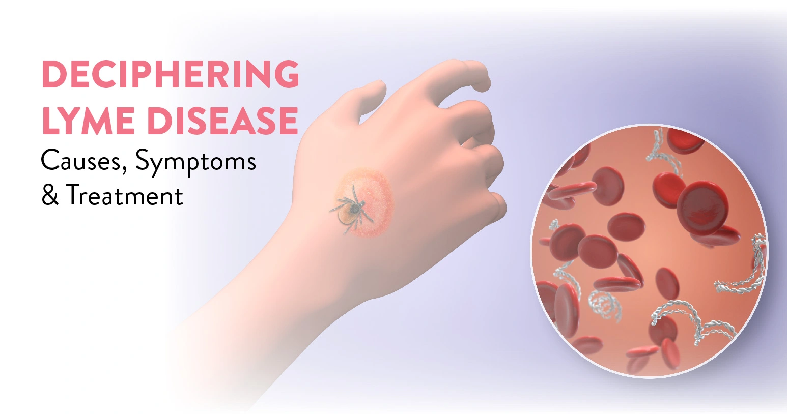
Lyme disease, a tick-borne illness caused by the bacterium Borrelia burgdorferi, presents a complex health challenge worldwide. With symptoms ranging from fatigue and joint pain to neurological issues, early diagnosis and treatment are crucial.
This blog explores the intricacies of Lyme disease: its symptoms, diagnosis, treatment options, prevention strategies, and the ongoing research aimed at better understanding this often misunderstood condition.
Table of Contents
Lyme disease stems from the bacterium Borrelia burgdorferi, transmitted through bites from infected deer ticks (also known as black-legged ticks). It’s important to note that common “wood ticks” and “dog ticks” do not transmit this infection.
The initial identification of Lyme disease, or borreliosis, traces back to 1975 when numerous cases of juvenile rheumatoid arthritis were diagnosed among children in Lyme, Connecticut, and nearby areas. It was later determined by researchers that the outbreak of arthritis was linked to bites from deer ticks carrying the infection.
Lyme disease progresses through three main stages if left untreated:
Early detection and antibiotic treatment are vital to prevent progression to later stages. Immediately seek medical assistance if you think you may have Lyme disease.
Early signs of Lyme disease often start with a distinctive rash called erythema migrans (EM), appearing as a red spot at the tick bite site. Here’s what to look for:
As Lyme disease progresses, symptoms may evolve into the second stage:
Untreated Lyme disease can lead to late-stage symptoms:
Recognizing these signs helps in the timely diagnosis and treatment of Lyme disease.
Lyme disease is caused by the bacterium Borrelia burgdorferi, transmitted through the bite of infected ticks like black-legged or deer ticks. Outdoor activities in wooded or grassy areas elevate the risk, exposing individuals to tick bites.
Lyme disease, caused by Borrelia burgdorferi transmitted through tick bites, can result in various complications if untreated. The disease progresses through these stages:
Early Localized Infection
Early Disseminated Infection
Late Disseminated Infection (Chronic Lyme disease)
The severity varies among individuals. Early diagnosis and antibiotic treatment are crucial for preventing progression and complications.
Also read:10 Signs of Breast Cancer During COVID-19
Lyme disease can be challenging to diagnose due to its symptoms overlapping with other illnesses. Healthcare providers start with a medical history and physical exam to spot signs like erythema migrans.
Key steps in Lyme disease diagnosis include:
Prompt treatment outweighs waiting for tick test outcomes in Lyme disease.
Treatment for Lyme disease varies depending on the infection’s stage:
Early Stages
Best treated early with 10- to 14-day oral antibiotics (e.g., doxycycline, amoxicillin, cefuroxime) to eliminate the infection.
Later Stages
In all cases, treatment duration and method are tailored to the severity and progression of Lyme disease. Early detection is crucial for successful outcomes.
Lyme disease peaks in late spring to early fall with active tick feeding. Deer ticks thrive in wooded and grassy areas, less so in sunny lawns.
Key points to prevent tick bites include:
After outdoor activities in risky areas:
Additionally, pets can carry ticks indoors. Consult a veterinarian for tick prevention strategies for your pet.
Lyme disease is a complex illness with varying symptoms and treatment challenges. Early detection is crucial for effective management. Awareness, prevention measures, and ongoing research are essential in combating this widespread health concern. It is always advisable to seek medical help from a general physician. Timely care and help can ensure an appropriate diagnosis and treatment of your condition.
At the CK Birla Hospital, we ensure patients get holistic medical support which includes treatment in a compassionate environment. This patient-centric approach not only helps patients heal better but also ensures they are aware of the preventive measures as well. In case you need to consult a general physician, reach out to us, or book a direct appointment with rheumatologist at the CK Birla Hospital.
Yes, Lyme disease can typically be cured with antibiotics if treated early. However, some individuals may experience lingering symptoms known as post-treatment Lyme disease syndrome (PTLDS).
Seek medical attention promptly if you notice symptoms like rash, fever, fatigue, or joint pain within a few weeks after a tick bite. Early treatment can prevent Lyme disease complications.
Lyme disease is not contagious among humans. It is transmitted only through the bite of infected ticks, primarily deer ticks (black-legged ticks) carrying the bacterium Borrelia burgdorferi.
Long-term effects of Lyme disease can include chronic joint inflammation, neurological symptoms (like nerve pain or cognitive difficulties), and in rare cases, heart problems or persistent symptoms known as post-treatment Lyme disease syndrome (PTLDS).
Pets can get Lyme disease from infected ticks. Dogs are particularly susceptible and can develop symptoms similar to humans, including fever, joint pain, and lethargy.

Autism, a complex neuro developmental condition, manifests uniquely in each individual, affecting communication, behaviour, and social interaction. Understanding autism requires dispelling common myths and embracing diverse perspectives.
This blog delves into the multifaceted world of autism, exploring its intricacies, challenges, and triumphs. Join us on a journey of discovery and acceptance, celebrating the rich diversity within the autism spectrum.
Table of Contents
Autism, now known as autism spectrum disorder (ASD), is a condition affecting brain development. ASD is a developmental disability resulting from variations in a child’s brain.
Individuals with ASD may demonstrate unique behaviours, interactions, and learning patterns compared to others. They may encounter challenges in social interactions and understanding and utilizing both verbal and nonverbal communication.
Autism (ASD) involves challenges with social skills, repetitive behaviours, speech, and communication. Subtypes are no longer distinct diagnoses; ASD is seen as a spectrum disorder with varying signs/symptoms and severity. The subtypes are:
Today, autism is diagnosed and understood based on a spectrum, with varying support needs and symptoms, emphasizing individual strengths and challenges rather than distinct subtypes.
Autism symptoms vary widely in severity, with each individual exhibiting unique characteristics. Recognizing signs of autism early is crucial to seek appropriate evaluation and support for your child. Consider the following indicators:
Social Interaction Challenges
Restricted/Repetitive Behaviours
If you notice these signs, consult your child’s healthcare provider promptly for further guidance and evaluation.
The exact cause of Autism Spectrum Disorder (ASD) remains unknown, with research indicating it likely stems from a combination of factors rather than a single cause. Among the possible risk factors for ASD are:
Autism frequently co-occurs with various physical and mental challenges, encompassing:
Obtaining an autism diagnosis involves a multifaceted process that relies on:
Though challenging, this process ensures a thorough understanding of a child’s development for accurate diagnosis and support.
Treatment for autism spectrum disorder (ASD) is tailored to individual needs and often combines various approaches, such as
Interventions include parent-mediated and sensory therapies, and music/art therapy. Education and support services are crucial for better outcomes. Early diagnosis and personalized plans enhance the quality of life.
While you cannot prevent autism, you can reduce the risk of having a baby with the condition by taking specific measures, which are:
Autism is a complex neurological condition that manifests uniquely in each individual. By fostering understanding, acceptance, and support, we can create inclusive environments that empower and celebrate autistic individuals’ strengths and contributions. It is always advisable to seek medical help from a developmental pediatrician. Timely care and help can ensure an appropriate diagnosis and treatment of your condition.
At the CK Birla Hospital, we ensure patients get holistic medical support which includes treatment in a compassionate environment. This patient-centric approach not only helps patients heal better but also ensures they are aware of the preventive measures as well. In case you need to consult a developmental pediatrician, reach out to us, or book a direct appointment with pediatric neurologist at the CK Birla Hospital.
Autism cannot be cured, but various therapies and interventions can help manage symptoms and improve quality of life. Acceptance, understanding, and support are crucial for individuals with autism.
No, scientific studies and experts confirm that vaccines do not cause autism. Extensive research has consistently shown that vaccines are safe and essential for public health.
Autism prevalence in India is estimated at around 1 in 100 children. However, accurate data can be challenging due to limited awareness, varied access to healthcare, and cultural factors affecting diagnosis.
Yes, adults can be diagnosed with autism spectrum disorder (ASD) through comprehensive evaluations by qualified professionals. Diagnosis can provide understanding, support, and access to appropriate interventions and services.
Common misconceptions about autism include viewing it as a disease or deficit, assuming all autistic individuals are alike, believing it’s solely a childhood condition, and thinking it’s caused by vaccines.
Yes, there are various support services for individuals with autism and their families, including therapy, educational programs, community resources, advocacy groups, and specialized healthcare professionals.

Gynecomastia, a condition characterized by the enlargement of breast tissue in males, can impact self-esteem and quality of life.
This blog explores the causes, symptoms, and treatment options for gynecomastia, providing valuable insights into this often misunderstood condition. Whether you’re seeking information for yourself or a loved one, join us as we delve into the intricacies of gynecomastia.
Table of Contents
Gynecomastia is a prevalent condition characterized by the enlargement of breast tissue in males. It can affect males of various ages, typically manifesting during infancy, puberty, and later in adulthood. Gynecomastia has multiple causes, with the most common being an imbalance between testosterone and oestrogen hormones.
Gynecomastia, the development of enlarged breast tissue in males, encompasses several types with distinct causes, such as:
Gynecomastia can be unilateral or bilateral, and identifying its type aids in diagnosis and treatment. Consulting a healthcare provider is recommended for evaluation and management.
The symptoms of gynecomastia can manifest as:
Additional symptoms may arise depending on the underlying cause. If you experience these symptoms, it’s important to consult your doctor for proper evaluation and diagnosis.
Gynecomastia, the enlargement of male breasts, often results from hormonal imbalances such as excess oestrogen or reduced testosterone. Here’s a breakdown of the causes:
Medical Conditions
Medications (Which affect hormone levels or metabolism)
Other Substances Linked to Breast Swelling
Gynecomastia, the enlargement of breast tissue in males, presents several complications, like:
Gynecomastia can signal hormonal imbalances, liver disease, and a slightly increased risk of breast cancer in men. It impacts intimacy and requires medical evaluation for diagnosis and treatment.
Your healthcare provider will evaluate symptoms, conduct a physical examination, and go over your medical and family background. They may suggest a blood test to assess hormone levels.
Since both gynecomastia and breast cancer can result in breast lumps, your provider might request:
Gynecomastia can be addressed through various treatments tailored to the individual’s needs, which are:
Also read: 10 Best Gynaecologists to Visit in Delhi-Gurgaon.
Gynecomastia can often be prevented or minimized by addressing underlying causes and making certain lifestyle choices. Here are some effective strategies:
Gynecomastia is a common condition with various causes and treatment options. Seeking medical advice is crucial for accurate diagnosis and personalized management, ensuring optimal outcomes and improved quality of life. It is always advisable to seek medical help from a plastic surgeon. Timely care and help can ensure an appropriate diagnosis and treatment of your condition.
At the CK Birla Hospital, we ensure patients get holistic medical support which includes treatment in a compassionate environment. This patient-centric approach not only helps patients heal better but also ensures they are aware of the preventive measures as well. In case you need to consult a plastic surgeon, reach out to us, or book a direct appointment with plastic surgeons at the CK Birla Hospital.
Also read:10 Best Gynaecologists to Visit in Delhi-Gurgaon
Gynecomastia can sometimes resolve on its own, especially during puberty. However, if it persists or is due to other causes, medical evaluation and treatment may be needed.
Hormonal changes during puberty often result in gynecomastia. It often resolves on its own as hormone levels stabilize, but medical evaluation may be needed if it persists.
Certain medications such as hormones, anti-androgens, and some antidepressants can cause gynecomastia by altering hormone levels. Consulting a doctor about medication side effects is advisable.
Recovery time after gynecomastia surgery typically ranges from a few days to a few weeks. Full recovery and final results may take several months, depending on individual healing.
Non-surgical treatment options for gynecomastia include hormone therapy, medications like tamoxifen or raloxifene, and lifestyle changes (exercise, weight loss) depending on the underlying cause. Consulting a healthcare provider is recommended.

Caliectasis refers to the dilation or enlargement of the calyces, the structures in the kidneys responsible for collecting urine. In this blog, we’ll explore the causes, symptoms, diagnosis, and treatment options for caliectasis, providing valuable insights into this condition and its impact on health.
Table of Contents
Caliectasis is a condition that impacts the calyces of the kidneys, which are the starting points for urine collection. Normally, each kidney has 6 to 10 calyces situated on its outer edges. In caliectasis, the calyces become enlarged and swollen due to an accumulation of excess fluid. This condition is typically triggered by an underlying kidney-related issue like a urinary tract infection (UTI). Caliectasis can only be identified through diagnostic testing, and many individuals with this condition are unaware of it until undergoing testing for other reasons.
Caliectasis, the dilation of renal calyces, encompasses various types with distinct etiologies, like:
Caliectasis itself typically does not present symptoms. However, symptoms may manifest from the underlying condition causing caliectasis.
Caliectasis typically results from conditions affecting the kidneys, such as:
If not addressed, conditions resulting in caliectasis may lead to serious complications such as kidney failure, where the kidneys sustain irreparable damage. Depending on the extent of damage, a kidney transplant or dialysis may be required. Additionally, caliectasis associated with a urinary tract infection (UTI) or obstruction (UTO) can heighten the risk of developing kidney disease.
Caliectasis is frequently identified alongside other kidney-related conditions. Initially, your doctor will inquire about any symptoms you are experiencing and conduct a physical examination to assess swelling and tenderness around your kidneys. Subsequently, they will likely utilise a diagnostic procedure such as:
Caliectasis is typically detected during one of these diagnostic tests.
Managing caliectasis varies based on its root cause. Approaches for addressing typical kidney issues involve:
Preventing caliectasis involves several key measures, such as:
Monitor medications, manage chronic conditions (e.g., diabetes, hypertension), and schedule regular health check-ups. Recognize symptoms like flank pain or changes in urination for early detection and treatment.
Caliectasis is a significant medical condition involving dilation of the renal calyces. Understanding its causes, symptoms, and management is crucial for timely diagnosis and effective treatment to prevent complications and promote renal health. Early intervention and ongoing monitoring are key to managing caliectasis and optimising patient outcomes. It is always advisable to seek medical help from a urologist. Timely care and help can ensure an appropriate diagnosis and treatment of your condition. At the CK Birla Hospital, we ensure patients get holistic medical support which includes treatment in a compassionate environment. This patient-centric approach not only helps patients heal better but also ensures they are aware of the preventive measures as well. In case you need to consult a urologist, reach out to us, or book a direct appointment with nephrologist .
Caliectasis can be serious depending on its underlying cause. It refers to the dilation of the calyces in the kidney. Diagnosis and management should address the specific underlying condition.
Yes, caliectasis can potentially cause kidney damage if it’s due to an underlying condition like kidney stones or obstruction. Timely diagnosis and treatment are important to prevent complications.
Risk factors for caliectasis include kidney stones, urinary tract obstructions (such as from tumours or strictures), congenital abnormalities of the urinary tract, and recurrent urinary tract infections.
Individuals with caliectasis may benefit from dietary modifications to prevent kidney stone formation, such as increasing fluid intake and reducing sodium and oxalate-rich foods. Speaking with a healthcare professional is advised.
Caliectasis can affect kidney function long-term if it leads to persistent obstruction or recurrent infections, causing damage to the kidney tissue over time. Regular monitoring and management are essential.

Measles, a highly contagious viral infection, continues to be a significant public health concern globally. Despite the availability of a safe and effective vaccine, outbreaks still occur due to gaps in immunization coverage and vaccine hesitancy.
Understanding the basics of measles, its symptoms, transmission, and the importance of vaccination is crucial for combating this preventable disease and protecting vulnerable populations.
Table of Contents
Measles (rubeola) is a highly contagious viral disease causing fever and rash, transmitted through airborne droplets from coughing or sneezing. No specific treatment exists; the virus must run its course and vaccination provides the best defense against it. Though less common due to vaccination, measles outbreaks persist due to declining immunization rates. Measles (or rubeola) differs from German measles (rubella).
Symptoms typically emerge approximately eight to 12 days following exposure to someone with measles, although there are instances where symptoms may appear as late as 21 days post-exposure.
The primary symptoms of measles often consist of:
Several days after these initial symptoms manifest, a distinctive red, blotchy rash develops, starting on the face and then spreading to other parts of the body. This rash typically persists for about seven to 10 days.
Additional symptoms of measles may include:
Measles is caused by a contagious virus (morbillivirus) spread through the air by breathing, coughing, sneezing, or talking. Airborne particles can infect others and linger up to two hours in a room, settling on surfaces for further transmission.
Measles can be transmitted through:
Measles can lead to various serious complications, especially affecting certain groups more than others, like:
Potential complications include:
Typically, healthcare providers can diagnose measles effectively through a comprehensive patient history, physical examination, and evaluation of the characteristic rash. Occasionally, they might request laboratory tests to detect the virus in samples obtained from:
In contrast to bacterial infections, antibiotics are ineffective against viral infections. Typically, viral infections, including measles, resolve on their own within about three weeks. Therefore, treatment for measles focuses on alleviating symptoms and reducing the risk of complications.
If you have been exposed to the measles virus, healthcare providers may take proactive measures even before symptoms appear, such as:
For managing acute symptoms like cough and fever, they may recommend:
To reduce the chance of getting measles and its complications, consider the following steps:
Vaccination
MMR Vaccine: Administered in two doses and it is highly effective (offers 97% protection)
Timing for Vaccination:
Who Should Avoid Vaccination:
Side Effects:
Other Prevention Methods
Herd Immunity: Critical to have ~96% population vaccinated to prevent measles spread.
Hand Hygiene: Wash hands frequently, especially before eating or touching your face.
Avoid sharing personal items and minimize contact with sick individuals.
Steps to Take if you Have Measles
Isolate Yourself:
Practice Respiratory Etiquette:
Maintain Hygiene:
Vaccination not only protects you but also contributes to community health by preventing the spread of measles.
Measles remains a serious public health concern despite available vaccines. Continued efforts in vaccination, education, and surveillance are crucial to prevent outbreaks and protect vulnerable populations from this highly contagious disease. It is always advisable to seek medical help from a general physician. Timely care and help can ensure an appropriate diagnosis and treatment of your condition.
At the CK Birla Hospital, we ensure patients get holistic medical support which includes treatment in a compassionate environment. This patient-centric approach not only helps patients heal better but also ensures they are aware of the preventive measures as well. In case you need to consult a general physician, reach out to us, or book a direct appointment with paediatrics at the CK Birla Hospital.
Measles is a serious illness that can lead to complications such as pneumonia, encephalitis, and death, especially in young children and those with weakened immune systems.
Yes, adults can get measles if they are not immune either through vaccination or previous infection. It is more severe in adults compared to children and can lead to complications.
The long-term effects of measles can include brain damage (encephalitis), hearing loss, and an increased susceptibility to other infections due to weakening of the immune system.
The measles vaccine is highly effective, providing about 97% protection after two shots. It significantly reduces the risk of contracting measles and also helps prevent severe complications and transmission of the virus.
It’s rare but possible to get measles after vaccination. The measles vaccine is very effective, but a small percentage of vaccinated individuals may still contract the disease if exposed to the virus.
If you’ve been exposed to someone with measles and are not immune or vaccinated, contact your healthcare provider immediately. They can advise on potential preventive measures or post-exposure vaccination.

Colorectal cancer is a significant health concern affecting both men and women worldwide. This type of cancer develops in the colon or rectum and can be life-threatening if not detected and treated early.
In this blog, we will explore the causes, symptoms, diagnosis, treatment options, and prevention strategies for colorectal cancer, providing valuable insights into this prevalent disease.
Table of Contents
Colon (colorectal) cancer originates in the colon, the large intestine, responsible for transporting digested food to the rectum.
It arises from specific polyps in the colon lining. Screening tests identify precancerous polyps, preventing their progression to tumours. Undetected or untreated, colon cancer can metastasize. Screening, early treatment, and innovative therapies contribute to reduced mortality rates from this disease.
Colon cancer encompasses various types originating from different cells and locations within the digestive tract, like:
Understanding these diverse origins is crucial for effective diagnosis and treatment strategies.
Colon cancer can develop without noticeable symptoms, making regular screenings essential. If symptoms arise, it’s important to discern whether they indicate colon cancer or another condition due to overlapping symptoms. Typical indicators to watch out for are:
Regular medical check-ups are crucial for prompt diagnosis and treatment.
Key causes and risk factors contributing to colorectal cancer include:
Colorectal cancer can lead to various complications if not diagnosed and treated promptly:
Managing these complications requires close collaboration with healthcare providers for optimal treatment and outcomes. Early detection is key to improving prognosis and reducing risks.
An early diagnosis of colon cancer is crucial for successful treatment.
Colon cancer is often asymptomatic in its early stages, making routine screenings vital for detection. Screening is normally advised from age 40 due to rising diagnoses in younger adults.
Colon cancer treatment primarily involves surgical procedures tailored to the specific needs of the patient. The following are a few typical procedures and therapies:
Additionally, healthcare providers may combine surgery with adjuvant therapies like:
Reducing your risk of colon cancer involves:
Awareness and early detection are vital in combating colorectal cancer. By staying informed, adopting healthy lifestyles, and undergoing recommended screenings, we can reduce its impact and save lives. Prioritising prevention and proactive health measures is important. It is always advisable to seek medical help from an experienced oncologist. Timely care and help can ensure an appropriate diagnosis and treatment of your condition.
At the CK Birla Hospital, we ensure patients get holistic medical support which includes treatment in a compassionate environment. This patient-centric approach not only helps patients heal better but also ensures they are aware of the preventive measures as well. In case you need to consult an oncologist, reach out to us, or book a direct appointment with oncologist .
Start screening for colorectal cancer at age 45, or earlier if there’s a family history or other risk factors. Discuss with your healthcare provider to determine the best screening method for you.
Yes, colorectal cancer is curable, especially if detected early. Treatment options include surgery, chemotherapy, and radiation therapy. Regular screenings can improve early detection and outcomes.
To lower colorectal cancer risk, increase fibre intake (fruits, vegetables, whole grains), limit red and processed meats, consume calcium-rich foods, maintain a healthy weight, and limit alcohol and tobacco use.
Colorectal cancer can spread (metastasize) to other organs like the liver, lungs, or lymph nodes. Early detection and treatment can help prevent or manage the spread.

In today’s fast-paced world, the silent epidemic of Post-Traumatic Stress Disorder (PTSD) continues to affect millions globally. Rooted in distressing experiences, PTSD transcends mere psychological turmoil, infiltrating every facet of daily life. From debilitating flashbacks to crippling anxiety, its ramifications are profound.
Understanding PTSD, its triggers, symptoms, and treatment modalities, is paramount for both those affected and the society at large. Let’s delve into this complex condition in this blog.
Table of Contents
PTSD (post-traumatic stress disorder) is a mental health condition that arises in some individuals after experiencing or witnessing a traumatic event. This event may be life-threatening or significantly threaten one’s physical, emotional, or spiritual well-being. Every age group is susceptible to PTSD.
Individuals with PTSD experience intense and intrusive thoughts and feelings related to the trauma long after the event has passed. PTSD symptoms include:
Significant anxiety and difficulties with day-to-day functioning are caused by these symptoms.
PTSD can be broken down into subtypes based on symptoms, known as condition “specifiers,” to aid in diagnosis and treatment.
With Derealization –
Also read: Behavioural problems in children: symptoms, causes, and treatment
To be diagnosed with PTSD, a person’s symptoms must be severe enough to interfere with everyday functioning or continue longer than a month. Four categories of PTSD symptoms exist:
These symptoms can be confused with ADHD, so it’s important to consult a specialist experienced in diagnosing PTSD in children.
Approximately 61% to 80% of people experience a traumatic event in their lives, with PTSD developing in about 5% to 10% of this population. The reasons for different responses to trauma are unclear, but studies indicate that people with PTSD show abnormal levels of certain neurotransmitters and hormones, and experience brain changes.
The following conditions are frequently observed in individuals with PTSD and can exacerbate its symptoms:
Additionally, people with PTSD face a higher risk of experiencing suicidal thoughts and attempts.
Also read:5 Common cancers affecting women
Diagnosing PTSD involves a comprehensive evaluation by a healthcare provider, as there’s no single test for it. The process includes:
It can be challenging to discuss the trauma. Bringing a loved one for support and additional details can be helpful. Additionally, your provider might conduct a physical exam and order tests to rule out other conditions.
Psychotherapy (talk therapy) is the main treatment for PTSD, especially forms of cognitive behavioural therapy (CBT). This therapy takes place with a trained, licensed mental health professional, such as a psychologist or psychiatrist. They provide support, education, and guidance to help you and your loved ones function better and increase well-being.
There are no FDA-approved medications specifically for PTSD, but healthcare providers may prescribe:
Also read: 9 Easy Ways for Desk Warriors to Stay Fit and Healthy
While you can’t always prevent a traumatic event, research suggests that certain actions might help mitigate the development of PTSD afterwards. Known as “protective factors” these behaviours consist of:
Understanding PTSD is crucial for providing effective support and treatment. By raising awareness and promoting empathy, we can help those affected lead healthier, more fulfilling lives. It is always advisable to seek medical help from a psychiatrist. Timely care and help can ensure an appropriate diagnosis and treatment of your condition.
At the CK Birla Hospital, we ensure patients get holistic medical support which includes treatment in a compassionate environment. This patient-centric approach not only helps patients heal better but also ensures they are aware of the preventive measures as well. In case you need to consult a psychiatrist, reach out to us, or book a direct appointment with psychiatry at the CK Birla Hospital.
Yes, PTSD can develop years after the traumatic event, often triggered by reminders or new stressors, reflecting the latent impact of unresolved trauma.
PTSD is not exclusive to military veterans; it can affect anyone who has experienced or witnessed a traumatic event, such as accidents, assaults, disasters, or abuse.
Children can develop PTSD after experiencing or witnessing traumatic events, such as abuse, natural disasters, or violence, impacting their emotional and psychological well-being.
Yes, untreated PTSD can lead to chronic mental health issues like depression, anxiety, substance abuse, relationship problems, and increased risk of physical health conditions.
PTSD can strain relationships through symptoms like irritability, emotional numbness, and withdrawal, leading to misunderstandings, reduced intimacy, and difficulties in communication and trust.
PTSD may not be completely cured, but effective treatments like therapy and medication can significantly reduce symptoms, helping individuals manage and lead fulfilling lives.
If you suspect someone has PTSD, encourage them to seek professional help, offer support and understanding, listen non-judgmentally, and help them connect with resources for treatment and support.

Kawasaki Disease, though rare, is a perplexing paediatric illness characterized by inflammation of blood vessels throughout the body. Its cause precise remains elusive, making early diagnosis crucial. Symptoms often mimic other common childhood illnesses, posing diagnostic challenges.
Without prompt treatment, Kawasaki Disease can lead to severe complications, including damage to the heart. Understanding its symptoms, diagnosis, and treatment is essential for timely intervention and improved outcomes.
Table of Contents
Kawasaki disease, also known as Kawasaki syndrome, is a rare form of vasculitis characterized by inflammation of blood vessels. Inflammation can cause blood vessels to weaken and expand, increasing the risk of tearing or narrowing. This restricts blood flow, limiting the nourishment of tissues and organs.
Kawasaki disease primarily occurs in children aged 6 months to 5 years. While it affects all arteries, the greatest concern is for the coronary arteries, which supply blood to the heart. Children with affected coronary arteries may experience heart complications.
With timely treatment, most children typically recover within approximately two months.
Kawasaki disease (KD) is characterized by distinct stages and seasonal patterns, typically emerging in late winter and spring, peaking in some Asian regions during summer.
Contact your doctor if your child shows these signs, especially if under 1 or over 5 years old. Such children, comprising 25% of KD cases, face an increased risk of heart complications.
The exact reason behind Kawasaki disease remains a mystery, although it tends to occur more frequently during late winter and early spring. Scientists are investigating potential causes such as infections, environmental elements, or genetic factors.
Kawasaki disease should be suspected in any child with a fever lasting over five days, especially if accompanied by symptoms like peeling skin.
Also Read: Cardiac Arrest vs. Heart Attack: Know the Key Differences & Signs
Treatment for Kawasaki disease typically involves several approaches to address its symptoms and prevent complications. Here’s what’s typically involved:
During treatment, your child will stay in the hospital to achieve the following treatment goals:
Cold compresses may also be applied to alleviate discomfort and reduce fever during treatment.
Kawasaki disease is a complex condition that requires prompt recognition and treatment. With ongoing research and awareness, we can improve outcomes for affected children and advance our understanding of this enigmatic disease. Early diagnosis remains pivotal in preventing complications and ensuring better long-term health for patients. It is always advisable to seek medical help from a paediatrician. Timely care and help can ensure an appropriate diagnosis and treatment of your condition.
At the CK Birla Hospital, we ensure patients get holistic medical support which includes treatment in a compassionate environment. This patient-centric approach not only helps patients heal better but also ensures they are aware of the preventive measures as well. In case you need to consult a paediatrician, reach out to us, or book a direct appointment with paediatrician at the CK Birla Hospital.
Although rare, adults can develop Kawasaki disease. It typically affects children, but adult cases can occur, often with different symptoms and outcomes compared to paediatric cases.
Kawasaki disease is not contagious; it is believed to be triggered by an abnormal immune response to certain infections, but it does not spread directly from person to person.
Kawasaki disease is distinct due to its inflammatory nature affecting blood vessels, causing fever, rash, and swollen lymph nodes. It can lead to coronary artery complications if untreated, unlike typical childhood illnesses.
Yes, Kawasaki disease can lead to long-term effects, primarily affecting the heart. Complications may include coronary artery abnormalities, aneurysms, and risk of cardiovascular problems, requiring ongoing monitoring and care.
Risk factors for developing coronary artery abnormalities in Kawasaki disease include delayed treatment, younger age, male gender, prolonged fever duration, and certain laboratory findings like elevated inflammatory markers and low albumin levels.
Currently, Kawasaki disease cannot be prevented as its exact cause is unknown. Early diagnosis and prompt treatment with intravenous immunoglobulin (IVIG) can help reduce complications and improve outcomes.

Rectal prolapse is a distressing condition where the rectum protrudes through the anus. This issue often causes discomfort, embarrassment, and challenges with daily activities. Factors like age, weakened pelvic muscles, chronic constipation, or childbirth can contribute to this condition.
In this blog, we’ll delve into the causes, symptoms, treatment options, and lifestyle changes that can help manage rectal prolapse effectively.
Table of Contents
The rectum, the final part of the large intestine before the anus, is where faeces gathers before exiting the body. Faecal arrival in the rectum signals the urge to defecate, with muscles aiding expulsion through the anus.
Rectal prolapse involves the rectum shifting downwards into the anal canal, sometimes protruding outside. Healthcare terms this as prolapse, where a body part descends from its original position due to weakened muscles. Muscle weakening, common with ageing, can be hastened by factors like childbirth, chronic constipation, or diarrhoea, impacting the rectum’s stability.
You can also read: 10 things all couples must do before becoming pregnant
There are three classifications of rectal prolapse, distinguished by the extent of rectal movement:
Rectal prolapse symptoms start slowly with a bulge sensation around the anus, resembling sitting on a ball. Over time, a reddish bulge may extend from the anus, visible with a mirror.
Key Symptoms to be Aware of include:
Moreover, rectal prolapse often coincides with chronic constipation (in 30-67% of cases) and diarrhoea (in about 15%).
Also Read: Abnormal uterine bleeding: symptoms, causes, diagnosis and treatment
Muscular weakness supporting the rectum can result in rectal prolapse, which can be influenced by various factors, such as:
Rectal prolapse can potentially result in serious complications, such as:
Your healthcare provider will review your medical history and conduct a rectal exam. They might ask you to contract your muscles. Additional tests may be used for diagnosis, like:
Rectal prolapse treatment options depend on symptom severity and impact on quality of life. The treatment is also impacted by:
For definitive treatment, these surgical options are often necessary:
Involves making an incision in the abdomen to reposition the rectum (abdominal rectopexy), sometimes done laparoscopically.
These surgeries are recommended for severe cases or when laparoscopic procedures are unsuitable due to constipation.
Preventing rectal prolapse is challenging, but you can lower your risk by prioritizing intestinal health. To minimize constipation:
Rectal prolapse is a challenging condition that requires prompt medical attention. Treatment options vary based on severity, with surgery often providing the most effective long-term solution. Early intervention is key to restoring quality of life. It is always advisable to seek medical help from an experienced general surgeon. Timely care and help can ensure an appropriate diagnosis and treatment of your condition.
At the CK Birla Hospital, we ensure patients get holistic medical support which includes treatment in a compassionate environment. This patient-centric approach not only helps patients heal better but also ensures they are aware of the preventive measures as well. In case you need to consult a general surgeon, reach out to us, or book a direct appointment with gastroenterologist at the CK Birla Hospital.
Rectal prolapse rarely resolves on its own and typically requires medical intervention such as surgery. Early diagnosis and treatment are essential for managing rectal prolapse effectively.
Rectal prolapse is uncommon in children but can occur, especially in those under 4 years old. It’s often associated with underlying conditions like cystic fibrosis or chronic constipation.
Risk factors include elderly individuals, women who have given birth multiple times, chronic constipation sufferers, and those with connective tissue disorders.
Seek medical advice if you notice symptoms of rectal prolapse, such as a protrusion from the anus, difficulty controlling bowel movements, or persistent rectal discomfort.