Filter :

Arthritis mostly affects a person at old age. Most people experience a variety of health concerns at this age. You may be undergoing changes in your skin, heart health, mobility, digestive health and whatnot. It is, therefore, possible to miss the early symptoms of arthritis.
Surprisingly, the prevalence of arthritis is higher than that of many popular ailments such as diabetes and cancer. This condition nearly 180 million people in India. The rising incidence makes it important to learn about the signs and symptoms of arthritis.
In this article, Dr Ashwani Maichand, a leading orthopaedic doctor in Gurgaon, discusses arthritis symptoms and treatment.
Table of Contents
Let us explore what arthritis is before we find out about arthritis symptoms.
Arthritis is a condition that causes inflammation of the joints. This ailment can impact people across ages, however, it is highly common in people over the age of 50. Arthritis in children and young adults is possible but rare.
Arthritis symptoms depend on the joint that has been affected. However, some common warning signs indicate this ailment:
Sometimes, arthritis can also cause non-joint related symptoms. These include:
There are more than 100 different types of arthritis depending on the joint they affect. However, two types of arthritis are most common among all – Osteoarthritis and Rheumatoid arthritis.
Osteoarthritis is the most common among all types of arthritis. It is considered as degenerative arthritis that impacts most patients.
The ends of your bones are protected by smooth cartilage. Osteoarthritis happens when this protective cartilage wears down and causes your bones to rub together. This situation leads to pain and inflammation in your joints that are at further risk of developing into other conditions.
Symptoms: The common symptoms of osteoarthritis include pain, inflammation, loss of flexibility, joint soreness and morning stiffness.
Osteoarthritis can affect any joint in your body. However, it mostly impacts the hands, knees, hips and spine. It is because these joints suffer from most wear and tear from bearing most of your body’s weight.
Rheumatoid arthritis is an autoimmune disease (when your immune system mistakingly attacks healthy cells in your body). It is also one of the common types of arthritis.
Women are three times more likely of being affecting by rheumatoid arthritis as compared to men. In this chronic disorder, your immune system targets your healthy joint tissues and causes pain and inflammation.
Symptoms: Common rheumatoid arthritis symptoms include tenderness and swelling in joints, morning stiffness, fatigue, fever and loss of appetite.
Rheumatoid arthritis majorly affects the joints that attach your fingers to your hands and your toes to your feet. This condition can also affect your eyes, skin, heart, kidneys, nerve tissues, lungs and more.
Lupus, Ankylosing spondylitis, infectious arthritis and reactive arthritis are more types of arthritis affecting people worldwide.
Read: Your comprehensive guide to joint pains and their treatment
There is no arthritis cure. Medical science has not yet discovered any curative measure for this condition. Although there have been no advancements in how to cure arthritis, effective measures are present to treat this condition.
Your doctor will indicate several diagnostic tests to confirm the type of arthritis. He/she will also discuss the severity of your symptoms during the preliminary exams.
Some of the common tests done before arthritis treatment are laboratory tests, X-rays, CT scans, MRI, ultrasound and more.
There are surgical and non-surgical interventions for arthritis treatment. Your healthcare provider will indicate the relevant treatment protocol based on your diagnosis. Your arthritis symptoms and treatment depend on several factors such as your overall health, type of arthritis, the severity of symptoms and more.
Most people feel relief from arthritis symptoms after medicinal support. Your doctor may indicate surgical joint repair, joint replacement or joint fusion as a remedy to your arthritis.
Arthritis prevention or management of arthritis symptoms is possible with the help of simple lifestyle changes.
Here are some steps you can take for arthritis prevention:
Albeit the fact that there is no arthritis cure, it is possible to live a healthy life and alleviate symptoms of this ailment. The early symptoms of arthritis allow us to take preventive measures and avail treatment at a sooner stage.
You can consult Dr Ashwani Maichand – best bone & joint specialist at the CK Birla Hospital to learn more about arthritis symptoms and treatment.
Also, read: Can you prevent osteoporosis by just increasing your calcium intake?

Most of us are familiar with the term Urinary Tract Infection or UTI. It is a condition that is more commonly associated with women. While women are more prone to developing urinary tract infections, men can also develop UTIs. In this article, Dr Shalabh Aggrawal – a leading urologist and andrologist in Gurgaon answers common questions about urinary tract infections or UTI in men, their symptoms, treatment and preventive measures.
Table of Contents
The male urinary system or urinary tract comprises of the kidneys, ureters, bladder, urethra and prostate gland. It is responsible for removing waste from the body and expelling it in the form of urine. Infections in any of these body parts are referred to as urinary tract infections. It is caused by bacteria that can get inside the urinary tract.
If the infection is limited to the bladder and urethra (the lower part of the urinary tract), it is referred to as a lower UTI or cystitis. In more severe cases, the infection can spread to rest of the urinary tract, impacting the bladder and the kidneys. This is known as pyelonephritis. It is much more serious and can cause irreversible damage to the kidneys.
The male urinary tract has a longer urethra as compared to women, which makes the passage of bacteria much more difficult. Hence, women are more prone to developing UTIs. The proximity of the anus to urethra also increases the risk of developing UTI in women.
UTIs are caused by bacterial buildup in any part of the urinary tract.
Infections in the urethra are called urethritis. It is most commonly caused due to sexually transmitted infections such as chlamydia or gonorrhoea. Other types of infections are less common as urine sterilises the urethra before it can get infected.
Bladder infections are less common in men due to the length of their urethra. Infections in the prostate is called prostatitis. It can be due to bacteria from the bladder or bloodstream. In severe cases, kidneys can also get affected.
Some men may have a higher risk of developing urinary tract infections. The risk factors of UTI include:
Also, read: Everything you need to know about prostate enlargement: Benign prostatic hyperplasia
Signs and symptoms of UTI in men include:
Diagnosing this condition in elderly patients may be challenging, especially if the patient is unable to express his/her issues due to age-related conditions such as dementia. In such cases, the patient may exhibit fever and confusion.
UTI is diagnosed on the basis of the patient’s medical history. The doctor will make a detailed note of all your symptoms and assess your risk of developing UTIs. Your urine sample will be tested for bacteria to confirm the diagnosis. If this is a recurrent problem, further tests may be done to identify the root cause.
Like any bacterial infection, UTI is also treated with antibiotics. The doctor may also recommend pain medication in case of severe discomfort.
Some causes of UTI may not be preventable. But with a little care, the risk of developing it can be reduced significantly. Preventive measures for UTI include:
Seek immediate medical attention if:
UTIs are seldom a cause of worry. However, recurrent or severe UTIs do need medical attention at the earliest. You can meet Dr Shalabh Aggrawal at the CK Birla Hosptial Gurgaon for treatment of all urological problems.
Also, read: Guide to identifying UTI and managing its risk

No single woman is exempt from the risk of developing breast cancer. Nevertheless, some women have a definitive increased risk. Your screening instructions differ when you have a high risk of breast cancer. In this article, Dr Rohan Khandelwal, a leading breast cancer expert at the CK Birla Hospital, sheds some light on the breast cancer screening guidelines for women with above-average risk.
Table of Contents
Some women are more vulnerable to developing cancerous cells in their breasts. There is a wide variety of risk factors that determine this predisposition. Some of the common risk factors of breast cancer include:
Breast cancer early detection is the ideal strategy for effective treatment. If you have an association with any of the above-given risk factors, your breast cancer screening guidelines are altered to offer an optimal diagnosis.
Breast cancer early detection is possible with routine screening tests. Breast cancer screening tests are a group of tests that help in the identification and detection of cancer cells in the breast at an early stage. Screening tests are used by healthcare providers to look for signs and symptoms of cancer cells.
Patients can avoid developing cancer to an advanced stage and receiving relevant treatment beforehand with the help of screening tests. These tests are only a way for detection and should not be misunderstood as breast cancer prevention options.
Some of the common types of breast cancer screening tests include:
Your doctor may indicate other screening tests such as CT scans, ultrasound, PET scans, biopsy and more if and when the need for them is suspected.
Watch the video as Dr Rohan Khandelwal, Breast cancer specialist – the CK Birla Hospital explains how to perform breast self-examination and how often it should be done.
The breast cancer screening guidelines differ for women depending on their risk factors. All women have an average risk of breast cancer, irrespective of their predisposition. However, only some have a higher risk.
Since breast cancer early detection is more crucial for women with an increased risk, the instructions for screenings are different for them.
Women with an average risk for breast cancer should implement the following protocol for screening:
Frequent and additional tests are indicated for women with high risk for breast cancer.
There are important differences in screening guidelines between average and high-risk women – increased frequency of tests and an early age for screening.
While knowing that you have a high risk of breast cancer can be overwhelming, it also paves way for early detection. Breast cancer screening guidelines need to be taken seriously whether or not you have an increased possibility for developing this ailment.
If you wish to learn about your risk of breast cancer, you can consult with Dr Rohan Khandelwal, breast cancer specialist at the CK Birla Hospital.
Also, read: Breast Cancer- Screening & Diagnosis

Rickets is a bone disorder that can be traced as far back as the 17th century. During this time it rose to infamy as “the English disease” due to its outbreak in 17th century England. Until the 1920s not much was known about the disorder or what causes rickets. With time, further research highlighted the role that diet, lifestyle, environment and genetic factors played in developing this condition. Rickets affects infants and children causing long-term developmental problems and stunted growth.
In this article, we will shed more light on this condition and ways to manage it.
Table of Contents
In the early days, rickets was mistakenly thought to have socio-economic roots. Today, that misconception has been cleared up and rickets is no longer considered to be a disease limited to lower economic countries or regions. So, what causes rickets?
Rickets can be inherited or be caused due to diet and lifestyle. The latter is called nutritional rickets. In some cases, underlying conditions can also increase the risk of developing rickets.
The most common cause of rickets is:
The importance of Vitamin D in our diet and lifestyle has come into light daily recently. Vitamin D is produced in our body when we are exposed to sunlight. This vitamin helps us absorb and process calcium. In adults, vitamin D deficiency can cause osteomalacia.
Rickets can occur in any child. Some children however are more prone towards developing this condition. Studies show that ethnicity can also increase the risk of developing rickets. Premature babies also have a greater tendency to develop this condition during their childhood as babies build up stores of vitamin D while they are inutero.
Today, children tend to spend more time indoors. This further decreases their exposure to healthy sunlight. This greatly contributes to the increasing incidence of rickets in children. You should encourage your child to spend some time outside regularly to ensure that they get their daily dose of the sun.experts recommend 10-15 minutes of sunlight exposure daily. Remember to avoid direct sunlight, use ample amount of sunscreen and protective clothing to keep them safe from harmful rays.
Delayed growth and delayed motor skills are some of the most obvious symptoms of rickets to watch out for. Read more on important growth and developmental milestones for your child.
Children and infants with rickets may also experience pain in the spine, pelvis and legs as well as exhibit muscle weakness.
Physical deformities such as bowed legs (commonly called knocked knees), thickened wrist and ankle joints as well as breastbone projection are common physical symptoms of rickets. Idetifying symptoms in infants can seem challenging. Babies with rickets are generally fussy and have soft skulls.
If these symptoms become too obvious and is becoming a cause of concern, consult a paediatric orthopaedic doctor immediately to form a treatment plan for arresting and correcting the deformity.
Long term impact of rickets includes an abnormally curved spine, failure to grow, bone and dental deformities and seizures in extreme cases.
If your child has certain underlying or preexisting health conditions, they have a greater risk of developing rickets. You would need to take special care in spotting symptoms early if you child has condition such as Celiac disease, inflammatory bowel syndrome, Cystic fibrosis or kidney problems. These conditions impact the way vitamin D is absorbed in the body, resulting in rickets.
In some cases, long term use of specific medications such as anti-seizure medication and antireteroviral medications can also interfere in the body’s ability to absorb vitamin D. In such cases, consult your child’s paediatrician to develop a plan for preventing rickets.
Depending on the severity of the condition and its root cause, treatment approaches can be medical, dietary or surgical. In case the condition is caused by some other underlying ailment, treating the primary disease is essential. Inhertited rickets requires a more specialised approach and it is best to consult a doctor experienced in managing rickets.
Extremely serious deformities such as extreme cases of knock-knees can also be surgically corrected.
Medical management of rickets includes annual injections of vitamin D (recommended if the child is unable to take oral supplements or has intestinal/liver disease), oral vitamin D & calcium supplements and dietary changes.
Also, read: Importance of Vitamin D
A paediatrician or a paediatric orthopaedic doctor will be able to provide more insight into your child’s health and help you create a customised diet that addresses any nutritional gaps your child has. You would need to take extra care if your child is lactose intolerant. In India, we get most of our calcium from dairy products. If your child is lactose intolerant, talk to your doctor to know more about dietary substitutes.
For more information, you can visit at the CK Birla Hospital, Gurgaon. Book your appointment today!
Also, read: Countering Vitamin D deficiency with little sunshine and right lifestyle

A woman becomes a mother from the moment she learns that she is carrying a child. The news of pregnancy irks the behaviour of motherhood. She encounters a variety of emotions ranging from joy, excitement, calmness and worry. However, for some women, the extent of worry goes overboard. It is because they have a possibility of a high risk pregnancy.
In India, nearly 20-30% gestational women go through a high risk pregnancy. Despite such high figures, there is little understanding of the management and prevention of high risk pregnancy.
In this article, Dr Deepika Aggarwal, the best obstetrician & gynecologist in Gurgaon for high risk pregnancy, provides an overview of this health condition.
Table of Contents
As the name denotes, a high risk pregnancy is the one that has higher risks of health complications for the newborn, the mother or both.
These health complications can arise anytime during, before or after pregnancy. There is special gynecologist for high risk pregnancy that offer additional monitoring and care.
Some women are more prone to endure a high-risk pregnancy than others. Several risk factors result in this condition. Some common factors include:
Maternal Age: If you are an expecting mother over the age of 35 years or younger than 17 years, you would have higher odds of going through a high-risk pregnancy. There are additional risks of genetic deformities and miscarriage in women over the age of 40.
Lifestyle: Your lifestyle choices directly impact your child’s life. The chances of high-risk pregnancy increase in women who lead a sedentary life. If you have a history of smoking, drinking alcohol or substance abuse, you would be more vulnerable to this condition. It is recommended that you discuss your medical history with your doctor so that preventive healthcare can be given to you.
Underlying health conditions: Pre-existing health conditions such as high blood pressure, thyroid, obesity, diabetes, asthma, epilepsy, cancer, autoimmune diseases, sexually transmitted diseases, blood disorders, fertility issues and more can cause serious health concerns to the unborn child.
Obstetrics history: Your previous pregnancies can also cause a high-risk pregnancy in the present. You should discuss your obstetrics history with your doctor to plan the care you need. Previous obstetrics medical conditions that can lead to high-risk pregnancy are-
Numerous complications can arise during pregnancy. Some of the common pregnancy complications are:
Among the top pregnancy complications that pave way for high risk pregnancy is the possibility of preterm labour. Preterm labour is the one that is induced before 37 weeks gestation.
One of the most common pregnancy complications in women is preeclampsia. It is a disorder that leads to a sudden rise in blood pressure. It mostly develops after the 20th week of gestation.
It is a specific type of diabetes that develops during pregnancy. In most cases, gestational diabetes can be treated post-delivery with the help of lifestyle-based treatment plans.
Numerous birth defects can affect a child. If the presence of such birth defects is seen during screening, there are higher odds of a high-risk pregnancy. In such cases, expecting mothers require closer medical care.
A baby is expected to grow at a moderate pace. If the baby’s growth is interrupted for some reason, the expecting mother would have to deliver pre-term. This situation raises the likelihood of a high-risk pregnancy.
There is a greater chance of undergoing a high risk pregnancy if the mother is expecting multiples. Birthing more than one baby can give rise to several complications.
If you are impacted by any of these risk factors, you should consult the best gynecologist in Gurgaon for high risk pregnancy.
You can avoid high-risk pregnancy with an attentive approach. Here is a list of measures you can take for the prevention of high-risk pregnancy:
You can book an appointment with Dr Deepika Aggrawal to learn more about how to prevent high risk pregnancy.
The incidence of high-risk pregnancy is very common for gynaecologists. Some specialised doctors help in the management of high-risk pregnancies.
Each pregnancy is unique and so is its prenatal care. Your gynaecologist for high-risk pregnancy will carefully monitor your health. Depending upon on your health conditions, he/she indicate certain tests or screenings. These include:
High-risk pregnancy treatment is varying in every case. The type of treatment offered depends upon the type of disease, its effect on the baby, the patient’s overall health condition and more. Most of the high-risk pregnancy complications can be treated effectively if the condition is highlighted in the early stages. This is why it is more important to have regular prenatal care.
You can get customised high-risk pregnancy treatment in Gurgaon at the CK Birla Hospital.
Undoubtedly, high-risk pregnancy is a critical time for expecting parents. You would have to practice extra prenatal care and look out for any problems.
You should immediately consult your healthcare provider in case you have:
Read: Vaginal Discharge: What’s normal and what is not?
High-risk pregnancy puts, both, the mother and child at risk of health complications. However, these complications can be minimised with the early intervention of a specialised gynaecologist and extra prenatal care.
A high-risk pregnancy is an ordinary phenomenon around the globe. Due to its high prevalence, it requires an increased amount of awareness. The above article is your complete guide for a high-risk pregnancy.
To know more about maternal and child health, you can book an appointment with Dr Deepika Aggarwal, the best gynecologist in Gurgaon for high-risk pregnancies.
You can also visit the CK Birla Hospital for more details.
Ques: How will a high-risk pregnancy alter my prenatal care?
With a high-risk pregnancy, you can expect to visit your obstetrician/gynaecologist more frequently. Your doctor will monitor your health more closely than for a normal pregnancy.
Ques: How do I know my pregnancy is high risk?
Your gynaecologist will be able to tell if you have a high-risk pregnancy. However, you can get a better idea by yourself by knowing the risk factors of a high-risk pregnancy.
Also, read: Tips to ensure healthy Pregnancy
“You are what you eat”
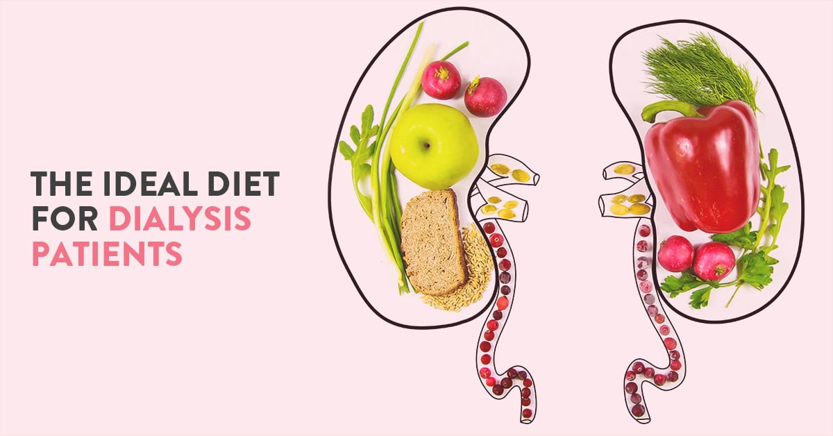
Notice how models and gym-goers emphasise on eating a well-balanced diet. One week of a salad-based diet can prepare them to feature on a magazine cover. What you eat literally acts as fuel for your body. A highly nutritious diet can do wonders for a healthy person. For a chronically ill person, it can alter the course of life. People living with chronic kidney diseases would understand the importance of consuming sufficient nutrients. However, planning a dialysis patient’s diet chart needs clinical help.
In this article, Dr Mohit Khirbat, a leading nephrologist at the CK Birla Hospital will help you design the best diet plan for dialysis patients.
Table of Contents
To understand the importance of diet for patients on dialysis, let us rewind some school-time biology. Dialysis is offered to patients suffering from chronic kidney diseases.
In human anatomy, kidneys are bean-shaped organs responsible for filtering waste generated by our body. However, sometimes the functioning of the kidneys can be disrupted due to various reasons. As the kidneys fail to function properly, the waste can get buildup and cause harm to the body, in general.
In such situations, healthcare providers offer dialysis to keep the body functioning. A dialysis machine helps to maintain the balance by managing the kidney’s functions. Dialysis treatment helps to remove extra waste, salt and water from the body.
However, even during dialysis some waste can fluid can buildup. A prolonged collection of these substances in the body can lead to heart and bone problems.
Dialysis patient diet comes in place when the patient is willing to benefit entirely from the treatment and avoid additional health problems. It is important to properly track and consume only sufficient nutrients. A dialysis patient is required to cautiously avoid specific food items as well to prevent any health complications.
It is important to understand that dialysis cannot replace the functioning of a kidney. Dialysis is a treatment that only offers support to patients with end-stage kidney diseases. Therefore, a conscious choice of food should be made to maintain health.
There is a range of foods good for a kidney dialysis patient. However, in a dialysis patient diet chart, the focus should be kept on four key areas:
Potassium is an essential mineral which is considered highly beneficial for dialysis patients. It is important for the healthy functioning of nerves and muscles including the heart.
An increased amount of potassium in the body can lead to heart problems such as irregular heartbeats and even cardiac arrest. On the other hand, a low potassium intake can cause muscle cramps and difficulty in breathing. The key is moderation.
Dialysis patients are required to monitor their potassium intake and restrict it to a moderate level.
Most fruits and vegetables are generally high in potassium. You can remove some amount of potassium from vegetables by the process of leaching. Leaching means removing potassium amount from raw vegetables by soaking them in the water at least two hours before cooking.
Other food products with a high amount of potassium are nuts, seeds, coconuts, wholegrain cereals, brown sugar and more.
Sodium is a necessary electrolyte for body functions that helps in the regulation of blood pressure. Sodium also helps in regulating acid and base balance in the body fluids.
Despite being necessary, an increased level of sodium can lead to multiple health problems in a kidney dialysis patient. These include hypertension, edema and shortness of breath among others.
Therefore, it is mandatory for dialysis patients to restrict their sodium intake. Sodium is most commonly found in salt. Minimising the levels of salt in your meals can be really helpful. Other items such as processed foods are some foods for a dialysis patient to avoid.
The energy that our body produces is stored with the help of phosphorus. It is an essential mineral required by our body to utilise other vitamins and minerals. Additionally, the strength of our bones and teeth are maintained by phosphorus and calcium.
Products rich in phosphorus are foods for a dialysis patient to avoid. It is because dialysis cannot manage the required level of this mineral in our body. A high level, on the other hand, can cause various complications such as renal bone disease, skin itching and more.
A patient who receives kidney dialysis needs to establish a strong control on his/her fluid intake. It is because fluid removal is an integral function of the kidneys. When the kidneys do not function properly, the excess water can get collected and move to the body cells. An excess buildup of fluids in cells can cause swelling of the feet and hands, commonly known as edema. More so, the extra volume of fluids can enter tissues present in the lungs and disrupt their functioning leading to breathlessness.
In such cases, a patient is only dependent on dialysis for the removal of fluids from the body. The best method to avoid the above-given health complications is to limit the intake of fluids.
Furthermore, even dialysis can remove a certain quantity of fluids, therefore a restriction on the consumption of fluids at the first hand is the best preventive measure.
Tips to limit your fluid intake –

Note: The requirements of each dialysis patient is different. The above-given information can help you custom-make your diet chart. You should ensure to restrict sodium and phosphorus intake while moderately managing the quantity of potassium. Sometimes food items have low sodium but are rich in calories. It is, thus, important to carefully design a plan that does not overwhelm you with one nutrient and drive you of another. You can discuss these tips with your healthcare provider to prepare a personalised dietary plan.
Dialysis diet plan becomes significant from the day you start taking treatment. The treatment of kidney disease is key for managing symptoms and removing waste from your body. It is up to you to make a conscious choice and live a healthy life during dialysis sessions. Dietary intake along with the following medications are vital components for the success of the treatment.
To learn more about kidney disease or plan your dialysis patient’s diet chart, you can book an appointment with Dr Mohit Khirbat, a leading nephrologist at the CK Birla Hospital.
Also, read: Common signs of Kidney Stones & steps to preventing Kidney Stones

Picture this: Your school football team has made it to the finales. You have to play in the big game tomorrow but you accidentally fall and sprain your ankle. Now before you stress out in such a situation, let us tell you some expert-approved ways on how to heal a sprained ankle as soon as possible.
A sprained ankle is a common occurrence among athletes and people who routinely exercise. It is also considerably easy to prevent common foot and ankle injuries.
In this article, Dr Anuj Chawla, best foot and ankle specialist in Gurgaon discusses some top tips for the prevention of ankle injuries and how to manage them.
Table of Contents
A sprained ankle happens when you hurt the ligaments in your ankles. Ligaments are flexible tissues that hold two adjacent bones together. You can sprain your ankle when you are rolling or twist it by mistake.
Common symptoms of a sprained ankle include:
Athletes would understand how recurring sprained ankles are. However, this injury can also be healed comfortably. The following remedies can be applied at home if a person suffers from an ankle sprain.
Tips on how to heal a sprained ankle-
The R.I.C.E approach is very popular and beneficial for a swift recovery. It is a traditional tried and tested healing method. The acronym R.I.C.E. stands for – Rest, Ice, Compression and Elevation, respectively.
Rest: If you suffer from any injury at any body part, the primary step is to give proper rest to the affected area. You should not engage in any activity that could worsen the swelling or pain.
Ice: Applying ice to the sprain has also been proved favourable in most cases. You should place an ice pack to the ankle to reduce swelling and pain. If an ice pack is not available, you can simply wrap some ice cubes around in a towel and use it.
Compression: Doctors recommend compressing the sprained ankle by using an elastic or crepe bandage. This helps in reducing the swelling and alleviates pain. It is also helpful in maintaining joint stability. While the bandage should be wrapped around firmly, you should not bind it too tight. If the bandage is excessively tight, it can obstruct blood circulation and cause numbness.
Elevation: The last step of the R.I.C.E. is to elevate the sprained ankle. Elevating the ankle helps to avoid the buildup of fluid in the joints. It is suggested that you should support the ankle by placing a pillow under it while sleeping. You should keep the ankle at an elevated level from the heart to help reduce swelling.
In severe ankle sprains which limit the ability of the person to walk, some form of protection is helpful to prevent worsening of the injury. Protection and immobilization can be in the form of a cast, pneumatic walker boots or ankle brace. In fact, the acronym for healing acute ankle sprain has been changed by many from RICE to PRICE so as to include protection.
Non Steroidal Anti Inflammatory Drugs (NSAIDS) like ibuprofen and Voveran (diclofenac) have been proven to be of significant benefit in early healing These medicines work by reducing the pain and swelling and hence hastening the recovery.
Exercises and rehabilitation play a paramount role in preventing stiffness, early recovery and prevention of recurrence in future. Physical therapy after an ankle sprain should be started after a brief period of rest of 5-7 days or after the plaster is removed. The rehabilitation for acute ankle sprain can be divided into 3 phases:
You can also go to a verified healthcare provider and choose for lightweight massage. You may also do a gentle massage at home yourself.
Besides effective management, ankle sprains can also be prevented. Since such injuries are frequent events in sports. Athletes need to learn about the prevention of ankle injuries.
Here are some useful tips to prevent foot and ankle injuries:
If your pain does not alleviate after implementing these tips, you should instantly seek medical support. You should consult your healthcare provider if:
A sprained ankle usually heals on its own without clinical intervention. The above-given tips can help you to encourage the healing process for a quicker recovery.
To learn more about muscle and joint injuries, consult Dr Anuj Chawla, best foot and ankle specialist at the CK Birla Hospital.
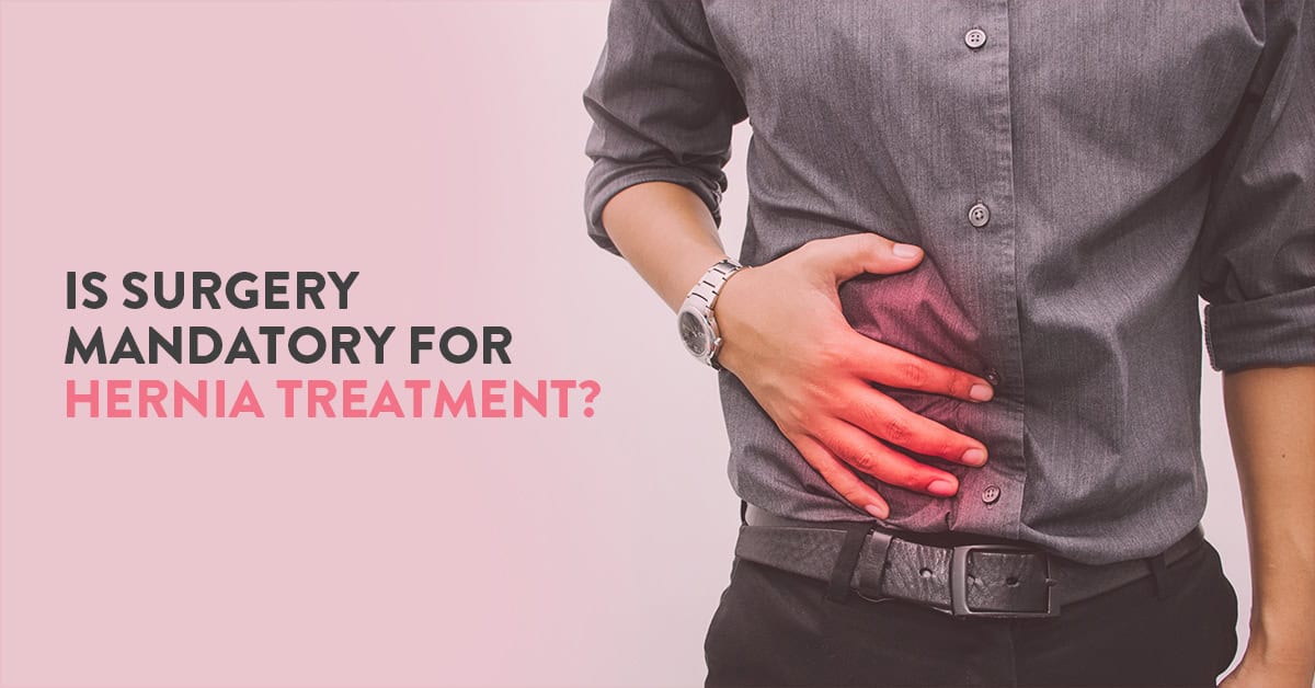
The incidence of hernia in India is very high. Men, in comparison to women, are more susceptible to developing hernias. Although this ailment can occur in people across ages, a hernia is most common among the elderly. In this article, Hernia surgeons in the CK Birla Hospital, explores what is a hernia about and when a hernia surgery required.
Table of Contents
A hernia is a condition in which a body organ pushes through weak muscles holding it together. A hernia can occur anywhere between the chest and the groin. It appears in the form of an abnormal bulge.
Apart from dull recurrent pain and discomfort, hernia mostly does not cause severe symptoms. You can expect to notice a lump or swelling that highlights this condition.
The hernia is of different types depending upon its location. Some major forms of hernia include inguinal, femoral, umbilical, hiatus and incisional among others. Inguinal hernias are the most common type.
There is no single cause attributed to the development of a hernia. Weak muscles that may be present since birth is one reason behind this condition. Excessive and repeated strain on abdomen and groin can also cause hernias. Simple health concerns such as physical exertion, coughing, obesity, constipation etc can unknowingly cause much abdominal strain.
Read: All you need to know about inguinal hernia | Symptoms and treatment
Accurate investigation of the ailment is necessary before hernia treatment. The diagnosis of hernia is fairly simple. At most times, the lump (hernia) is visible and even felt. Your healthcare provider will assess the bulge and ask about your symptoms.
Sometimes, your doctor may also indicate screening tests such as ultrasound and CT scans to visualise the state of the hernia.
Hernia treatment is varied for each patient. Several factors help your healthcare provider decide which treatment protocol to follow. Some of these factors include:
At most times, surgery for a hernia is the relevant method for improving your health.
There are majorly two treatment types – watchful waiting and hernia surgery. If your symptoms are mild and do not interrupt your life, your healthcare provider may suggest active surveillance as hernia treatment. You will be frequently examined for changes in symptoms. Your doctor will provide medicinal support to relieve pain and discomfort.
A hernia repair surgery is when your general surgeon cuts open the area of the bulge and push the muscles back into their original place. Hernia surgery is an effective remedy and provides complete relief for an extended period.
Hernia surgery is a safe procedure when performed by a qualified surgeon. It does not have severe side effects and has high success rates.
Call +911244882200 to know the hernia operation cost in Gurgaon.
Hernia repair surgery is done in two ways – traditional surgery and advanced laparoscopic hernia surgery.
Laparoscopic hernia surgery is a minimally invasive procedure and outscores the benefits of open hernia surgery. The patient experiences lesser pain during keyhole surgery. The amount of blood loss and scarring is also minimal.
Also, read: How safe is Laparoscopic surgery?

Surgery for a hernia is a safe and common procedure. It is a low-risk method for a permanent solution for a hernia. However, both open and laparoscopic surgery for hernia has certain side-effects.
Common risks associated with a hernia repair surgery are infection, pain, headache, nausea, bladder injury, nerve damage and blood clots.
Sometimes, a patient may experience recurrence of the hernia. However, disease recurrence is extremely rare post-surgery.
The time taken to recovery from hernia surgery depends on the type of hernia, patient’s overall health and type of surgery. Most patients can return home the same day.
The estimated time for recovery from open hernia surgery is at around 3 weeks. You should, however, avoid strenuous physical activities for up to 6 weeks. The recovery from laparoscopy is much less than open surgery.
You should follow your surgeon’s guidelines for proper postoperative care:
In case of any complications, consult your healthcare provider instantly.
Hernia affects every person uniquely. If you have severe symptoms, surgery may be the ideal fix for your condition. An untreated hernia can cause several complications as it grows.
To learn more about hernia surgery, you can book an appointment with best hernia surgeon at the CK Birla Hospital. You can also call +911244882200 to know the Hernia operation cost in Gurgaon.
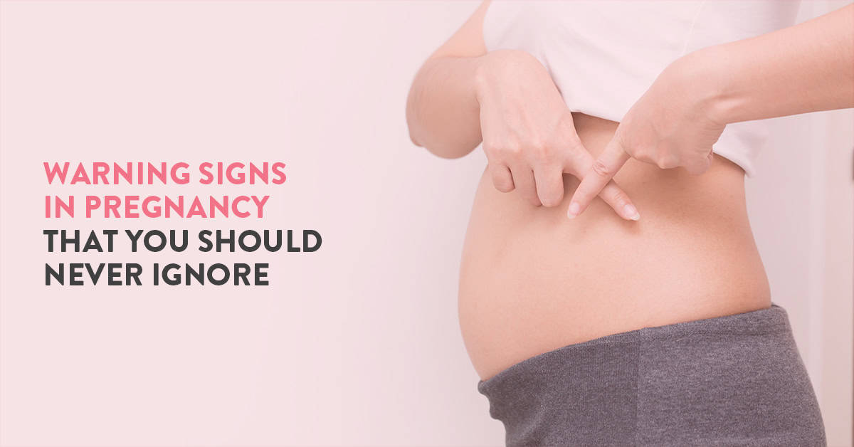
Congratulations on your pregnancy! You can now prepare to go through a rollercoaster of emotional and physical changes during the next 9 months. While pregnancy is mostly a joyous life event, it is also a time that calls for caution. You need to stay informed and practice watchfulness at every step. It is, therefore, important to learn about the warning signs of pregnancy.
In this article, Dr Astha Dayal, one of the best obstetricians in Gurgaon explains the most common warning signs during pregnancy.
You can expect to feel the effects of pregnancy as growing a baby inside your belly is no easy task. However, sometimes, a woman may experience bad pregnancy symptoms. These symptoms should not be mistaken for ordinary body changes.
You should proactively monitor your health and stay alert of the following warning signs during pregnancy:
There are diverse reasons that cause bleeding during pregnancy. During the first trimester, vaginal bleeding could be a sign of ectopic pregnancy. An ectopic pregnancy is when the embryo is implanted outside the uterus. Ectopic pregnancies can lead to complications for the mother and child.
Bleeding during pregnancy, in the second trimester, may indicate a loss of pregnancy or miscarriage.
While bleeding during the final trimester is a sign of placental abruption. At any stage of gestation, bleeding should be considered a warning sign. You should consult your gynaecologist even if you experience spotting and not heavy blood flow.
Severe back pain should not be taken lightly at any stage during pregnancy. The several changes in your body can lead to some level of discomfort. Nonetheless, persistent back pain during pregnancy requires clinical care.
Unlike popular opinion, fainting during pregnancy is not common. You can expect to feel a certain amount of light-headedness. However, fainting or feeling extremely dizzy in pregnancy is not a healthy sign.
It is highly common to feel nauseated and vomit during the first trimester of pregnancy. These symptoms are popularly called ‘morning sickness’.
However, if these symptoms persist for a longer time, it could be a cause of concern. This warning of pregnancy can also lead to a condition called hyperemesis gravidarum. Excessive nausea and vomiting could dehydrate your body and harm fetal health.
Your hormones may be a reason for frequent headaches and abdominal pain during pregnancy. However, if your aches are persistent and unbearable, you may need to see your gynaecologist.
Consistent pain is among the bad pregnancy symptoms. It may indicate high blood pressure and lead to the development of preeclampsia (a fatal pregnancy complication).
A sudden change in vision or blurry vision is one of the top pregnancy symptoms you should never ignore. This warning sign is an indicator of preeclampsia or gestational hypertension.
If you feel a sense of pain or burning during urination, you might have contracted a urinary tract infection (UTI). If not treated, this warning sign can lead to other serious infections or even preterm labour.
One of the most serious pregnancy symptoms you should never ignore is itching all over the body. It could be a sign of cholestasis during pregnancy.
Cholestasis is a liver condition. In this illness, the production of bile juice from your gall bladder is either stopped or disrupted. Cholestasis does not form any kind of rash.
Skin dryness and itching are not unusual for pregnant women. Your skin stretches to accommodate your baby and can become dry over time. Nonetheless, if the itching is extreme and beyond control, you should immediately consult your healthcare provider.
Early contractions in the uterus could be a sign of preterm labour. Preterm labour is the one that starts before 37 weeks of pregnancy. Preterm labour can cause multiple birth complications.
Sometimes, a pregnant woman may confuse these contractions as false labour. You should ask your OB-GYN to explain the difference between true and false labour.
Through the 9 months of gestation, you will become accustomed to the different types of movements made by your baby. You can begin to feel these movements between 18 and 25 weeks of pregnancy.
Your baby will turn, kick and flutter. If you feel a decline in this activity, you should meet your healthcare provider. It is essential to keep a track of these movements over time.
You can lie down and notice how long it takes for your baby to kick, roll or flutter 10 times. You can eat a light snack and lie down again if you do not notice any movement for over an hour.
If you feel a rush of fluid flowing down your vagina before 37 weeks, you should consider it a warning sign during pregnancy. This phenomenon indicates an early rupture of membranes.
Undoubtedly, you will gain weight during pregnancy, However, the weight gain is associated with swelling in legs and hands could be a sign of preeclampsia.
Some of the above-given symptoms are misinterpreted for normal events during pregnancy. This is why it is significant to educate yourself and your partner about the bad pregnancy symptoms.
You should not panic if you experience any of these atypical symptoms. Simply contact your OB-GYN and seek clinical intervention. Your healthcare provider will assess your situation and offer the relevant treatment.
An expecting mother should actively monitor her health. You can maintain a notebook to highlight any warning signs of pregnancy. You should also have a detailed discussion with your gynaecologist on what are the warning signs of pregnancy.
If you wish to learn more about maternal health during pregnancy, you can book an appointment with Dr Astha Dayal, obstetrician and gynaecologist at the CK Birla Hospital.
Also, read: Tips to ensure a healthy pregnancy

