Trust img


Filter :

Blogs
बंद नाक का कारण, लक्षण और इलाज | Nose block in Hindi
Dec 8, 2022|Dr Anish Gupta

बंद नाक का कारण, लक्षण और इलाज | Nose block in Hindi

नाक बंद होने की समस्या सर्दी और गर्मी दोनों ही मौसम हो सकती है। इसका कारण सामान्य एलर्जी से लेकर साइनस में इंफ्केशन या नाक की हड्डी टूटना आदि हो सकते हैं। नाक बंद यानी जाम होने के कारण आपको अनेक परेशानियां जैसे की नाक में सूजन, गले में खराश सांस लेने में तकलीफ, सर्दी, खांसी और बुखार आदि का सामना करना पड़ सकता है।

बंद नाक के कारण

नाक बंद यानी जाम होने के कई कारण हो सकते हैं जिसमें एलर्जी या सामान्य संक्रमण के कारण नाक के उत्तक (टिशू) में सूजन आना या द्रव भर जाना शामिल है। अधिकतर मामलों में यह गंभीर स्वास्थ्य समस्याओं के कारण नहीं होता है। हालाँकि, यह देखा गया है कि ज्यादातर मामलों में साइनस क्षेत्र में इंफेक्शन इसका मुख्य कारण होता है।

इन सबके अलावा, बंद नाक के कारणों में निम्न शामिल हो सकते हैं:

  • इंफेक्शन या एलर्जी होना
  • पराग ज्वर से ग्रसित होना
  • नाक की हड्डी टेढ़ी होना
  • नेजल पॉलिप्स की शिकायत होना
  • रसायन पदार्थों के संपर्क में आना
  • क्रोनिक साइनस की शिकायत होना
  • वातावरण में उपस्थित कणों के संर्पक में आना

साथ ही, कभी-कभी गर्भवती महिलाओं में भी बंद नाक की शिकायत हो सकती है। गर्भवती महिला के शरीर में हार्मोनल परिवर्तन होने पर रक्त का प्रवाह तेज हो जाता है जिसके कारण नाक बंद यानी जाम हो सकता है।

बंद नाक के लक्षण

नाक बंद होने पर आप खुद में नाक जाम होने के साथ-साथ दूसरे भी लक्षणों को अनुभव कर सकते हैं जैसे कि:

  • खांसी होना
  • गले में खराश होना
  • नाक में सूजन होना
  • सांस लेने में तकलीफ होना
  • कभी-कभी नाक से पानी आना
  • सांस फूलने जैसा महसूस करना

इंफेक्शन यानी संक्रमण के कारण नाक बंद होने पर आप निम्न लक्षणों को अनुभव कर सकते हैं:

  • जुखाम होना
  • बुखार होना
  • सर में दर्द होना
  • दांत में दर्द होना
  • सांस से बदबू आना
  • थकान महसूस करना
  • कमजोरी की शिकायत होना

हालाँकि, ये लक्षण सामान्य कारणों से नाक बंद होने के कारण भी हो सकते हैं। डॉक्टर लक्षणों के आधार पर जांच की मदद से नाक बंद होने के सटीक कारण की पुष्टि कर सकते हैं।

बंद नाक का इलाज

बंद नाक का इलाज उसके कारण और गंभीरता पर निर्भर करता है। इस समस्या का उपचार करने के लिए डॉक्टर कुछ विशेष टैबलेट और सीरप निर्धारित कर सकते हैं। हालाँकि, नाक बंद होने का कारण संक्रमण, नाक की हड्डी टूटने आदि पर डॉक्टर सर्जरी या दूसरे प्रकार के उपचार की सहायता ले सकते हैं।

निष्कर्ष

धूल, गर्मी, सर्दी या बदबू के प्रति एलर्जी, साइनस में संक्रमण या नाक में सूजन और दूसरे अनेक कारणों से नाक बंद होने की शिकायत हो सकती है। अधिकतर मामलों इसके कारण सामान्य होते हैं। यह हर मौसम में हर उम्र के व्यक्ति को प्रभावित कर सकता है।

बंद नाक का कारण अगर कोई सामान्य समस्या जैसे की धूल, बदबू या ठंड के प्रति एलर्जी है तो कुछ ख़ास घरेलू नुस्खों से इसे दूर किया जा सकता है। दूसरे मामलों में दवाओं की आवश्यकता होती है। अगर बंद नाक का कारण कोई गंभीर समस्या है तो ऐसे में डॉक्टर सर्जरी या दूसरे माध्यम का चयन करते हैं।

अगर आप भी बंद नाक से परेशान हैं तो तुरंत नाक के विशेषज्ञ डॉक्टर से परामर्श करना चाहिए। समय पर उचित उपचार और देखभाल से इस समस्या को आसानी से दूर किया जा सकता है।

How to deal with Rotator Cuff Tear?
Dec 1, 2022|Dr. Praveen Tittal

How to deal with Rotator Cuff Tear?

Our body is divided into different muscle groups for different bodily functions. The rotator cuff is one such group of muscles and tendons, surrounding the shoulder joint. It is responsible for keeping the head of your upper arm bone within the socket of your shoulder. A rotator cuff injury causes a dull ache in the shoulder. 

This is a group of four muscles and tendons. Their main function is to help you do heavy lifting and rotate your arms. So if there is a rip in the muscles it is called a rotator cuff tear. These rotator cuff tears are of three types.

  • Partial tear: This is when one of the fours muscles of the rotator cuff is frayed or damaged. 
  • Complete tear: This one is where the tendon is pulled off the bone.
  • Degenerative tear: This happens over time when the muscle wears off. 

Rotator cuff injuries like these are quite common, especially in sports involving a lot of arm action, like baseball or tennis. It can also happen to people who have jobs that involve a lot of arm activity like cleaning windows. Over time with normal wear and tear also this can happen, especially if the same movement is repeated over and over or due to the impact of a fall. Let us understand the causes better. 

What are the causes of Rotator cuff tear?

These injuries are very common around us and once they happen the pain can bother you, especially at night. Unless treated properly, with every future hand action trauma it will be triggered again and the injury will then tend to worsen over time. These injuries are mainly caused by 

  • progressive wear and tear of the tendon in the muscle set 
  • repeated overhead activity of the arm
  • prolonged sessions of heavy lifting 
  • accidents or sudden fall on the respective muscle group

Based on the above causes here are the people who have the highest risk of having a rotator cuff tear: 

  • People above the age of 60 years. Aging will make your muscles weak. A sudden heavy lifting can trigger the injury too. 
  • Lack of blood supply or poor circulation can also cause this problem. Due to lack of blood small tears might appear over time which can be hard to repair. 
  • Bone spurs or overgrowth of bone in the shoulder can happen with age, tearing the cuff tissues. 
  • Occupations demanding repeated overhead arm action, for example: house painting, carpentry, etc. 
  • Sports persons in games like tennis, rowing, cricket, weight-lifting and baseball. The tear can happen due to stress on the muscle group.  
  • Those with a family history of rotator cuff tears are definitely more likely to get it. 

What are the symptoms of Rotator cuff tear? 

The most tell tale sign of a rotator cuff tear is a dull pain in the area of the muscles and tendon tissue. But it will also be accompanied with other symptoms. To summarise, these are: 

  • Disturbed sleep
  • Difficulty to reach your hand to your back
  • Difficulty in combing your hair
  • Difficulty wearing clothes by raising your arm 
  • Overall weakness of the arm 
  • Dull persistent pain deep in the shoulder

While some injuries don’t come with pain, if you are having discomfort in your shoulders, it is advisable to reach out to a physician and get it checked. If ignored, these problems will keep persisting leading to permanent loss of motion of the arm, frozen shoulder, arthritis or persistent weakness.  

How is a rotator cuff tear diagnosed?

Once you reach out to a doctor or sports injury specialist, they will feel the region and affected parts. Test your arms mobility and strength, as to whether you can lift your arm or not. Based on the physical assessment the doctor will prescribe one or more of these tests to form a clear picture:

  • X-ray of the arm: A muscle tear is less likely to show in an x-ray, but this will help rule out any bone growth or arthritis. 
  • Ultrasound: This will help understand the level of tissue damage or muscle damage that has happened. Comparing between the healthy and injured shoulder will help gauge the extent of damage. 
  • MRI: This will provide a more detailed view of the shoulder structure to isolate the injury. 

Based on the findings from the tests above, a treatment plan will be drawn, where the orthopaedic will decide whether this needs physiotherapy or surgery. Traditional treatment includes rest, therapy and icing the region. But if the damage is severe, then surgery is the ultimate option.

Types of rotator cuff surgery

  • Arthroscopic tendon repair: Here the orthopaedic surgeon will insert a tiny camera called arthroscope and tools, via small incisions to help the torn tendon reattach to the bone
  • Open tendon repair: When an open tendon repair is a better option, your surgeon will operate through a larger incision to help the damaged tendon reattach to the bone
  • Tendon transfer: If the tendon damage is beyond repair, your orthopaedic surgeon will use a nearby tendon as a replacement. This replacement will be done without impairing any other muscle group
  • Shoulder replacement: Massive tears will ask for a shoulder replacement surgery by replacing the damaged joint with an artificial one through reverse shoulder arthroplasty

Post surgery, you’ll keep your shoulder on a sling to assist healing. Your surgeon will also advise the following: 

  • Several times a day take off the sling and move your arm to get better blood flow. 
  • For pain and swelling use an ice pack for 20 mins.
  • Avoid lifting your arm unless the orthopaedic gives a green signal

How to prevent shoulder muscle tear?

To look after your muscles you must exercise. But this does not mean rigorous exerting exercise. By this we mean exercises that improve your blood circulation to make sure blood and oxygen reaches each muscle group. Good circulation is the best form of healing that will repair the tissues that are damaged over time. 

Sport physios all over recommend focusing on the front muscles of the chest, upper arm,and your shoulder, including the back. Strengthening these muscle groups will help even out the stress on your shoulder muscles that leads to this injury in the first place. 

In conclusion, rotator cuff injuries are common to sports persons, but can happen to anyone. Your motor mechanic can get it, and if you suddenly fell down and your shoulder broke your fall, then even you can get it. The best way out is to not panic but immediately seek medical help. The quicker an injury is attended to, the faster is the route to recovery. 

At the CK Birla Hospital, we have a special set of orthopaedic surgeons, dedicated to treat sports injuries like the Rotator Cuff Tear and provide you treatment or surgery that will involve minimal stay but complete recovery in the quickest possible time. So that you can go and play your favourite sport again. If you have a pain in your shoulder and want an expert opinion on it, then do walk in to any of our hospitals or book an appointment .

How does COPD happen?
Nov 30, 2022|Dr. Kuldeep Grover

How does COPD happen?

What is chronic obstructive pulmonary disease?

Chronic obstructive pulmonary disease, popularly called COPD, is a chronic lung disease that obstructs the airflow in our lungs. This is by far the most complex respiratory disorder and so far without a cure. The disease can surface with breathing problems, coughing fits, wheezing and mucus (sputum) production.

The coughing in this case is usually that of a smoker’s cough. Therefore smoking is one of the biggest contributors to the rise in COPD cases followed by long-term exposure to particulate matter, and lung irritants. That’s not all, those who are diagnosed with COPD have a high risk of developing heart disease or lung cancer. Truly misery loves company when it comes to COPD.

COPD is a combination of two incremental conditions, Emphysema and Chronic Bronchitis. Both these conditions must be present to lead the situation past control in the form of COPD. These two conditions mostly occur together, however, they vary in severity for different COPD patients. Consider this as the villain’s sidekicks.

  • Chronic Bronchitis: When the inner lining of the bronchial tubes is inflamed it leads to this type of bronchitis. These tubes are responsible for transporting fresh air to the lungs and dispensing stale air via tiny air sacs or alveoli. It gives a daily coughing fit with ample mucus (sputum) production. Over time the air sacs have a tendency to develop pus. 
  • Emphysema: In this case, the alveoli of the lungs are destroyed as a result of near-fatal exposure to cigarette smoke, particulate matter and other irritants. 

COPD is a progressive disease. The conditions seldom improve and tend to get worse over time. Having said that COPD is treatable. With proper management, most patients can control the symptoms and improve their quality of life while living with the disease.

Is COPD different from Asthma?

It has been observed that childhood asthma increases your chances of having COPD in adult life. Many children tend to outgrow their asthma, however, their lungs don’t mature well and work as they should in some cases. This is noticed in children who had persistent childhood asthma, i.e., children who have breathing trouble almost every day. This directly translates to lower lung capacity and restricted lung growth in their youth.

Asthma and COPD, both block your airways and have common symptoms. But the major difference is that in the case of asthma you get tightness in your chest and persistent wheezing problems. Whereas, COPD symptoms are more constant, and include coughing fits which bring up a lot of phlegm. But in case of flare-ups, asthma can subside with medication, but COPD will not. Both are severe respiratory disorders, but COPD is worse in terms of recovery. 

What causes COPD? 

COPD happens due to blockage in the airways. What causes these blocks is the actual reason why we develop the problem. In the case of a healthy adult, air goes down the trachea into the lungs via two large tube-like structures called bronchi. These tubes branch out into smaller tubes, ending in tiny air sacs called alveoli.

The alveoli have thin walls full of capillaries. The capillaries carry the oxygen you inhale into the bloodstream. Similarly, carbon dioxide is exhaled in the reverse flow. To ensure this oxygen and carbon dioxide exchange is properly done, there needs to be a natural elasticity in the tubes. But with COPD they over expand and the air gets trapped in the lungs every time you exhale.  

While the main culprit that robs the elasticity of bronchial tubes is prolonged exposure to smoke, here are the major causes of COPD: 

  • Exposure to smoke: COPD occurs in people exposed to fumes in industrial set-ups, eg: exposure to blast furnaces or places where there is exposure to fumes and particulate matter. This can also be observed indoors in the form of burning fuel for cooking and heating in poorly ventilated homes.
  • Smoking tobacco: COPD is directly attributed to tobacco smoking. The smoke in this case is directly inhaled by the patients over a period of time. Once COPD conditions start developing, the more the patient smokes the more fatal the condition becomes. Chain smokers develop weaker lung function or unique complications which might even be misdiagnosed as COPD.
  • Congenital reasons: Weak lung function can be genetic as well. This means that the slightest concerns will trigger these conditions for the person. Even exposure to second-hand smoke will trigger these issues. So they need to pay more attention in their day-to-day life and use as much prevention as possible. Alpha-1-antitrypsin deficiency is one such congenital cause. This protein, when deficient, can cause lung and liver disorders.  

Signs and symptoms of COPD

Unlike other life-threatening diseases, COPD has very significant and easily identifiable symptoms. However, they surface only when the damage has gone too deep. Identifying these signs helps make it easier to create a customised treatment plan. These signs and symptoms are: 

  • Shortness of breath: During physical activities, if suddenly one notices that they are quickly running short of breath then it is a cause of concern. 
  • Wheezing: A typical symptom of COPD is wheezing. These are the noises that you make when breathing. Usually, high-pitched noisy breathing, especially when you exhale. 
  • Chest tightness: This is an associated feeling with shortness of breath. Chest tightening is an indication that the condition is flaring up and one must get checked for COPD. 
  • Mucus production: Chronic cough-producing bouts of mucus (sputum) with every coughing fit. It might be clear white, yellow or greenish in colour. 
  • Frequent respiratory infections:  When the inner layers of the lungs are compromised it keeps showing up in the form of respiratory infections. This is the body’s way of alerting that there might be bigger concerns underneath. 
  • Lack of energy: Due to the presence of the above symptoms, there is an onset of fatigue and lethargy. It takes extra effort to breathe comfortably, tiring the patient out for other operations. 
  • Sudden Unintended weight-loss:  If the disease has reached an advanced stage, the body will undergo sudden weight loss since the energy is fully diverted to fighting off the disease. 

COPD patients also experience exacerbations, during which the symptoms get worse and persist for several days. In the worst cases, patients’ lips and nail beds start turning blue. This is an emergency indication and one must be rushed to the hospital. 

Diagnosis and tests for COPD

Wondering when to see a doctor? Whenever any of the above symptoms start showing and show no signs of improvement with treatment or otherwise. The pulmonologist will suggest one or more of the following tests:

  • Lung (pulmonary) function test (PFT)
  • Spirometry test 
  • Treadmill test
  • Pulse oximetry test 
  • Chest X-ray 
  • CT Scan

Other than these normal pathological tests might be carried out, reports of which will help assess the severity of the condition. Based on the reports the COPD patient can be categorised into 4 stages: 

Stage I (Early) | Stage II (Moderate) | Stage III (Severe) | Stage IV (Very Severe)

Treatment & Management of COPD

COPD can be treated. Effective therapy can control symptoms, slow progression, manage complications and exacerbations, and overall improve your quality of life.

The first step is to quit smoking. The more oxygen enters your body the better your chances are of managing COPD. The next step is medication. Several kinds of medications are used to treat COPD apart from antibiotics, such as: 

  • Bronchodilators: Usually taken with inhalers they relax your airway muscles, and relieve coughing and shortness of breath. 
  • Inhaled steroids: These are effective in reducing airway inflammation and preventing exacerbations. Taken as an inhaler, these are best suited for people with frequent exacerbations.
  • Phosphodiesterase-4 inhibitors: This drug reduces inflammation in people with severe COPD and chronic bronchitis. 

Lung therapies, oxygen therapy and breathing exercises are some other ways in which people in different stages can work their way around COPD.

To summarise, your lungs depend on how healthy you keep them. With more oxygen and healthy practices, you can not only make sure the lungs are fully functional, but also deliver the much-needed oxygen effortlessly throughout your body. However, if you are prone to lung infections and can’t kick the habit of smoking, it is advisable to reach out to a professional pulmonologist, at the CK Birla Hospital and get yourself checked for COPD. Book an appointment now.

How to prevent Osteoperosis?
Nov 29, 2022|Dr Ashwani Maichand

How to prevent Osteoperosis?

What is Osteoporosis?

Every part of our body is made of cells and tissues. The bone tissue, like all other tissues, keeps decaying and regenerating to keep our bones strong. The moment this process slows down, new bone tissues can’t keep up with the old decaying tissues, leading to the bones becoming fragile. This happens in the case of men and women, however, post-menopausal women are more susceptible to getting this than any other person. Osteoporosis is also linked to vitamin D deficiency, which also increases the fragility of our bones. 

This disease is a silent assassin. The condition progresses internally without any symptoms or pain, and the only time it surfaces is when the bones undergo a fracture. But it can be prevented with timely treatment and diagnosis. But before we reach treatment options let us understand the causes and symptoms of osteoporosis.

What are the symptoms of Osteoporosis? 

Osteoporosis arrives silently and leaves colossal damage behind. In the initial stage, there are no distinguishable symptoms of bone or tissue loss. But over time when the bones become fragile  the following symptoms start surfacing: 

  • Persistent back pain due to a fractured/collapsed vertebra bone
  • A gradual decrease in the height of the person
  • Stooped body posture
  • Bones break very easily with the slightest nudge

The symptoms are very basic, but the impact is deeply rooted as bone tissue takes time to repair in normal circumstances, with a reduced regeneration time, it takes twice the time to heal and repair, adding a greater burden on the patients.

What causes osteoporosis? 

The insides of a healthy bone resemble a sponge and are called trabecular bone. An outer hard shell of bone covers the inner spongy layer forming what is called the cortical bone. During osteoporosis, these sponge holes inside grow in number & size making the insides weak. These bones have a bigger role to support the body, protecting vital organs, and storing calcium and other minerals. When the body requires calcium, it breaks down the bone tissue and rebuilds it. Supplying the calcium needed for the body’s skeleton to stay strong.

To understand the causes behind osteoporosis we need to identify the people who are at a greater risk of developing the problem.

  • Women who go through early menopause are at the highest risk.
  • People who have consumed corticosteroids for several months are also at high risk.
  • An existing family history of fractures (eg: hip fracture) can increase the risk of others in the family carrying the risk congenitally.
  • Those in their old age are also susceptible to this due to slow-down cell regeneration.

Now that we know the high-risk groups that can get the disease, our focus will move to the root causes that lead to osteoporosis.

  • Lower bone mass: Those who have smaller body frames can get osteoporosis. Osteoporosis translates to porous bones. This means that while the bone looks healthy and strong from the outside, it may be decaying from the inside. As we have discussed earlier, the bone tissue is under constant renewal. Old bone decays and new bone tissue replaces it to keep the bones strong. When we are young, this cell regeneration is fast. Doctors suggest that a full-grown adult’s bone mass keeps growing and attains its peak at the age of 30 years. 

Gradually with time, level of activity and age this mass keeps decaying faster than it is regenerated. Your likelihood of developing osteoporosis depends on the bone mass attained by your body in its youth. So the higher mass the lower the decay when you age. 

  • Low sexual hormone Levels: Osteoporosis can develop due to hormonal imbalance also. Lowered sex hormones weaken bones. Lowered oestrogen levels in a woman on the brink of menopause are a strong risk factor for developing osteoporosis. Women in general have a higher tendency to bone decay than men. Hence they must pay special attention after the age of 30. 

Men undergoing prostate cancer treatment and women undergoing breast or gynae cancer treatments have reduced testosterone and oestrogen levels which are bound to accelerate bone mass loss drastically.

  • High thyroid hormone level: Those suffering from thyroid problems also undergo drastic bone loss. This is usually seen in cases of hyperthyroidism or hypothyroidism with excessive hormone medication. Apart from this overactive adrenal or parathyroid gland secretions also bring about osteoporosis. 
  • Dietary problems: Multiple dietary irregularities can also lead to osteoporosis. 

– Deficiency of calcium and Vitamin D will invite the disease. Lower calcium intake gives a diminished bone density, increased risk of fractures and early bone mass loss. 

– Anorexia or bulimia like eating disorders can also lead to this. Severely restricting food intake weakens the bones.

– Steroids, especially corticosteroids when consumed over a prolonged period will lead to weaker bones. 

– Bariatric or gastrointestinal surgery will reduce the size of your stomach leaving less surface area to absorb nutrients like calcium. This happens in rare cases since these corrective surgeries are done to rectify other gastro concerns.

  1. Pre-existing comorbidities: Those with some pre-existing medical problems also attract the risk of having osteoporosis. Patients with celiac disease, IBS, Kidney or liver dysfunction, rheumatoid arthritis, cancer and myeloma ( Myeloma doctors )tend to develop fragile bones. 
  2. Other medical causes: Different medications may also lead to osteoporosis. These are mainly associated with problems like seizures, acid reflux, cancer medications, etc. 
  3. Poor lifestyle choices: Not all problems are due to bodily functions, some are man-made due to poor day-to-day choices. Such as:

– Sedentary lifestyle: Those who have to sit around all day gradually develop a lot of bone and vertebrae problems, increasing the risk of osteoporosis. More oxygen and circulation can prevent this. 

– Too much consumption of alcohol & tobacco consumption in any form will also make your bones porous. Anything more than 2 alcoholic drinks in a day is not desirable. 

How is osteoporosis diagnosed?

Once you reach out to an orthopaedic, they will suggest the following tests based on the severity of your conditions and complaints. 

Bone Density Scan: Since the problem lies in the internal decay of bones, the first step towards diagnosis is by running a bone density test. This is a painless test where low-intensity x-rays determine the mineral content in your bone. This is the most comprehensive test for the full body.

CT scan: Another internal imaging test that will give the physician a clearer picture of what is happening inside. 

Bone Ultrasound: This is an imaging test done on specific bones in the body. This will help us understand exactly how deep the problem is. 

The results from these imaging tests will help the orthopaedic form an opinion about the patient’s current condition and suggest a treatment route.

How to prevent osteoporosis?

While most of our height and bone growth are attributed to genetics, we do not have much control over bone mass. However, we can make some necessary lifestyle adjustments to ensure our body steers clear of the disease.

  • Quit smoking. This is a major success factor in maintaining stronger bones as smoking is known to slow down cell regeneration.
  • Watch where you walk. Since the disease is internal, we do not know we are in trouble until we fall and break something. If you have a family history of fractures, wear strong-grip shoes to prevent falls and avoid drinking alcohol. 
  • Have a calcium and Vitamin D-rich diet. We need at least 1,000 milligrams of calcium every day till the age of 50. So load up your diet with low-fat dairy, green leafy vegetables, omega-rich fish, soybean, and of course orange juice. 
  • Spend more time in the sun. Especially if you spend most of your time indoors. Early morning sun is rich in Vitamin D which helps absorb calcium faster in our bodies. 
  • Exercise. With different levels of intensity, aerobic exercises always help keep your bones strong. Improved circulation and weight management help keep your bones healthy longer and regular activity keeps them agile.

In conclusion, the older we get the more our bones decay. The best way out is a healthy and disciplined lifestyle in the long run. However, not all of us can do this which is why ailments like osteoporosis start dictating our life and lifestyle. There is a way out and relief is just one consultation away. Reach out to the best Orthopaedic Surgeons in Delhi NCR, who will not only compassionately treat your pain points but also ensure you get a better quality of life for as long as you live. To book an appointment, call +911244570112.

गायनेकोलॉजिकल कैंसर के प्रकार और लक्षण – Types of Gynecological Cancer in Hindi
Nov 29, 2022|Dr. Archana Pathak

गायनेकोलॉजिकल कैंसर के प्रकार और लक्षण – Types of Gynecological Cancer in Hindi

महिलाओं के प्रजनन अंगों में होने वाले कैंसर को गायनेकोलॉजिकल कैंसर कहते हैं। शोध के मुताबिक, दुनियाभर में कैंसर के कारण लाखों महिलाओं की मौत होती है, लेकिन अधिकतर मामलों में उनकी मौत का मुख्य कारण गायनेकोलॉजिकल कैंसर होता है।

कैंसर एक जनलेवा बीमारी है जिससे पीड़ित मरीजों की संख्या लगातार बढ़ रही है। कैंसर शरीर के किसी भी हिस्से में हो सकता है। जब शरीर की कोशिकाएं अनियंत्रित रूप से विभाजित और विकसित होने लगती हैं तो कैंसर की समस्या पैदा होती है। इस ब्लॉग में हम गायनेकोलॉजिकल कैंसर के प्रकार और लक्षण के बारे में विस्तार से बात करने वाले हैं।

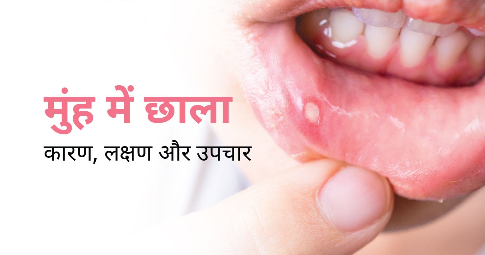
यह भी पढ़े: मुंह में छाला होने के कारण, लक्षण और उपचार | Mouth Ulcers in Hindi

गायनेकोलॉजिकल कैंसर के प्रकार – Types of Gynecological Cancer in Hindi

गायनेकोलॉजिकल कैंसर के मुख्यतः 6 प्रकार होते हैं:

  1. गर्भाशय कैंसर
  2. गर्भाशय ग्रीवा कैंसर
  3. वल्वर कैंसर
  4. सर्वाइकल कैंसर
  5. योनि का कैंसर
  6. गर्भकालीन ट्रोफोब्लास्टिक ट्यूमर

गायनेकोलॉजिकल कैंसर के कारण – Causes of Gynecological Cancer in Hindi

गायनेकोलॉजिकल कैंसर कई कारणों से होते हैं जिसमें मुख्य रूप से खान-पान, जीवनशैली और शारीरिक स्वास्थ्य से जुडी स्थितियां आदि शामिल हैं। निम्न कारकों से गायनेकोलॉजिकल कैंसर का खतरा बढ़ता है: 

  • कम उम्र में पीरियड्स शुरू होना (12 साल से कम उम्र होना)
  • डायबिटीज से ग्रसित होना
  • धूम्रपान करना
  • मोटापा से ग्रसित होना
  • प्रतिरक्षा प्रणाली कमजोर होना
  • ब्रेस्ट (स्तन) कैंसर होना
  • ह्यूमन पेपिलोमावायरस संक्रमण होना
  • परिवार में गायनेकोलॉजिकल कैंसर का इतिहास होना
  • ओरल बर्थ कंट्रोल या सेक्सुअल दवाओं का अत्याधिक इस्तेमाल करना
  • फैट से भरपूर भोजन करना

इन सबके अलावा, दूसरे भी अनेक कारण हैं जो एक महिला में गायनेकोलॉजिकल कैंसर का खतरा बढ़ाते हैं।

गायनेकोलॉजिकल कैंसर के लक्षण – Symptoms of Gynecological Cancer in Hindi

  • गर्भाशय कैंसर के लक्षण

गर्भाशय कैंसर होने पर एक महिला खुद में अनेक लक्षणों को अनुभव कर सकती है। हालाँकि, इसके मुख्य लक्षणों में मेनोपॉज के बाद योनि से रक्तस्राव होना, पेट के निचले हिस्से में दर्द, पीरियड्स के बाद अत्यधिक रक्तस्राव और शारीरिक संबंध बनाते समय उसे दर्द महसूस होना आदि शामिल हैं।

  • गर्भाशय ग्रीवा कैंसर के लक्षण

इसे ओवेरियन कैंसर के नाम से भी जानते हैं। गर्भाशय ग्रीवा कैंसर के मुख्य लक्षणों में शामिल हैं भूख नहीं लगना, भोजन करने में परेशानी होना, अचानक वजन घटना, बार-बार पेशाब लगना, श्रोणि क्षेत्र में दर्द और बेचैनी होना एवं प्रभावित क्षेत्र में अत्यधिक सूजन होना आदि।

  • वल्वर कैंसर के लक्षण

यह अधिकतर मामलों में अधिक उम्र की महिलाओं को प्रभावित करता है। वल्वर कैंसर होने पर महिला खुद में कुछ लक्षणों में पेशाब करते समय दर्द और जलन होना, पीरियड के अलावा गंभीर रक्तस्राव होना, घाव या अल्सर होना और लाल, गुलाबी या सफ़ेद रंग के मस्से जैसी सतह आदि शामिल हैं।

  • सर्वाइकल कैंसर के लक्षण

इस कैंसर से पीड़ित महिला को खुद में कुछ लक्षण अनुभव हो सकते हैं जैसे कि योनि से असामान्य रक्तस्राव, योनि से सफेद बदबूदार पानी का स्राव होना, पेट के निचले हिस्से में दर्द होना, भूख कम लगना, वजन कम होना, कमजोरी होना, थकान महसूस करना, अनीमिया और बॉन फ्रैक्चर की शिकायत होना और पेशाब करने में दिक्कत होना आदि।

  • योनि का कैंसर के लक्षण

यह एक दुर्लभ प्रकार का कैंसर है जो ज्यादातर 50 से अधिक उम्र की महिलाओं में होता है। इस कैंसर के लक्षणों में शारीरिक संबंध बनाते समय और बाद में दर्द महसूस होना, असामान्य स्क्त्स्राव होना, मेनोपॉज के बाद रक्तस्राव होना, मल या मूत्र के साथ खून आना, बार-बार पेशाब लगना और कब्ज की शिकायत होना आदि।

  • गर्भकालीन ट्रोफोब्लास्टिक ट्यूमर के लक्षण

गर्भकालीन ट्रोफोब्लास्टिक ट्यूमर गर्भावस्था से जुड़ा एक तरह का ट्यूमर है। यह एक दुर्लभ प्रकार का गायनेकोलॉजिकल कैंसर है। इससे पीड़ित महिला को अनेक लक्षण अनुभव हो सकते हैं जैसे कि:

  • डिलीवरी या गर्भपात के बाद 6 सप्ताह से अधिक समय तक रक्तस्राव होना
  • गर्भाशय का आकार बढ़ना
  • श्रोणि में दबाव या दर्द होना
  • गंभीर रूप से मतली और उल्टी आना
  • ब्लड प्रेशर बढ़ना और सिर में दर्द होना

इन सबके अलावा, महिला को अनीमिया की शिकायत हो सकती है जिसके कारण उसे साँस लेने में दिक्कत होना, दिल की धड़कन तेज होना, चक्कर आना और थकान महसूस करना आदि की शिकायत भी हो सकती है।

गायनेकोलॉजिकल कैंसर का इलाज – Treatment For Gynecological Cancer in Hindi

गायनेकोलॉजिकल कैंसर का इलाज कई तरह से किया जाता है। इसका उपचार करने के लिए सर्जरी, कीमोथेरेपी, रेडिएशन थेरेपी, हार्मोन थेरेपी, इम्यूनोथेरेपी, स्टेम सेल ट्रांसप्लांट, टार्गेटेड थेरेपी और दवाएं आदि शामिल हैं। अधिकतर मामलों में गायनेकोलॉजिकल कैंसर का उपचार सर्जरी से किया जाता है।

हालाँकि, गायनेकोलॉजिकल कैंसर के प्रकार और गंभीरता, महिला की उम्र और समग्र स्वास्थ्य को ध्यान में रखते हुए कैंसर विशेषज्ञ उपचार के प्रकार का चयन करते हैं। कई बार विशेषज्ञ ऊपर दिए उपचारों में से एक या उससे अधिक का सहारा ले सकते हैं।

अगर आप खुद में गायनेकोलॉजिकल कैंसर के लक्षणों को अनुभव करती हैं या इस बीमारी से पीड़ित हैं तो हमारे कैंसर विशेषज्ञ के साथ अपॉइंटमेंट बुक करके उनसे विस्तृत परामर्श कर सकती हैं। अपॉइंटमेंट बुक करने के लिए आप इस पेज के ऊपर दिए गए Book Appointment Form का इस्तेमाल कर सकती हैं।

सर्वाइकल (गर्भाशय ग्रीवा) कैंसर क्या है – कारण, लक्षण, जांच और इलाज (Cervical Cancer in Hindi)
Nov 29, 2022|

सर्वाइकल (गर्भाशय ग्रीवा) कैंसर क्या है – कारण, लक्षण, जांच और इलाज (Cervical Cancer in Hindi)

सर्वाइकल (गर्भाशय ग्रीवा) कैंसर क्या है – Cervical Cancer in Hindi

जब शरीर की कोशिकाएं अनियंत्रित रूप से बढ़ने लगती हैं तो कैंसर का जन्म होता है। कैंसर का नाम हमेशा उस हिस्से के आधार पर तय किया जाता है जहाँ वह शुरू होता है – भले ही बाद में वह शरीर के दूसरे हिस्सों में फैलता हो। 

जब कैंसर गर्भाशय ग्रीवा में होता है तो उसे सर्वाइकल कैंसर या गर्भाशय ग्रीवा का कैंसर कहते हैं। गर्भाशय ग्रीवा को अंग्रेजी में सर्विक्स कहते हैं। यह योनि को गर्भाशय के ऊपरी भाग से जोड़ता है। 

दुनियाभर में महिलाओं में होने वाले कैंसर में सर्वाइकल कैंसर सबसे आम प्रकार का कैंसर है। शोध के मुताबिक, यह कैंसर मध्य जीवन में अधिक होता है। लगभग आधी महिलाएं जिनमें सर्वाइकल कैंसर का निदान किया गया है – उनकी उम्र 35-55 वर्ष के बीच है।

ये भी पढ़े: गर्भाशय कैंसर के कारण, लक्षण और उपचार (Uterine Cancer in Hindi)

सर्वाइकल (गर्भाशय ग्रीवा) कैंसर के प्रकार (Types of Cervical Cancer in Hindi)

जांच की मदद से सर्वाइकल कैंसर के प्रकार की पुष्टि करने में मदद मिलती है। कैंसर के प्रकार और गंभीरता के आधार पर डॉक्टर उपचार, उसकी तकनीक और पद्धति का चयन करते हैं। सर्वाइकल कैंसर के मुख्यत दो प्रकार होते हैं:

  • स्क्वॉमस सेल कैंसर
  • ग्रंथिकर्कटता या एडिनोकार्सिनोमा 

गर्भाशय ग्रीवा (सर्वाइकल) कैंसर के चरण (Stages of Cervical Cancer in Hindi)

टेस्ट के परिणाम के आधार पर यह तय किया जाता है कि सर्वाइकल कैंसर किस स्टेज में है। स्टेज से डॉक्टर को यह समझने में मदद मिलती हैं कि कैंसर किस हद तक फैला है। सर्वाइकल कैंसर के निम्न स्टेज हैं:

  • स्टेज 0 – इस दौरान, सर्विक्स में कैंसरयुक्त कोशिकाएं नहीं होती हैं, लेकिन कुछ जैविक परिवर्तन हो चुके होते हैं जिससे भविष्य में कैंसर होने का खतरा होता है। इसे कार्सिनोमा इन सीटू या सर्वाइकल इंट्राएपिथेलियल नियोप्लाजिया कहते हैं।
  • स्टेज 1 – इस स्थिति में कैंसर सर्विक्स में ही मौजूद होता है।
  • स्टेज 2 – कैंसर सर्विक्स के आसपास के उत्तकों में फ़ैल जाता है। लेकिन श्रोणि की लाइनिंग (परत) या योनि के निचले भाग तक नहीं पहुंचा होता है।
  • स्टेज 3 – कैंसर योनि के निचले हिस्से और या श्रोणि की लाइनिंग तक पहुँच चुका होता है।
  • स्टेज 4 – कैंसर आँतों, मूत्राशय या अन्य अंगों जैसे कि फेफड़ों तक पहुँच जाता है।

सर्वाइकल (गर्भाशय ग्रीवा) कैंसर के कारण (Causes of Cervical Cancer in Hindi)

जब कोशिकाएं असामान्य होकर अनियंत्रित रूप से विभाजित होने और बढ़ने लगती हैं तो कैंसर की समस्या पैदा होती है। सामान्य कोशिकाएं एक निर्धारित समय के बाद मर जाती हैं, लेकिन असामान्य कोशिकाएं मरती नहीं हैं और समय के साथ विभाजित होती रहती हैं।

जब असामान्य कोशिकाएं एक जगह एकत्रित होती हैं तो ट्यूमर बनता है। सर्विक्स में असामान्य कोशिकाओं के बनने की स्थिति को सर्वाइकल कैंसर कहते हैं। हालाँकि, कुछ ऐसे कारक हैं जो सर्वाइकल कैंसर का खतरा बढ़ाते हैं। इसमें शामिल हैं:

  • ह्यूमन पेपिलोमा वायरस (एचपीवी) से ग्रसित होना
  • असुरक्षित यौन संबंध बनाना
  • सिगरेट का सेवन करना
  • प्रतिरक्षा प्रणाली कमजोर होना
  • लंबे समय तक तनाव से ग्रस्त होना
  • बहुत छोटी उम्र में गर्भधारण करना
  • लंबे समय तक गर्भनिरोधक दवाओं का सेवन करना
  • बार-बार गर्भधारण करना (आमतौर पर तीन से ज्यादा बच्चों को जन्म देना)
  • यौन संचारित बीमारियां जैसे कि क्लैमाइडिया, सुजाक या उपदंश से पीड़ित होना

सर्वाइकल (गर्भाशय ग्रीवा) कैंसर के लक्षण (Symptoms of Cervical Cancer in Hindi)

आमतौर पर सर्वाइकल कैंसर की शुरुआत में इसके लक्षण दिखाई नहीं देते हैं। लेकिन जैसे-जैसे यह गंभीर रूप लेता है – लक्षण दिखाई देने शुरू हो जाते हैं। सर्वाइकल कैंसर के लक्षणों में निम्न शामिल हो सकते हैं:

  • योनि से असामान्य ब्लीडिंग होना
  • योनि से अनियमित ब्लीडिंग होना
  • संभोग के दौरान दर्द महसूस होना

इन सबके अलावा, सर्वाइकल कैंसर से पीड़ित महिला खुद में दूसरे भी लक्षणों को अनुभव कर सकती है। अगर आप खुद में नीचे दिए गए लक्षणों को अनुभव करती हैं तो आपको तुरंत विशेषज्ञ डॉक्टर से परामर्श करना चाहिए:

  • योनि से असामान्य ब्लीडिंग (खासकर दो नियमित पीरियड्स के बीच)
  • संभोग के बाद योनि से खून आना
  • संभोग के समय योनि में तेज दर्द होना
  • पेशाब के दौरान तेज दर्द अनुभव होना
  • पेल्विक दर्द जो पीरियड से संबंधित नहीं है
  • पेल्विक एक्ज़ाम या मेनोपॉज के बाद ब्लीडिंग होना
  • योनि से भारी या असामान्य रक्तस्राव होना (यह पानी जैसा या गाढ़ा हो सकता है और इससे तेज बदबू आती है)

हालाँकि, ये लक्षण स्वास्थ्य संबंधित दूसरी समस्याओं के कारण भी हो सकते हैं। समय पर उचित जांच और उपचार से सर्वाइकल कैंसर को दूर किया जा सकता है।

सर्वाइकल (गर्भाशय ग्रीवा) कैंसर की जांच कैसे करे (Diagnosis of Cervical Cancer in Hindi)

सर्वाइकल कैंसर के शुरुआती स्टेज में इसका निदान होने पर उपचार के सफल होने की संभावना अधिक होती है। इस बीमारी का निदान करने के लिए डॉक्टर आमतौर पर कुछ जांचों का सुझाव देते हैं।

एचपीवी डीएनए टेस्टिंग के दौरान, इस बात की पुष्टि की जाती है कि महिला किसी प्रकार के एचपीवी संक्रमण से ग्रसित तो नहीं है। इस दौरान, सर्विक्स की कोशिकाओं को जमा करके प्रयोगशाला में जाँच के लिए भेजा जाता है।

अगर कोई महिला खुद में सर्वाइकल कैंसर के लक्षणों को अनुभव करती है या पैप स्मीयर टेस्ट में असामान्य कोशिकाएं दिखती हैं तो अन्य जांच का सुझाव दिया जाता है जिनमें निम्न शामिल हो सकते हैं:

  • बायोप्सी
  • कॉल्पोस्कोपी
  • कोन बायोप्सी
  • एलएलईटीजेड
  • ब्लड टेस्ट
  • सीटी स्कैन
  • एमआरआई
  • पेल्विक अल्ट्रासाउंड
  • एनेस्थीसिया के प्रभाव में योनि और सर्विक्स की जाँच

गर्भाशय ग्रीवा (सर्वाइकल) कैंसर का इलाज (Treatment For Cervical Cancer in Hindi)

सर्वाइकल कैंसर का उपचार मुख्यतः चार तरह से किया जा सकता है जिसमें सर्जरी, रेडिएशन थेरेपी, कीमोथेरेपी और टार्गेटेड थेरेपी शामिल हैं। हालांकि, कुछ मामलों में कैंसर की गंभीरता को ध्यान में रखते हुए इन उपचारों में से दो या अधिक को एक साथ किया जा सकता है।

अक्सर पूछे जाने वाले प्रश्न

प्रश्न 1. सर्वाइकल कैंसर का सबसे प्रमुख कारण क्या है?

सर्वाइकल कैंसर के प्रमुख कारणों में ह्यूमन पेपिलोमा वायरस (एचपीवी), सिगरेट का सेवन, कमजोर प्रतिरक्षा प्रणाली, गंभीर तनाव, छोटी उम्र में गर्भधारण करना और यौन संचारित बीमारियां शामिल हैं। हालांकि, इसके अन्य कारण भी हो सकते हैं। 

गर्भाशय कैंसर के कारण, लक्षण और उपचार (Uterine Cancer in Hindi)
Nov 29, 2022|Dr Keerti Khetan

गर्भाशय कैंसर के कारण, लक्षण और उपचार (Uterine Cancer in Hindi)

गर्भाशय (बच्चेदानी) का कैंसर क्या है – Uterine Cancer in Hindi

महिलाओं के प्रजनन अंगों में कई तरह के कैंसर होते हैं, गर्भाशय का कैंसर भी उन्हीं में से एक है। इसे एंडोमेट्रियल कैंसर, बच्चेदानी में कैंसर या यूटेराइन कैंसर के नाम से भी जाना जाता है। जब गर्भाशय की आंतरिक परत में मौजूद कोशिकाओं में आनुवंशिक बदलाव आता है तो वे असामान्य रूप से विभाजित और विकसित होने लगती हैं।

कोशिकाओं के असामान्य रूप से विभाजन होने और बढ़ने के कारण गर्भाशय में ट्यूमर बनने लगता है। यह ट्यूमर आगे जाकर कैंसर में बदल जाता है। लोग अक्सर गर्भाशय के कैंसर और गर्भाशय ग्रीवा के कैंसर को एक समझ लेते हैं। हालाँकि, यह दोनों एक दूसरे से भिन्न यानी अलग प्रकार के कैंसर हैं। इस ब्लॉग में आगे हम गर्भाशय कैंसर के प्रकार, चरण, कारण, लक्षण और उपचार के बारे में विस्तार से जानने की कोशिश करेंगे।

ये भी पढ़े: सर्वाइकल (गर्भाशय ग्रीवा) कैंसर क्या है – कारण, लक्षण, जांच और इलाज (Cervical Cancer in Hindi)

गर्भाशय (बच्चेदानी) में कैंसर के प्रकार – Types of Uterine Cancer in Hindi

गर्भाशय कैंसर मुख्यत दो प्रकार के होते हैं जिन्हें यूटराइन सार्कोमा और एंडोमेट्रियल कार्सिनोमा के नाम से जानते हैं।

  • यूटराइन सार्कोमा (Uterine Sarcoma)

यह गर्भाशय की मांसपेशियों की परत यानी एंडोमेट्रियम या आसपास की उत्तकों में होने वाला कैंसर है।

  • एंडोमेट्रियल कार्सिनोमा (Endometrial Sarcoma)

यह गर्भाशय की भीतरी परत में होने वाला कैंसर है। गर्भाशय के लगभग सभी कैंसर इसी प्रकार के होते हैं।

गर्भाशय (बच्चेदानी) में कैंसर के चरण – Stages of Uterine Cancer in Hindi

गर्भाशय कैंसर को उसकी गंभीरता के आधार पर चार चरणों में बांटा गया है जिसमें निम्न शामिल हैं:

  • चरण 1: जब कैंसर केवल गर्भाशय में होता है तो उसे पहले चरण में रखा जाता है।
  • चरण 2: इस दौरान, कैंसर गर्भाशय और गर्भाशय ग्रीवा में फ़ैल चूका होता है।
  • चरण 3: इस चरण में कैंसर गर्भाशय के बाहर जैसे कि श्रोणि लिम्फ नॉड्स में फ़ैल चूका होता है, लेकिन मूत्राशय या मलाशय में नहीं होता है।
  • चरण 4: जब कैंसर पेल्विक क्षेत्र के बाहर फ़ैल जाता है और मलाशय, मूत्राशय एवं शरीर के अन्य हिस्सों को भी संक्रमित करने लगता है तो उसे चौथे यानी आख़िरी चरण में रखते हैं।

ये भी पढ़े: पीरियड कम आने के नुकसान: कारण, प्रभाव और इलाज

गर्भाशय (बच्चेदानी) में कैंसर के कारण – Causes of Uterine Cancer in Hindi

जब एंडोमेट्रियम की कोशिकाओं में आनुवंशिक परिवर्तन होता है तो वे कोशिकाएं असामान्य हो जाती हैं। असामान्य होने के बाद, ये कोशिकाएं तेजी से बढ़ने लगती हैं जिससे ट्यूमर का निर्माण होता है। यह ट्यूमर एक समय के बाद कैंसर में बदल जाता है। अब ख़ास बात यह है कि एंडोमेट्रियम की कोशिकाओं में यह आनुवंशिक परिवर्तन क्यों होता है – इसके कारण की पुष्टि अभी तक नहीं हो पाई है। कुछ विशेषज्ञों का मानना है कि एस्ट्रोजन का स्तर बढ़ने पर गर्भाशय कैंसर यानी बच्चेदानी में कैंसर होने का खतरा बढ़ जाता है।

एस्ट्रोजेन और प्रोजेस्टेरोन अंडाशय यानी ओवरी में बनने वाले सेक्स हार्मोन हैं। जब इन दोनों के संतुलन में बदलाव आता है तो एंडोमेट्रियम में भी बदलाव आ सकता है। शोध के मुताबिक, अगर एस्ट्रोजेन के स्तर में वृद्धि हो, लेकिन प्रोजेस्टेरोन के स्तर में वृद्धि न हो तो एंडोमेट्रियम की परत मोटी हो जाती है जो कैंसर का कारण बन सकती है।

कुछ ऐसे कारक भी हैं जो गर्भाशय कैंसर के खतरे को बढ़ाते हैं। इसमें निम्न शामिल हो सकते हैं:

  • उम्र 60-70 वर्ष से अधिक होना
  • जिन्हें मेनोपॉज आ चूका है
  • हार्मोन रिप्लेसमेंट थेरेपी
  • मेनोपॉज देर से आना
  • पीरियड्स जल्दी शुरू होना
  • निःसंतानता या कभी प्रेगनेंसी नहीं होना
  • हार्मोनल परिवर्तन होना
  • वजन बढ़ना या मोटापा होना
  • शुगर और हाई ब्लड प्रेशर की शिकायत होना

ये भी पढ़े: पीरियड में कम ब्लीडिंग होने के कारण और उपचार

गर्भाशय (बच्चेदानी) में कैंसर के लक्षण – Symptoms of Uterine Cancer in Hindi

गर्भाशय कैंसर से पीड़ित महिला खुद में कुछ लक्षणों को अनुभव कर सकती है। इसके मुख्य लक्षणों में निम्न शामिल हो सकते हैं:

  • योनि से असामान्य रक्तस्राव या स्पॉटिंग होना
  • मेनोपॉज (रजोनिवृत्ति) के बाद योनि से सफेद पानी आना
  • मासिक धर्म के बीच रक्तस्राव होना
  • पीरियड्स का सामान्य से अधिक समय तक जारी रहना
  • योनि से अत्यधिक रक्तस्राव होना
  • लंबे समय तक रक्तस्राव होना
  • उम्र 40 से अधिक होने के बाद भी अधिक रक्तस्राव होना
  • पेट के निचले हिस्से में दर्द होना
  • सेक्स के दौरान दर्द महसूस करना

हालांकि, ये लक्षण दूसरी स्वास्थ्य समस्याओं के कारण भी हो सकते हैं। इसलिए यह आवश्यक है कि आप खुद में इन लक्षणों को अनुभव करते ही जल्द से जल्द विशेषज्ञ डॉक्टर से परामर्श कर इनके सटीक कारण की पुष्टि कराएं। विशेषज्ञ का कहना है कि गर्भाशय कैंसर का निदान इसकी शुरुआती स्टेज में होने पर उपचार के सफल होने की संभावना अधिक होती है।

ये भी पढ़े: पीसीओडी का कारण, लक्षण और उपचार (PCOD in Hindi)

गर्भाशय (बच्चेदानी) में कैंसर का इलाज – Treatment of Uterine Cancer in Hindi

गर्भाशय कैंसर का उपचार कई तरह से किया जा सकता है जिसमें सर्जरी, रेडियोथेरैपी, हार्मोनल थेरेपी और कीमोथेरेपी शामिल हैं। आमतौर पर सर्जरी से गर्भाशय के कैंसर का उपचार किया जाता है।

हालाँकि, कैंसर की गंभीरता के आधार पर डॉक्टर एक या एक से अधिक उपचार का भी इस्तेमाल कर सकते हैं। महिला की उम्र, समग्र स्वास्थ्य और दूसरे कई कारक गर्भाशय कैंसर की उपचार में मुख्य भूमिका निभाते हैं।

ये भी पढ़े: दिल के स्वास्थ्य के लिए हानिकारक खाद्य पदार्थ

अक्सर पूछे जाने वाले प्रश्न – FAQs

प्रश्न 1. गर्भाशय कैंसर का आमतौर पर पता कैसे लगाया जाता है?

दूसरी बीमारियों की तरह गर्भाशय कैंसर के भी कुछ सामान्य लक्षण होते हैं जिनकी मदद से इसका पता लगाया जा सकता है। महिला को गर्भाशय कैंसर है या नहीं इसका पता लगाने के लिए डॉक्टर लक्षणों के आधार पर कुछ जांच करने का सुझाव देते हैं जैसे कि पेल्विक जांच, पैप टेस्ट, ट्रांसवैजिनल अल्ट्रासाउंड और बायोप्सी आदि।

प्रश्न 2. क्या गर्भाशय का कैंसर ठीक हो सकता है?

हाँ. गर्भाशय के कैंसर को ठीक किया जा सकता है। इस बीमारी का इलाज करने के लिए सर्जरी, रेडियोथेरेपी, हार्मोनल थेरेपी और कीमोथेरेपी आदि उपलब्ध हैं। आपके लिए उपचार का कौन सा तरीका बेहतर है, यह गर्भाशय कैंसर के प्रकार, चरण और आपकी उम्र एवं समग्र स्वास्थ्य पर निर्भर करता है। अगर गर्भाशय कैंसर का निदान इसकी शुरुआती चरण में होता है तो उपचार के सफल होने की संभावना अधिक होती है।

How to avoid Influenza?
Nov 29, 2022|Dr. Kuldeep Grover

How to avoid Influenza?

What is Influenza (Flu)?

Influenza is a contagious respiratory illness, caused by the influenza virus, which infects our nose, throat, and lungs. This can be mild or severe, and if not treated timely can even lead to death. Due to its frequency and contagious nature, it is advised by most physicians to opt for a flu vaccine to stay protected. 

Why is it so contagious? ENT experts say that the flu virus spreads in tiny droplets that come out every time an infected person coughs, sneezes, or talks. These droplets are easily airborne and land on the faces, especially the mouth or nose area, of people in close vicinity. If a person touches a surface with the flu virus on it and then touches their face with it, they can also get the virus. This was the same way the COVID-19 virus had spread, only the common flu or influenza virus is not so fatal if treated right.

Who is at risk of getting influenza?

Any healthy person can catch the flu virus. Some people are at a greater risk than others of catching it. Complications from the flu virus can happen at any age, but the following are more susceptible to getting it:

  • Children below the age of 5 years with low immunity.
  • People above the age of 65 years.  
  • People with pre-existing chronic medical conditions. 
  • (eg: asthma, diabetes, coronary disease) 
  • Expectant mothers and those planning pregnancy.
  • Patients in nursing homes and care facilities.
  • Those with weak immune systems.
  • Those with a BMI of 40 or more

Now that we understand what influenza is, why it is contagious and who is at a greater risk of getting it, let us understand the causes of influenza. 

What are the causes of influenza? 

The influenza virus is responsible for causing the flu. Influenza A, B and C are the most common types of flu viruses that infect people. While Influenza A & B are seasonal i.e., mostly occur around winter, Influenza C is not seasonal. However, the symptoms found under cases of Influenza A & B are much more severe compared to Influenza C. 

Like all viral infections, when transmitted it only grows when our immunity is at its weakest. This is a highly contagious illness since it can be spread by touch and proximity. Any exchange of respiratory fluids will be able to trigger the virus in another person’s body. In most cases, we ignore the initial irritation that develops into full-grown symptoms of the flu. The key lies in proper prevention and maintenance of hygiene.

But how will you know which type has caused your illness? It will be found through a pathological test where a sample of your mucus will be collected from your nose. A long stick with a soft swab tip will be inserted in your nose to collect the mucus. The results will be given in 48 hours. This is similar to the RT PCR test done for COVID-19. This test is usually prescribed by your physician to identify the right course of treatment.    

What are the symptoms of influenza?

Flu symptoms arrive suddenly and multiply swiftly. If someone is sick, they can automatically spread the virus by touch or proximity. However, one can also spread the virus before they are aware that they have contracted the virus. It has been seen that people with the virus are most contagious in the first 72 hours from the onset of the illness. Having said that, people can transfer the virus even after 5 to seven days of contracting it. In this case, those who are infected are children or adults with weakened immune systems.

The symptoms start showing from the 2nd or 3rd day of catching the virus. Mostly the symptoms are similar to the common cold. But one or more of these are observed in flu patients too. Such as: 

  • Fever or feverish chills
  • Cough or sore throat
  • Shortness of breath
  • Runny or stuffy nose
  • Headaches, muscle and body aches
  • Fatigue due to the above
  • Vomiting and diarrhoea in children but rarely 

If the flu is not severe, then it will not be accompanied by fever. Fever is an indication that the virus has spread all over the respiratory system.  

What are the preventive measures?  

Flu starts small but can lead to very complicated and fatal outcomes if not treated properly. So the first step towards ensuring this is properly done, is to make sure we have some clear preventive measures in place.

  • Get a flu vaccine annually: With the help of a flu vaccine we can reduce the recurrence of the illness every time there is a seasonal change. The flu vaccine helps reduce the risk of serious flu complications that can lead to hospitalisation. 
  • Social distancing: This was the main mantra during COVID-19 which also had flu-like symptoms. It is best to stay away from people who are sick or are having coughs and sneezes. 
  • Practise good hygiene: As we know that the virus can spread from touching infected surfaces and people, we must get into the habit of frequent hand washing for at least 20 seconds to slow down the spread of the flu. In case you are an infected person, try to cover your mouth and nose when coughing or sneezing, and don’t leave your used tissues astray. Get into the habit of using sanitizers in absence of soap or water.  
  • Use protective gear: Apart from this, we can wear N95 masks to protect our mouths and nose in places like hospitals and welfare centres where people come for medical help. This is particularly applicable to healthcare staff. 

What are the lifestyle and home remedies for flu?

If you are down with the flu or a loved one is down with it, and it is not a severe case, then you may do the following at home:

  • Increase your fluid intake, drink water and stay hydrated. Apart from the water, you can also have juice or warm soup to avoid dehydration
  • Take rest and sleep as much as possible. Your immune system will fight the infection better in your sleep as all your energy will be concentrated on your immunity
  • Take doctor-prescribed pain relief medication in case of body aches caused by the virus
  • Wear a mask and wash your hands often to prevent it from spreading to others in your family

How to control the spread of infection? 

The influenza vaccine is helpful but not 100% effective. Therefore, it is important to take special measures to reduce the spread of infection. 

  • Wash your hands frequently for up to 20 seconds and sanitise in absence of soap.
  • If already infected, practise restraint in touching your face, eyes, nose and mouth.
  • Cover your face when you cough or sneeze, with a tissue or your elbow. 
  • Clean regularly touched surfaces to prevent the virus from spreading by touch. 
  • Flu is a communicable disease, hence avoiding crowds and large gatherings is desirable. If you must, then wear a mask and avoid touching surfaces. 
  • If you are sick, maintain social distancing and avoid places like child care centres, schools, offices, public transportation, etc. to lower the chances of spread. 
  • If you are sick, don’t share food or utensils used by you.  
  • Include vitamins and antioxidants in your diet to keep your immunity strong. 

To conclude, it is best to be prepared and protected during flu season. Adopt safety and hygiene practices to protect yourself and those around you. If you feel any of the symptoms mentioned above, seek proper medical attention to prevent the fatality and complications that may arise with severe flu. You can always reach out to the CK Birla Hospital or book an appointment with our ENT, Dr Kuldeep Grover to get treatment for all your respiratory health concerns.

How do we get Pneumonia?
Nov 29, 2022|Dr. Kuldeep Grover

How do we get Pneumonia?

What is pneumonia?

Pneumonia is an infection born out of inflammation in the air sacs of your lungs called alveoli. Alveoli are found at the end of the bronchioles or the joining air tubes of the lungs, where the circulating blood in our body exchanges oxygen and carbon dioxide every time we breathe in and out. When these are inflamed, in one or both of our lungs, these sacs start filling with pus or fluids. It leads to bouts of cough with phlegm or pus, often accompanied by fever or chills. Over time this blocks the air sacs causing breathing difficulty.

Who is at risk?

Pneumonia can vary between mild to life-threatening. However, it can be serious for :

  • Children up to 5 years of age: Due to weakened immune systems.
  • Seniors around the age of 65 years: Due to weakened immune systems, existing chronic diseases, hospitalisation, etc.
  • Those hospitalised for long periods and have machine-assisted breathing. Also, those who are undergoing daycare or OPD treatments.
  • Those with pre-existing problems like asthma, COPD and cardiac diseases. 
  • Chain smokers, since their respiratory and circulatory systems, are weak, making it ideal for infections to grow.

What causes pneumonia?

This is an infection that spreads over an inflamed surface. This is the perfect breeding ground for a variety of organisms, like bacteria, viruses and fungi, which can all cause pneumonia. Of all the germs, airborne bacteria and viruses are the most common causes.

Our immune system is capable of dealing with this infection, but sometimes these germs can also overpower it despite having good health. We can classify pneumonia based on the germ that causes the infection and the place where it is acquired from.

Germ-Based Classification:

Bacteria or Bacteria organisms: One lobe-inflamed pneumonia caused by streptococcus pneumoniae bacteria is called lobar pneumonia. Those experiencing illness, poor nutrition, old age, or impaired immunity are more susceptible to this. When both lobes are affected by the mycoplasma pneumonia bacteria it is called bronchial pneumonia. This is mild and does not require bed rest, hence referred to as Walking Pneumonia.

Virus or virus-like organisms: Various viruses can cause this, but almost 1/3rd of all cases are traced back to the flu (influenza) virus. Bacterial pneumonia can also develop in an existing viral infection. With the recent onset of the COVID-19 virus, pneumonia was a closely followed occurrence in the majority of cases. Viral pneumonia is very common in children.

Fungi: Fungal infection is common for people having chronic health problems and weak immune systems. It is also found in people with proximity to higher concentration of fungal matter. For example Fungi rich soil or bird droppings.

Location-Based Classification:

Community-acquired pneumonia: This is a community-based spread of the infection and is concentrated in areas where more people are suffering from the infection. This was a phenomenon we observed during the COVID-19 outbreak. Containment zones depicted where the infection was spreading more.

Hospital/ Healthcare acquired pneumonia: This can happen in two ways: during prolonged hospital stays due to or during daycare or outpatient treatments.

In the first case, the infection is acquired during hospital stays for ongoing treatment. The infection develops a higher resistance to antibiotics, due to pre-existing comorbidities for which the patient is undergoing a prolonged hospital stay. Usually, these patients have to be put on ventilation devices from which the infection travels.

In the second case, it is acquired from daycare procedure devices. For example, Dialysis or OPD treatment, assisted living facilities. These usually transmit from commonly used equipment.

Other ways of acquiring pneumonia:

When you inhale any type of food or drink, bodily fluids like the vomit or saliva of an infected patient into your lungs can also get pneumonia.

What are the symptoms of pneumonia?

The symptoms are dependent on the germ causing the Pneumonia, your age and your level of immunity. While a newborn’s prominent symptom will be fever & vomiting, an older person might show increased weakness and lower body temperature. So every case will be different. The overall symptoms common to all forms of pneumonia are:

  • Green/ yellow/ bloody mucous heavy cough. 
  • Difficulty in breathing, gasping, shallow or rapid breathing. 
  • Fever or cold sweats with chills
  • Sharp or deep chest pain that increases with deep breaths or bouts of cough
  • Low energy and fatigue due to coughing fits
  • Nausea and vomiting primarily in children
  • Sudden disorientation post a coughing fit primarily in older people

Bacterial pneumonia is more serious than others. Here symptoms surface with very high fever, excessive sweating, rapid change in pulse rate and pale or bluish lips and nail beds due to lack of oxygen. Lack of oxygen also makes these patients delirious.

Viral pneumonia develops over several days. Initial symptoms are like flu, which gets worse, with increased coughing, breathlessness and muscle pain. Fever will follow if the symptoms are not attended to.

How is pneumonia diagnosed?

Your recent health and travel history might be enough to diagnose pneumonia. In addition to this, an ENT or Pulmonologist will perform a physical examination. After assessing these the following tests will be performed:

  • Thoracic cavity X-ray: To get proper imaging of the internal tissues, bones, and lungs.
  • Blood tests: This will reveal if the infection has spread to your bloodstream and the level of oxygen in it. 
  • Sputum culture: Your saliva or sputum is collected in a swab and tested to check the extent of infection. 
  • Pulse-oximetry: A small device clipped to your finger that checks the level of oxygen in your body. During the COVID outbreak, this has been an indispensable test. 
  • Chest CT Scan: A more detailed imaging than x-rays, often suggested when there is a pre-existing chronic disease.   
  • Bronchoscopy: This is prescribed in severe cases, where one tries to evaluate the extent of respiratory blockages and take out samples of tissue or fluid to assess whether the pneumonia is just an indication of a bigger fatality like cancer.

How is pneumonia treated?

Treatment is mostly given with home care and over-the-counter medicines. Bacterial pneumonia is treated with antibiotics, but viral pneumonia doesn’t have any specific treatment. Your doctor will prescribe medicines, rest, proper diet and fluid intake to bring your immunity back.

In severe cases, however, the doctor will ask you to get hospitalised for increased observation and targeted treatment. But prevention of pneumonia can help us avoid the disease at its source. Vaccines and flu shots are the best way to prevent pneumonia. A pneumococcal vaccine will protect you from bacterial pneumonia. These are a must for high-risk age groups. 

What are the complications of pneumonia?

Pneumonia affects each person differently. Some respond well to treatment, but others might be suffering from a much more fatal infection. In severe cases, the body develops complications which mixed with weak immunity, high blood sugar or cirrhosis can create further complications.

  • Pneumonia complications might lead to acute respiratory distress syndrome (ARDS), a severe form of respiratory failure.
  • Pleural effusion or fluid accumulation in the lungs between the tissue lining and chest cavity. This needs to be drained else it leads to organ failure. 
  • Lung abscesses might be found all over the lungs. These tiny pockets of pus can be the starting point of COPD or lung cancer. 
  • Total respiratory failure due to heightened breathlessness and gasping can occur in severe cases. The patient needs life support through a ventilator.
  • Sepsis is the most fatal complication, as this implies that the infection has spread to the patient’s blood completely leading to multiple organ failures.

FAQs

Q1. What are the 4 stages of pneumonia?

  1. Congestion: First 24 hours of acquiring the infection. Here the patient experiences coughing & deep breathing.
  2. Red Hepatization: The next two to three days after congestion. Here the air sacs of the lungs will be completely congested. The lungs look red and packed, resembling a healthy liver.
  3. Grey Hepatization: The next two to three days after red hepatization the lungs will appear to be a greyish brown or yellow colour and look pale and dried.
  4. Resolution: Complete recovery.

Q2. Do all patients with COVID-19 get pneumonia?

It has been observed that most patients who underwent a serious COVID diagnosis also developed pneumonia-like symptoms and further in fatal cases it led to severe complications similar to pneumonia which resulted in increased fatalities.

Q3. How long does it take for the lungs to heal after pneumonia?

It all depends on your level of immunity, the severity of pneumonia, your age and the time taken for recovery. In severe cases, it is very taxing upon the body to get back to its former glory since severe respiratory infections leave the person very weak.

In normal cases of pneumonia, which can be treated with home care and over-the-counter medication, the lungs recover back to health within two weeks.