Filter :

Tendons are the fibrous tissues that connect the body’s muscles to the bones.
Tendons are very flexible and allow us to move our limbs in all directions. The human body contains over a thousand tendons from head to toe. They vary in shape and size depending on where they are in the body.
Tendons also save the bones and muscles from injury by absorbing most of the impact. Additionally, when a body is engaged in physical activities like running, jumping, climbing, etc., the tendons stretch flexibly with the muscles making the movements smoother.
Table of Contents
Tendonitis, also referred to as tendinitis or tendinopathy, is the inflammation and irritation of the tendon. Depending on the degree of tendonitis, the pain and tenderness caused by it can vary.
Anyone, irrespective of age and gender, can get tendonitis while doing strenuous activities and even some common everyday activities like gardening, bathing, playing, etc. Sometimes, maintaining an incorrect posture for extended durations can also trigger this condition.
Tendonitis and tendinosis are often confused with each other. While these conditions have similar causes and symptoms, tendinosis refers to chronic tendonitis. With time, tendinosis can become more serious and extremely difficult to treat.
Depending on the seriousness of the tendon injury, the symptoms can vary. However, some common tendonitis symptoms are:
Some of the common causes of tendonitis are:
Depending upon which tendon of the body has been affected, tendonitis can be majorly divided into 5 types:
Achilles tendonitis affects the Achilles tendon, which connects the heel bone to the calf muscles. It is a very common sports injury caused due to extensive strain on the calf muscles. With proper care, it can be treated in 1-2 weeks.
The inflammation and irritation in the supraspinatus tendon lead to supraspinatus tendonitis or shoulder tendonitis.
The supraspinatus tendon is in the shoulder and is one of the common locations of tendon rupture. Repetitive strenuous activities and ageing are two of its main causes.
The wrist tendons connect the muscles in the forearms to the bones in the hand. There are a total of 6 tendons here that control the movements of the wrist, hand, and finger.
Lifting heavy loads for long hours and impactful injury can cause wrist tendonitis.
In trigger finger tendonitis, the tendon surrounding the finger becomes inflamed, keeping the finger locked in a bent position. It can be painful and highly inconvenient.
It is also referred to as tennis elbow or lateral epicondylitis. In this condition, a person experiences pain they bend the elbow outwards. If left untreated for a long, the pain can travel to the shoulders and wrist.
Usually, minor tendonitis injuries can heal with 3-4 days of rest. However, if your pain does not go away, visit a doctor as soon as possible.
Neglecting persistent tendonitis pain can lead to the development of chronic tendonitis. A proper diagnosis will allow you to know the extent of your injury and take steps for treatment accordingly.
When you visit the doctor, they will ask you to explain how you got the injury. Make sure you provide all the information to the doctor as accurately as possible. They might also ask you about your medical history to devise an informed treatment plan.
The doctor might recommend some physical exams such as X-rays and joint aspiration. To take X-rays, radiation is used to create a scan of your tendons, allowing the doctor to catch internal injuries. In joint aspiration, a needle is inserted neat the inflamed tendon to collect fluid from the joint and check it for infections.
Some of the common treatment plans for tendonitis are:
There are some simple steps you can follow to prevent tendonitis:
It’s crucial to visit a doctor if your tendonitis symptoms are not going away even after resting for a week or two. Make sure you reach out to a specialist orthopedic since identifying the pain point will be crucial. If you are struggling with tendonitis, you can always reach out to our Department of Orthopedics at the CK Birla Hospital(R) or book an appointment with Dr. Praveen Tittal
How long will recovery from tendonitis take?
You can recover from tendonitis if you get proper rest for one or two weeks. You can also take other measures, like hot/cold compression, pain medication, etc., to help you recover faster. However, if the pain persists, visit a doctor as soon as possible.
What triggers tendonitis?
Prolonged strenuous physical exercises can trigger tendonitis. Some other factors, like ageing, diabetes, rheumatoid arthritis, etc., can also contribute to tendonitis.
What can a doctor do for tendonitis?
Depending on the severity of the tendonitis, the treatment your doctor recommends might vary. They may administer pain medicines to minimise discomfort. Sometimes physical therapy is also recommended to treat severe tendonitis.
Does tendonitis go away on its own?
You may recover from tendonitis with proper rest for a week or two. However, if your symptoms do not improve, we recommend visiting a doctor as soon as possible.
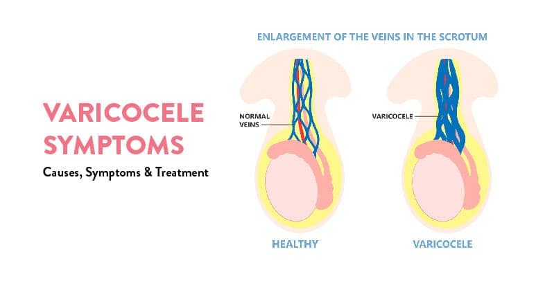
Many men with fertility problems and reduced semen quality are diagnosed with varicocele, an enlargement of the veins in the scrotum.
This potentially painful condition can cause infertility, so it’s important to understand what symptoms you may experience and why your varicocele was diagnosed in the first place.
Table of Contents
Varicocele is a condition that affects the veins in the scrotum.
The scrotum is the sac that holds the testicles. The veins in the scrotum are responsible for carrying blood away from the testicles. When these veins become enlarged, it’s called varicocele.
Many people with varicoceles do not experience any symptoms. However, when present, symptoms may include pain or discomfort in the testicle area (less common), low-grade fever (less common), and a general feeling of heaviness or discomfort in the scrotum (more common).
In some cases, there may be tenderness on one side of the scrotum due to blockage of venous drainage by an enlarged vein on that side of the body.
The exact varicocele causes are unknown. However, it’s thought that they may be caused by abnormal development of the veins in the testicles. This can happen during fetal development or after puberty.
In some cases, varicoceles may be caused by an injury to the testicles. Other possible causes include a clotting disorder or testicular torsion (when the testicle twists within its protective sac).
Men who have a history of kidney stones should also talk with their doctor about the risk for this condition.
If a man has had one episode of kidney stones, he has a 15% chance of developing them again. For men who have had two episodes, the likelihood increases to 30%.
Kidney stones are more common among men because they produce less urine than women. Therefore, there is more concentrated urine sitting in the bladder and collecting bacteria than would otherwise be present if there were more frequent urination as with women.
The most common varicocele symptom is a dull ache in the testicle on the affected side. The testicular pain may worsen with prolonged standing or sitting.
Other symptoms may include:
It’s important to see a doctor even if you don’t have any symptoms because varicoceles can lead to serious complications later on.
The scrotum will be visually inspected and touched to assess the possibility of a varicocele. The examination is typically conducted while you’re standing and while you’re lying down.
As you stand, your health care provider may ask you to take a deep breath, hold it and bear down, similar to how you do during a bowel movement. The Valsalva maneuver can make examining a varicocele easier.
A few different varicocele treatments are available, depending on the severity of the condition.
In some cases, no treatment is necessary. For milder cases, treatment may involve wearing special support stockings or taking over-the-counter pain relievers. For more severe cases, surgery may be necessary to remove the varicocele.
Doctors might sometimes recommend a surgery called varicocelectomy. A surgeon makes an incision in the groin area and removes one or both of the enlarged veins that are causing the problem.
If only one vein is removed, it’s typically done by ligation (tying off) because removing it surgically can lead to complications such as bleeding or scarring. If both veins are removed, they’re usually tied off so they can heal before removal from circulation.
One type of surgery used to correct varicoceles is laparoscopic varicocelectomy. Doctors insert a small tube with a camera attached through an incision near the navel and make another incision near the testicles.
They then cut away excess tissue from inside the scrotum and repair any abnormal connections between arteries and veins. Afterwards, surgeons seal off each end of the abnormal vessel connections using heat-activated clamps known as sutures.
Another procedure performed to stop blood flow coming from the varicocele is percutaneous embolization. With this technique, doctors inject tiny particles into the larger veins that supply blood to the varicocele.
The particles block blood flow, and pressure in the vessels decreases until they shrink away completely. Endovascular procedures have been found to have higher success rates than other types of treatments, but there are still risks involved with these techniques.
In most cases, the varicocele surgery can be done on an outpatient basis, which means you won’t have to stay in the hospital overnight. Recovery from the surgery is usually quick, and you can expect to return to normal activities within a week or so.
However, there are some possible side effects of surgery that you should be aware of before deciding whether or not to undergo this procedure.
For example, a low sperm count might result from removing the affected veins, although it’s rare for it to happen with just one operation.
Additionally, if you’re not completely cured after one operation (which happens in about 5% of cases), then additional procedures might be necessary down the line.
Varicoceles are often harmless and don’t require treatment. However, in some cases, they can cause fertility problems. They may also increase the risk of testicular torsion (twisting of the spermatic cord) or testicular rupture.
Surgery is sometimes recommended for men who want to conceive a child but have varicoceles. If surgery is not an option, the male partner should avoid contact sports or any activity that might lead to trauma during sex.
The female partner should undergo tests to ensure she doesn’t have a condition like pelvic inflammatory disease that would make it more difficult for her to get pregnant.
Both partners will need regular checkups as well as consultation with specialists in reproductive medicine.
If you are experiencing any varicocele symptoms, visit the CK Birla Hospital or book an appointment with .We will help you develop a treatment plan that is best suited for your condition, besides providing you with information on the causes and symptoms of varicoceles.
Can Varicocele be Cured?
Treatment for varicocele includes surgery to remove the swollen veins that are in the scrotum. Other treatment options include injecting certain drugs to destroy the enlarged veins or radiation therapy.
What happens if a Varicocele is left Untreated?
If left untreated, the varicocele may cause infertility in men due to damage to the spermatic cord and blood vessels by persistent pressure on them. It also may lead to chronic pain, swelling and lower back pain.
Can Varicoceles disappear naturally?
Rarely, but it could happen over time as the body compensates.
Most of the time, this condition doesn’t go away without treatment and will get worse over time. Not only do the varicose veins become more dilated, but there is an increase in the connective tissue around the veins, which does not allow them to return to their normal size.
What is the natural way to Cure Varicocele?
There is no natural way to cure varicocele. However, preventive methods include wearing supportive underwear and avoiding high-intensity exercises like biking, running, weightlifting or heavy lifting.
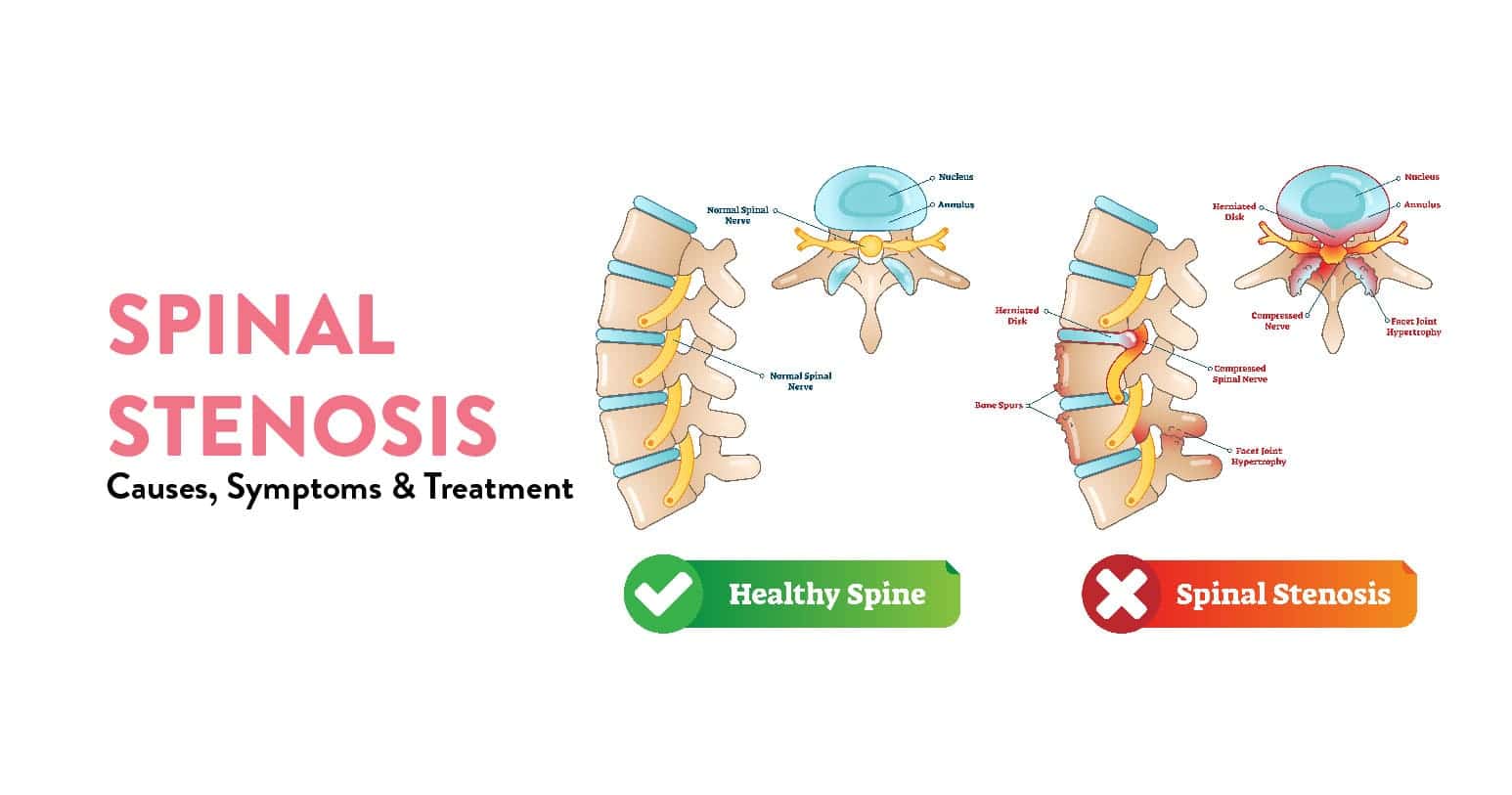
Spinal stenosis is a medical condition in which pressure on the spinal nerves causes various symptoms, including muscle weakness, numbness and tingling, leg pain, and back pain. Most often, the symptoms are low back pain and leg pain that worsens with activity.
The spinal cord is located within the bony spine (spinal column) and carries messages between the brain and body. Nerves branch out from the spinal cord to bring these messages to various parts of your body.
The spinal canal is a hollow space that runs through your spine. It protects your spinal cord and nerves. Too much pressure on the spinal cord or nerves can cause problems with movement and sensation (called neuropathy). If there’s too much pressure on the nerves in your back, it may also cause pain.
Table of Contents
If you have spinal stenosis, you may have trouble moving your legs or arms and lose strength or stamina when walking or climbing stairs. You may feel like something is pulling on one side of your body when you bend over.
The symptoms of spinal stenosis can vary depending on where they are located in the spine:
Further, the symptoms may vary from person to person but commonly include:
The causes of spinal stenosis can be divided into two categories:

The most common cause of herniated discs is ageing, which can also happen after a back injury or a fall. You may have one or more discs in your spine affected by this problem.
The bulging disc puts pressure on the nerves that travel through your spine and cause pain and weakness in your legs and arms.
Spinal stenosis also occurs due to abnormal narrowing (stenosis) of one or more spaces within the spinal canal that contain spinal nerves or spinal discs, resulting in pressure on these structures.
The bones forming this area may be abnormally shaped or misshapen, causing them to rub against each other as you move.
The following risk factors may increase your chances of developing spinal stenosis:
Spinal stenosis is diagnosed based on your signs and symptoms, imaging tests and sometimes a physical examination, which will check for tenderness in the lower back and thighs, leg strength and reflexes.
Your Spinal Stenosis doctor may use one or a combination of these tests to help diagnose your condition:
These are the most recommended options for spinal stenosis treatment:
This option is usually considered when pain has not responded to conservative treatment. The goal of surgery is to remove the narrow portion of bone or tissue that is compressing the nerve root.
Possible surgical treatments include:
Steroids are often injected into the spinal canal to reduce inflammation in people with spinal stenosis who do not respond to medications or want faster relief from symptoms.
Steroids may be injected directly into the spinal canal, which relieves pressure on the nerves inside it, or into the epidural space between the bones of your spine and your dura mater (the outermost layer of your spinal cord).
Epidural steroid injections usually have few side effects but can sometimes cause bleeding or infection at the injection site, as well as headaches and dizziness — especially when standing up quickly.
Recovery from spinal stenosis surgery usually takes two to three months. The length of time it takes to recover varies from person to person and depends on many factors, including age, health, and duration of symptoms.
You will have to avoid strenuous activity and heavy lifting during this period. The first three to four days after surgery are the most critical, so your doctor may request you to stay in the hospital during this time.
The pain from spinal stenosis can be mild to severe and debilitating. It is important to visit a doctor and get a check-up to explore your treatment options. Visit the C.K Birla hospital near you or book an appointment with for a consultation.
1. Where does spinal stenosis occur?
Spinal stenosis occurs most often in the lower (lumbar) spine. It can also occur in the neck (cervical).
2. Who gets spinal stenosis?
Spinal stenosis most often affects people over age 50, although it can occur at any age.
3. Can spinal stenosis cause permanent paralysis?
The answer to this question is both yes and no. Some patients with spinal stenosis may present with permanent paralysis. However, in most cases, the paralysis is temporary, lasting for a few weeks or months.
4. When is spinal stenosis surgery considered?
Surgical treatment of spinal stenosis is usually recommended for patients who have pain and other symptoms that affect their daily activities. Surgery may also be used to relieve compression on nerves in cases where there is no improvement with nonsurgical treatments.
5. Is spinal surgery safe?
In rare cases, surgery is not considered safe because of other medical conditions or because the patient might be too frail from age or illness to tolerate the procedure well.
You may also be recommended not to have surgery if you have cancer or another serious condition that could be worsened by the procedure.
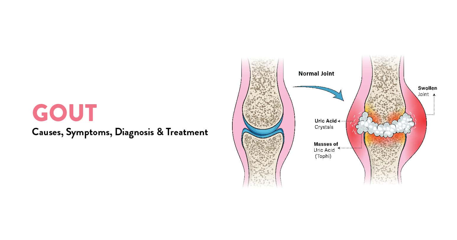
Gout is a painful arthritis condition that results in pain, tenderness, and inflammation in the joints. It can happen to anyone and at any time. While gout pain is generally felt in the hallux or the big toe, it can affect other areas too. Its vital to understand the implications of gout in more detail to identify and tackle this disease in a timely and efficient way.
Table of Contents
Gout is caused by excess uric acid in the body. Uric acid results from the breakdown of purines, which are naturally present in the body and also found in certain foods. The body generally removes the excess uric acid via the kidneys. The issue arises when the body fails to maintain the required levels of uric acid by either producing too much or failing to eliminate the excess quantity.

When uric acid levels become too high, sharp and hard crystals begin to form and deposit in various joints. You may not even be aware of this build-up happening over time.
Gout disease can present various symptoms, which can also vary in severity. The symptoms can feel quite severe during the flare phase of the disease. On the other hand, during the remission phase, you may not experience any symptoms at all.

Some of the most prevalent gout symptoms during an attack are:
Certain risk factors make you more vulnerable to hyperuricemia, or high uric acid, which in turn could lead to gout disease. Here are the ones to be aware of:

It’s worth noting that not everybody with high uric acid levels will experience gout.
If you’ve been feeling sudden and intense joint pain, it’s time to visit your general physician or rheumatologist for a consultation. You should share your symptoms in detail with your doctor, who’ll examine the affected joint and may order the following tests to diagnose gout:

Once your doctor has confirmed a gout diagnosis, the next step is to devise a suitable treatment plan.
There are two types of medications used for gout treatment. While certain medicines are advised to manage the pain and inflammation symptoms of gout, other medicines may be prescribed to reduce the uric acid levels in the body.
You must take medicines under the supervision of your doctor. Work with them to outline a treatment plan that works for your specific case.
Seeking timely treatment for gout disease is essential. If not treated, the frequency of gout flares can increase, and the disease may even start affecting other joints. Continuous gout attacks can deteriorate your arthritis over time (known as gouty arthritis). From cardiovascular problems to joint erosion, there can be many harmful consequences.

If uric acid levels continue to remain high, the crystals may start building up in the urinary tract as well, thus leading to kidney stones. As gout disease worsens and reaches the advanced stage, you may develop tophi, which are hard deposits of the uric acid crystals under the skin. Tophi can form in heels, toes, knees, elbows, fingers, etc. Even though tophi aren’t painful, they can disrupt daily activities and damage joints.
Gout attacks can feel debilitating and hamper many aspects of your life. However, through diet and lifestyle improvements, you can bring the disease as well as its negative impact under control. Here are some effective practices and remedies for gout pain that you can adopt:

While gout is an incurable condition, there are many ways to manage it to promote healthy living. Through medicines and self-care, you can minimise the impact of the disease on your life. Not only is it possible to reduce the frequency of gout attacks, but it’s also possible to reduce the pain and intensity of these attacks.
If you or someone you know is struggling with gout, then visit the nearest CK Birla Hospital or book your appointment with Dr. Rakesh Mattoo to get the best gout treatment and care.
Q 1. When should I see a doctor for gout?
A. If you’ve been experiencing symptoms commonly associated with gout, you should see your doctor immediately. In case there’s another medical issue causing the gout-like symptoms, like a joint infection, you want to get ahead of that promptly.
Q 2. How can I get quick relief from a painful gout attack?
A. Some quick remedies for gout pain include elevation of the impacted area and application of an ice pack. Talk to your doctor about medication for pain relief.
Q 3. What are some common warning signs of gout to watch out for?
A. If you feel severe joint pain (especially at night) that is sudden and extreme at the beginning and subsides after a few hours, it could be a sign of gout. Your joint is also likely to feel tender to even the slightest touch.
Q 4. How long does a gout attack last?
A. A typical gout attack can affect you for 1-2 weeks. The pain is worst during the first few hours of the attack. Treatment can help reduce the duration.
Q 5. What are some of the foods to avoid with gout?
A. Drinks and foods to avoid with gout include alcohol, organ meats, red meats, some seafood like sardines and scallops, fructose-rich products, etc.
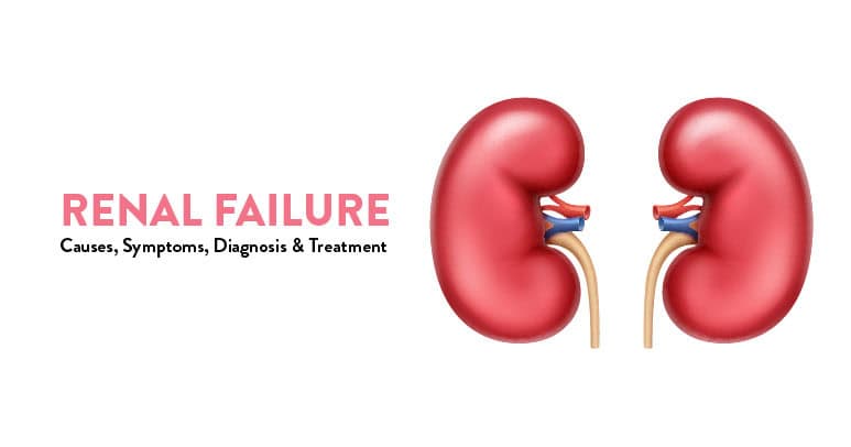
The kidneys are located on either side of your spine in the lower back region. They are bean-shaped organs and filter the waste products from your blood. They also produce vital hormones and balance crucial elements in your body like calcium, potassium, and sodium.
The proper functioning of your kidneys is critical to your health. You need to be aware of signs that there might be something wrong with your kidneys in the case of renal failure.
Table of Contents
There are two types of renal failure – acute renal failure or acute kidney injury (AKI) and chronic kidney disease (CKD).
AKI occurs suddenly, accompanied by acute pain. On the other hand, the symptoms of CKD appear gradually, and you may not realise it initially.
AKI occurs as soon as the kidney abruptly stops functioning normally. Renal failure symptoms of AKI are:
CKD advances slowly and reveals little or no symptoms in the early stages. You may not feel much until your kidney function reduces to 20% or less.
By this time, the following renal failure symptoms might appear:
Also Read: Healthy Diet Chart for High Blood Pressure Patients
The most common causes of renal failure are uncontrolled high blood pressure and diabetes, among other reasons.
If your kidneys suddenly deteriorate, your kidney disorder would come under the category of acute kidney injury.
You can suffer this type of renal failure due to the following:
Renal failure diagnosis is performed by observing how the kidneys are filtering impurities in the body. The filtering function of the kidneys is facilitated by tiny blood vessels called ‘glomeruli.’
To measure the rate of blood filtration by the glomeruli, the glomerular filtration rate (GFR) readings are taken.

A normally-functioning kidney typically should filter blood at 100 millilitres per minute. The value fluctuates with age and gender. Certain formulae, considering age and gender, are applied to get accurate and relevant GFR readings.
Another means of detecting renal failure is to check the creatinine levels through a blood test. Creatinine is a waste product that is excreted through urine. High creatinine levels in the blood indicate malfunctioning kidneys.
You cannot prevent chronic kidney disease. However, if you are diagnosed with renal failure, there is much you can do to restore your kidney function.
Suppose you adopt a healthy lifestyle and take certain preventive measures to manage renal failure. In that case, you can slow down the deterioration of your kidney function by observing the following:
Renal failure treatment depends on the extent of the condition. If you receive timely treatment for chronic kidney disease, you can delay the progression of the disease.
One or multiple measures may be required for proper management of renal failure.
Your doctor appointments might include routine blood pressure checks and blood tests. You will also receive a regime of medication that you should follow strictly post-renal failure.
In case of complete renal failure, you will probably have to undergo dialysis regularly. Dialysis is a process where your blood gets filtered by a machine that does the job of your kidneys.
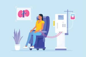
Alternatively, kidney transplant surgery is an extreme but effective method used by doctors to deal with the complications of renal failure. A kidney transplant operation involves replacing a defective kidney with the healthy one of a deceased or living donor.
If you start to experience any of the renal failure symptoms mentioned above, waste no time visiting a doctor.
When renal failure occurs, it can happen suddenly or gradually. If you get help immediately, you can arrest the progress of your medical condition. You don’t want to reach a stage where you will need dialysis.
To avail of expert advice and treatment, visit the C. K. Birla hospital or book an appointment with our doctors, who will recommend the best line of renal failure treatment for you.
With renal failure, you may experience swelling in your hands and feet. You may feel tired because your blood isn’t getting purified as well as it should.
If your kidneys stop functioning altogether, your doctor will recommend dialysis. Extreme cases will call for kidney transplant surgery.
If your kidneys stop functioning normally, your urine will appear dark brown due to excessive waste. You may find that you need to urinate less frequently. You might also observe foaming in your urine.
This is caused by increased protein levels in the urine. Increased protein in the urine is a sure sign of malfunctioning kidneys.
There are multiple causes of renal failure, but the leading cause is diabetes. Uncontrolled blood pressure can also take a toll on the kidneys.
Kidney disease, hereditary or otherwise, can also cause renal failure. Besides that, it can also stem from urinary tract issues, an enlarged prostate, kidney stones and various types of cancers.
All types of renal failure are serious and should be dealt with immediately. In extreme cases, it can be fatal. However, if you take timely action, you can reverse the effects of kidney failure, especially in the case of acute kidney injury (AKI).
There is no fixed formula for kidney failure recovery. However, if you are healthy and relatively young, the prognosis for total recovery can be good.
On the other hand, if you have comorbidities (existing medical conditions), there could be multiple complications of renal failure. The line of treatment would be more complex and extensive. Full recovery in such a case can prove to be more challenging.
The three early signs of renal disease are as follows:

Abdominal pain in children is a common complaint. It can be caused by various conditions, including constipation, gas, indigestion, abdominal muscle strain, and infections.
Sometimes, the cause is not apparent even after tests are done. In these cases, it’s essential to find out what’s causing your child’s symptoms so you can help your child feel better as quickly as possible.
Children can experience pain in the right lower abdomen to the upper abdomen and sometimes even as far as the chest (although this is rare). This type of pain can be:
Table of Contents
Common symptoms of children’s abdominal pain include:
The best way to help your child explain their pain symptoms is to discuss them. It’s vital for you and your child to understand the problem so you can discuss it with your doctor, who can devise a treatment plan.
Discussing pain with children can be challenging because they often don’t know how to describe their feelings. The following tips may help:
If your child is younger than six months old, contact a doctor right away if their belly looks swollen or distended (like a balloon being blown up). This could be from an intestinal blockage or other serious problem.
If your child is six months or older, call the doctor if:
These symptoms may be signs of appendicitis.
As a rule of thumb, you should see your doctor if your child:
A doctor diagnoses abdominal pain in children and babies after a thorough physical examination, history and investigations.
Diagnosing abdominal pain starts with a medical history and physical examination. Your doctor will inquire about the timing and location of the pain and how long it lasts.
They’ll ask questions about recent nausea, vomiting or diarrhoea your child has experienced. Your doctor will also examine your child’s abdomen to look for signs indicating an underlying problem.
Paediatricians use special techniques to pinpoint the cause of stomach pain in children. Some of these techniques include:
The most common cause of abdominal pain in children is a viral infection. The pain can be severe and usually go away in a few days.
If your child has pain in the lower abdomen, here’s what to do:
Abdominal pain is a common problem among children, and multiple potential causes of acute, recurrent, or chronic abdominal pain exist.
If your child complains of abdominal pain unrelated to food poisoning or constipation, consult a pediatrician at the CK Birla Hospital, or book an appointment with Dr Anukalp Prakash to learn more about treatment options.
It’s essential to understand the different types of lower abdominal pain in children and what they mean so that you can determine if your child needs urgent medical care.
Ask them the following questions:
Tummy aches are common in children. It’s essential to watch for signs (like a fever) that could signal a more serious problem and get help if needed.
If you don’t know when the fever started or how high it got (or if it came on suddenly), call your doctor immediately since it might be an infection that needs treatment.

Table of Contents
Diabetes insipidus is a disorder in which the body produces too much urine and is not able to retain water.
People with this condition pass urine frequently and drink large amounts of water. They tend to feel thirsty all the time since their body finds it difficult to retain water.
Diabetes insipidus can be a chronic condition or a temporary one. It may be mild or severe, depending on what is causing it. It is usually caused by an issue with the antidiuretic hormone (ADH).
Symptoms of diabetes insipidus may be mild or severe, depending on your specific condition. The main symptoms are as follows:
Other diabetes insipidus symptoms you may experience include:
Diabetes insipidus is caused by issues with your body’s ability to retain water and regulate fluid levels. Your kidneys filter out excess water and fluid from your body and remove waste products.
This condition is usually caused by problems with the ADH hormone (also called vasopressin), which helps your kidneys balance the fluid levels in your body. It can also be caused by issues with the part of your brain that stimulates thirst. The diabetes insipidus causes differ based on different types.
Sometimes, there is no certain cause of diabetes insipidus. In certain rare cases, the disorder may be caused by an autoimmune condition or response that makes the immune system attack the cells that make vasopressin.
There are 4 different types of diabetes insipidus. They are as follows:
This is caused by damage to the brain, which affects the release and regulation of ADH in the body.
This is caused by a structural defect in the kidneys that affects their response to ADH.
This type of diabetes insipidus is rare and occurs during pregnancy. It is caused when an enzyme released by the placenta starts to destroy ADH in the mother.
This is caused by damage to the part of the hypothalamus (in the brain) that regulates thirst.
If left untreated, diabetes insipidus can lead to more serious complications. These include:
Diabetes insipidus is diagnosed through different tests and methods.

The main tests for diabetes insipidus diagnosis include the following:
You will be asked not to drink any fluids for several hours. This stimulates your body to produce ADH. The doctor will then measure your ADH levels to determine if your body is producing enough ADH. The doctor may also measure other factors such as urine output.
Your doctor may suggest a blood test to check your ADH levels. Your blood and urine may also be tested for glucose (blood sugar), calcium, and potassium.
An MRI scan is used to check for abnormalities in the pituitary gland and hypothalamus in the brain.
If factors in your family suggest a genetic cause, your doctor may recommend genetic screening.
Diabetes insipidus treatment differs based on the type. The treatments are as follows:
If you have mild diabetes insipidus, increasing your water intake may be sufficient, and medication may not be required.
If the condition is caused by an abnormality in the pituitary gland or hypothalamus, your doctor will treat the lack of ADH. It is usually treated with desmopressin, a hormone that works similarly to ADH. It is taken as an injection, a pill, or a nasal spray.
It is usually treated with diuretics, which reduce the urine produced by your kidneys. It may also be supplemented with medication to reduce your urine volume.
There is no specific treatment for dipsogenic diabetes insipidus. However, if it is caused by an underlying issue such as a brain-related condition, treatment can target the cause.
Treatment for people with gestational diabetes insipidus is often administered in the form of the hormone desmopressin.
Certain factors can contribute to your risk of developing diabetes insipidus. These factors are as follows:
Diabetes insipidus can be uncomfortable and difficult to live with. If the condition is not addressed, it may lead to severe dehydration and other complications.
If you are facing any symptoms such as excessive thirst and frequent passing of urine, it is best to get it checked by a medical specialist.
To avail of the best diagnosis and treatment for diabetes insipidus, visit the CK Birla Hospital or book an appointment with our Endocrinologist.
The 4 types of diabetes insipidus are as follows:
1) Central diabetes insipidus (problems with ADH in the body)
2) Nephrogenic diabetes insipidus (structural defect in the kidneys)
3) Gestational diabetes insipidus (occurs during pregnancy)
4) Dipsogenic diabetes insipidus (problem with thirst function)
Diabetes insipidus and diabetes (diabetes mellitus or Type 1 or Type 2 diabetes) are two distinct conditions.
Diabetes mellitus happens because your body is not producing enough insulin or is not able to use it well. Diabetes insipidus occurs because your body is not producing enough antidiuretic hormone (ADH) or your kidneys are not able to use it properly.
You can be diagnosed with diabetes insipidus at any point in your life.
Central diabetes insipidus can often be diagnosed in infants as it is related to a brain condition that is present, affecting the hypothalamus and pituitary gland.
Nephrogenic diabetes insipidus may also be diagnosed in infancy or early in life as it is often caused by a genetic condition.
Gestational diabetes insipidus is diagnosed when you are pregnant.
Diabetes insipidus may be a chronic condition or a temporary one. It can be treated if it is caused by a treatable underlying condition.
However, if it is not a treatable condition (which is often the case with dipsogenic diabetes insipidus), then it may be a chronic condition.

Going through a miscarriage can be emotionally, mentally, and physically draining for an expecting mother, especially as it is completely out of your control. While there are some safety measures and precautions you can take to ensure a full-term pregnancy, miscarriages are often sudden.
You Can Also Read: A Guide to Increasing Baby Weight When 9 Months Pregnant
Table of Contents
Miscarriage refers to the loss of the embryo or foetus before the 20th week of pregnancy. A normal pregnancy lasts about 40 weeks or 9 months. The total period of the pregnancy is divided into three trimesters.
The first trimester is from week 1 to 12, the second trimester is from week 13 to 28, whereas the last and the third trimester goes from week 29 to 40. Miscarriages can happen during any of the three trimesters, but most occur before the 12th week of pregnancy, i.e., the first trimester. Hence, the first trimester is often very crucial, requiring regular monitoring.
Going through a miscarriage does not necessarily signify something wrong with you. Reasons for miscarriage can range from chromosomal abnormalities, hormonal imbalances, uterine abnormalities, malnutrition, accidental trauma, etc.
Miscarriages are very common; almost 1 in 8 pregnancies end with a miscarriage. Often, mothers might miscarry even before they become aware of their pregnancy. Additionally, a lot of women struggle with repeated miscarriages. Depending on the medical situation of each woman, miscarriages are broadly divided into 5 major types:
In a missed miscarriage, there are no typical signs and symptoms. However, when an ultrasound is performed, it shows an embryo without a heartbeat or an empty embryo sac.
If a woman undergoes three recurrent pregnancy losses, she is diagnosed with the risk of a recurrent miscarriage.
In complete miscarriage, there is a total loss of pregnancy with vaginal bleeding. All the foetal tissue passes out of the vaginal opening.
As the term ‘threatened’ suggests, in a threatened miscarriage, there is fear of miscarrying during the entire pregnancy due to risk factors like abnormal vaginal bleeding and abdominal pains. Around 50% of threatened miscarriages end in normal pregnancies, whereas the other 50% end in pregnancy loss.
An inevitable miscarriage occurs when there is excessive bleeding and the cervix begins to open. The embryo’s chances of survival in such a case are zero.
It is a misconception that routine activities like exercising, sexual activities, dancing, etc., are reasons for miscarriage. An expecting mother can continue all her regular activities in moderation while following safety precautions.
If you have underlying medical conditions, your doctor will advise you accordingly. The actual causes of miscarriage are several:
Oftentimes, expecting mothers might not even be aware that they are miscarrying. Having said that, some miscarriage symptoms and signs to watch out for are as follows:
Some risk factors for miscarriage are:
If a woman undergoes a complete miscarriage, the foetal tissue emerges via the vaginal opening. In such a case, no further treatment is required. However, in other cases, if a miscarriage has occurred and the tissues have not exited the body, the doctor will have to remove them.
Often doctors opt for the dilation and curettage procedure in which the cervix is dilated, and all the remaining tissues are removed using a surgical tool. Nowadays, medication is also available to remove the remaining tissue from the uterus. If a miscarriage occurs in the later stages of the pregnancy, when the foetus has been completely formed, the doctor will induce labour and deliver a stillborn baby.
In cases like recurrent miscarriages, the doctor will closely monitor your pregnancy. They might routinely schedule tests like a pelvic ultrasound, hysteroscopy, hysterosalpingogram, etc., to make sure the pregnancy is progressing well.
A miscarriage does not imply that you cannot get pregnant again. You will have to be cautious and follow more precautions and safety measures. 85% of women go on to have normal pregnancies after miscarrying for the first time.
In case of recurrent miscarriages, with proper advice from the doctor, it is possible to become pregnant again. You can schedule tests like chromosome tests, hormone tests, ultrasound, and blood tests to understand all the risk factors closely and proceed accordingly.

It can take a few weeks for your body to recover from the stress of miscarrying. After a miscarriage, you might also need time to heal yourself emotionally. You might experience strong feelings of loss and grief.
After a harrowing miscarriage experience, many couples find it difficult to plan their next pregnancy. Health conditions also make this difficult. Keep in mind that there is no rush to plan your next pregnancy; you can take your time to deal with your emotions.
Are you struggling with a tumultuous miscarriage and wish to find a professional and experienced health practitioner to help? Visit the CK Birla Hospital near you or book an appointment with Dr. Aruna Kalra who will help you learn about your condition and provide you with the best treatment options available.
Q1. How long does a miscarriage last?
A miscarriage can last from a few hours to a few weeks. Symptoms like abdominal pain and vaginal bleeding are normal during pregnancy, but the moment you notice something abnormal, you are advised to visit a doctor as soon as possible.
Q2. What happens first during a miscarriage?
If a woman is miscarrying, she might notice some initial symptoms like spotting, vaginal discharge, and bleeding. The cramps might also get worse with time. Symptoms differ for each woman, but the common symptoms are usually cramping and bleeding.
Q3. How do miscarriages start?
The start of the miscarriage varies for each woman. Normally miscarriages start with abdominal cramping and vaginal bleeding. With time, the symptoms worsen. It is thus advisable to visit a doctor as soon as possible.
Q4. What should you not do after a miscarriage?
Refraining from putting anything in your vaginal opening for the next few weeks after miscarriage. Avoid using period products like tampons and menstrual cups. Give yourself time to heal emotionally and mentally.

Personal hygiene is a crucial part of staying healthy. Excess humidity, a warm climate, and lack of personal hygiene are enhancing factors of UTIs (urinary tract infections), which include Chlamydia trachomatis.
However, unlike yeast overgrowth or fungal infections, the transmission of chlamydia happens through sexual contact.
Chlamydia is a silent spreader because there are no visible symptoms during its incubation period (lasting between a few weeks to six months). Besides, individuals having active and unprotected sex life are most vulnerable to contracting it.
Table of Contents
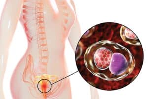
Named after its causative agent, Chlamydia trachomatis, chlamydia is a bacterial infection that affects the reproductive tract in both genders.
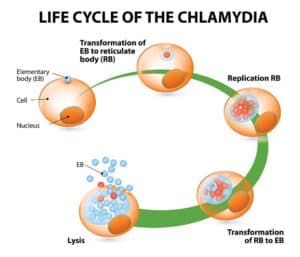
Chlamydia is the most prevalent STI, spreading rapidly often without the knowledge of the carrier, given the dormant character of the bacterium.
This condition can stay undetected until intense physical discomfort (pain during intimacy, difficulty in passing urine, or pelvic pain) post-incubation. Besides, chlamydia isn’t incurable like HIV. Seeking prompt treatment can cure this bacteria-borne STI.
Chlamydia infection spreads through sexual contact (both oral and penetrative). It can also infect a healthy individual using sex toys or indulging in self-pleasure with a silent carrier. Prevalent pathways include:

We understand the stigma associated with STI patients, especially since most are incurable. While chlamydia is curable, some things that don’t lead to the spreading of the bacteria are as follows:
Chlamydia affects irrespective of gender, making both men and women silent carriers.
Symptoms displayed are different for both genders. It’s difficult to self-diagnose because they resemble PID (pelvic inflammatory disease) and cervical issues (cervicitis).
Here’s what chlamydia in men and women looks like:
Chlamydia is a UTI (urinary tract infection). It can affect the urogenital tract, secreting discharge from the penis. The following symptoms suggest chlamydia in men:
Besides, most men often report being asymptomatic for a long time. If you are having oral sex with a suspected Chlamydia trachomatis patient, you might develop throat issues rather than contamination around the urinogenital tract.
Chlamydia infection in women often stays hidden, devoid of symptoms immediately after contracting this STI. Prevalent symptoms show similarities with ovarian problems like PCOS and ovarian torsion, worsening the issue if the patient attempts to self-diagnose.

Here’s what to look for in chlamydia among women:
Women are prone to be silent carriers of chlamydia infection. It can also infect the rectum (anal penetration) and throat (oral sex), making it deadly enough to affect various parts of your body at once.
Contracting chlamydia requires:
Chlamydia trachomatis can only spread through sexual contact between a healthy person and a potential silent carrier. People at risk of contracting it include:
Here are the risks associated with having chlamydia in men:
While chlamydia in women has serious side effects for both the mother and her unborn baby, complications include:
Chlamydia is a curable illness. Although chlamydia prevention is the best choice, antibiotics are the preferred treatment (since chlamydia trachomatis is a bacterium).
Your physician may prescribe you the following antibiotics:
Besides treatment, preventive behavior is the best course for chlamydia prevention. It includes:
Before making an appointment with your physician, know the chlamydia symptoms well or look for its gender-based side effects. You should make an immediate appointment if you are:
It’s crucial to maintain sexual distinction to prevent the spread of chlamydia infection. Never hesitate to reach out to your physician if you show suspected symptoms similar to chlamydia.
Not sure where to discuss your chlamydia symptoms with privacy? Visit the CK Birla Hospital near you or book an appointment with Dr. Astha Dayal to learn more about chlamydia treatment.
Q1. Is chlamydia a serious STD?
Chlamydia is a curable STD, making it not a severe condition, unlike HIV. However, if left untreated, it worsens to affect both the testis (reducing fertility) and fallopian tubes (risking ectopic pregnancy).
Q2. How do I know if I have chlamydia?/How to know if you have chlamydia?
Chlamydia symptoms, like a burning sensation during urination or smelly discharge from the urethra/vagina, show suspected signs of underlying chlamydia infection.
Q3. How long does chlamydia last?
Chlamydia infection can last for years when neglected and left untreated. Being a bacterial infection, it will keep on worsening the afflicted area unless treated with antibiotic therapy.
Q4. Does chlamydia have a smell?/Do chlamydia symptoms have a characteristic smell?
The most characteristic chlamydia symptoms include smelly and sticky discharge from the urethra and vagina. Besides, patients experience pelvic inflammation (women) and intense pain in the testis (men).
Q5. Can chlamydia go away without treatment?/Can chlamydia go away naturally?
Chlamydia is a bacterial infection contracted sexually, meaning it’s unlikely to go away naturally unless reduced through a preventive lifestyle or cured through antibiotic therapy.