Filter :
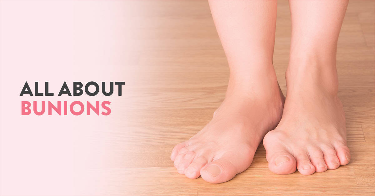
A bunion (or hallux valgus) is a deformity on the metatarsophalangeal (MTP) joint. Bunions can be unsightly and even painful in severe cases. It can develop in people of all ages. Dr Praveen Tittal leading orthopaedic surgeon and one of the best foot specialists in Gurgaon sheds more light on the causes of bunions, their treatment and how can you prevent this painful deformity.
The metatarsophalangeal joint (also known as the MTP joint) connects the toes to the main part of your foot. In some cases, the bones, ligaments and tendons in this joint can move out of place causing the big toe to get pulled towards the smaller toes. This misalignment disrupts the equal distribution of your weight on the joint resulting in instability and deformity in the joint.
The exact causes of bunions are still unclear. However genetic factors, underlying conditions and trauma or stress to the foot are known to contribute towards bunion formation. In the following section, these factors are explained in further detail.
Bunions might run in families. However, having a family history of bunion does not necessarily mean you will develop them as well. However, there is a higher risk of bunion formation due to inherited poor foot mechanics or shape. In such cases, extra care needs to be taken to prevent bunions from forming.
In extremely rare cases, babies may be born with bunions. This condition is also referred to as congenital hallux valgus. The causes of this are thought to be genetic. In mild presentations, it can be treated non-surgically with treatments such as corrective footwear. In more severe presentations, surgical treatment options are recommended. It is sometimes detected during prenatal ultrasound scans.
Conditions that can cause inflammation and pain the joints such as rheumatoid arthritis can also contribute to bunion formation. Diabetic people are also more prone to developing this deformity.
Sudden trauma to the foot or constant stress on the foot for a prolonged period of time (years) can also result in misalignment of the MTP joint. Experts also believe that prolonged use of ill-fitting or high heeled shoes can also put you at a greater risk of developing bunions, especially if you are already prone to it.
Read: 12 everyday tips for foot care in diabetes
The first and foremost step to treat bunions is to relieve the pressure on the MTP joint.
The most basic way to relive the pressure is to use the right kind of shoes. There are three factors to consider while selecting shoes for daily use. The form, fit and function.
The form is the visual aspect of the shoe and is more or less inconsequential to foot health. Fit and function on the other hand are vital to ensuring healthy feet.
Fit indicates how the shoe accommodates the shape of your foot. While ensuring that the shoe fits your foot lengthwise is not tough, finding shoes that are not too loose or tight breathwise can be challenging. This is also due to the fact that our feet are prone to changing their shape as we age.
Function refers to the shoe’s purpose. In many cases, shoes are designed especially for a particular activity. For example, sandals may not be the right shoes for a basketball game.
Also, read: Learn more about how to select the right pair of shoes
Another aspect of choosing the right shoe is ensuring the right amount of support and cushioning to the feet. Choose a wide, flexible sole with a sturdy heel counter. You can use stretchers to reshape narrow shoes if they feel tighter with time. For patients requiring extra care like diabetic patients, custom-made shoes can be a good option.
If you are experiencing mild discomfort or pain due to your bunions, you can use gell filled pads and over-the-counter painkillers. Patients also experience some relief by warm soaks, icepacks and massages.
In extreme cases, doctors can also recommend surgical correction of the deformity. For milder cases, a procedure called bunionectomy is performed. It involves shaving of the prominent bump along with realigning the soft tissues around the big toe. For moderate to severe deformities, an osteotomy which involves cutting the bone, realigning and fixing it back with screws or plate is performed. The outcome of successful surgery is improved function, a well-positioned big toe and pain relief.
If treated at a lesser stage, bunions can be corrected with minimally invasive surgery. This procedure is associated with lesser pain and quicker recovery.
Taking the right care of your foot after bunion surgery is an important deciding factor of the outcome of the procedure. After the procedure, the doctor holds the toe in place using a firm dressing. Keeping the toe in place is essential for the realignment of the toe. You would be advised to keep your foot dry and clean. Remember not to remove or change your dressing without consulting your doctor.
After the surgery, patients are advised to keep their feet in an elevated position for a few days. Keep pillows or cushions under your foot to keep it elevated. You would also have to wear special shoes that are designed to redirect the pressure of your body to the heel instead of your toes.
In some cases, you will be allowed to walk right after surgery. However, you would be required to wear special surgical shoes to protect the correction.
While bunions rarely cause complications that inhibit our daily lives, they can be irritating and painful. Prevention is better than cure. Remember to use the right kind of shoes, maintain healthy body weight and perform specialised exercises focusing on your toes.

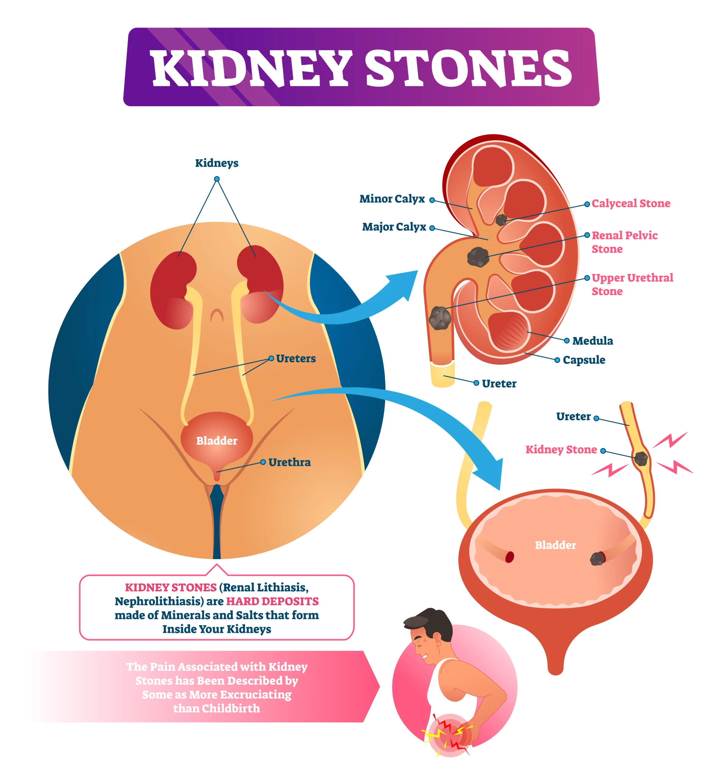
Children are funny creatures with strange interests. They like playing in mud or eating dirt. We are all guilty of falling for these games at some point in time. As little kids, we were told that our bellies will fill with stones if we eat impurities. Apparently, this make-believe story has an aspect of real life. Kidney stones are tiny rock-like objects that get collected in the kidneys.
The development of kidney stones is a very common condition. Nearly 1 out of every 10 individuals are affected by signs of kidney stones. This article explores kidney stones symptoms, signs and prevention in detail.
Dr. Mohit Khirbat, a leading nephrologist in Gurgaon, provides his insights on how to prevent kidney stones naturally.
Kidney stones are crystal-like substances that form inside the kidneys. They develop in varying sizes and shapes and range from minuscule like a grain of salt to large stones like a chickpea. They are made up of small substances that are usually found in urine.
Kidney stone prevention can be done without medical intervention also. However, it is necessary to first acknowledge its signs and symptoms.
Some stones are microscopic in mass. Due to this aspect, the symptoms usually go unnoticed. However, even these mini objects can expand later. They may also move within the organ causing pain and discomfort. Therefore, it is important to learn about the symptoms of kidney stones.
Here is a list of the most common sign and symptoms of kidney stones:
Apart from the above-given symptoms, the following are also signs of kidney stones-
It is advised that you seek immediate medical help, if:
Kidney stone pain is located at the lower abdomen or groin, along one side of your body and below the ribs.
There is no exact cause of kidney stones. It is known that certain substances in your urine lead to the production of kidney stones. The various types of kidney stones determine their individual causes.
While kidney stones can develop in anyone, some people are at a greater risk than others.
Risk factors of kidney stones include:
Read: Kidney transplant during the COVID pandemic
The strategy that ‘prevention is better than cure’ applies to kidney stones as well. In fact, kidney stone prevention is rather a matter of lifestyle changes. According to nephrology experts, here are some ways you can avoid kidney stones.
As mentioned earlier, kidney stones are not always diagnosed unless they cause health issues. Sometimes, patients live with small-scale stones for years without knowing. Therefore, the diagnosis is often delayed. The patient is informed about the condition after it has progressed. In such cases, both treatment and prevention can be offered to get the desired clinical results.
Prevention of kidney stones with the use of medicines depends on various factors. There are distinct types of kidney stones. For instance, calcium stones, uric acid stones, cystine stones, struvite stones and more. Doctors prescribe medications for condition-specific issues.
Kidney stones are not a modern disease. As preventive healthcare is gaining popularity, protection has become the key aspect. However, we cannot focus on preventing a condition unless we recognise its existence first. Thereby, a basic understanding of kidney stone symptoms is a must.
Afterwards, a focus on prevention is chief to avoiding kidney stones. A healthy way of living that includes sufficient hydration, a nutritious diet and exercise is the ultimate solution. For information or personalized guidance, you can book an appointment with Dr. Mohit Khirbat at CK Birla Hospital.
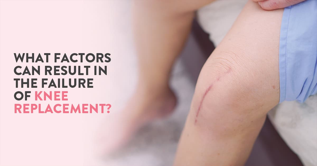
Even the thought of going through surgery is overwhelming. Imagine experiencing the complications from the said surgery. Though there has been revolutionary growth in healthcare, this field is not exempt from failures. A common example of such blunders is the prevalence of knee replacement surgery failure.
Knee replacement surgery, also known as knee arthroscopy, is a conventional and effective surgical treatment for conditions like knee arthritis. The average span of the impact of this treatment is about 15-20 years. However, some risk factors of knee replacement surgery can lead to its failure.
In this article, Dr Ashwani, one of the best knee replacement surgeon in Gurgaon, provides his insights on why a knee arthroscopy fails?
Table of Contents
During knee arthroscopy, surgeons cut open the affected knee for repair. They remove the damaged surfaces from the joint and cap the joint surface with metal and plastic implants.
There are numerous factors why do knee replacements fail. In some cases, more than one factor is responsible for the impairment. Here is a list of the top causes behind knee replacement surgery failure:
There is a variety of reasons why a knee arthroscopy damages. Some of these causes are beyond the patient’s control. However, most of these risk factors of knee replacement surgery failure can be avoided by practicing vigilance.
Here are a few ways knee replacement surgery recovery tips that can be maintained to avoid failure:
In most cases, knee replacement surgery failure occurs after 5-8 years of the procedure. After this gap, it is not very likely for patients to ignore the signs at the surgical area. The symptoms of knee replacement failure include:
Read: The risk of delaying Knee Replacement Surgery
Knee replacement surgery failure can be further treated successfully. Needless to say, the patient is required to take exceptional care if a revision treatment is recommended.
If and when a knee replacement surgery failure happens, revision surgery is done. It is similar to the original knee arthroscopy but aims at eliminating the factor that has lead to the failure.
In a revision surgical procedure, surgeons remove the pre-installed implants and replace them.
A revision knee replacement surgery failure is typically divided into three phases:
The recovery process of revised knee arthroscopy is similar to knee replacement surgery. Primarily, the patient would be kept under observation to keep a check on further improvements or complications.
Recovery tips for a corrective knee arthroscopy consist of:
Note: The length of the recovery period varies among patients.
Read: Quality of life after Total Knee Replacement Surgery
Knee replacement surgery failure is an ordinary medical complication. In the recent past, there has also been a rise in the number of revision surgeries.
For information or personalised guidance on knee arthroscopy, you can book an appointment with Dr Ashwani, best knee replacement surgeon in Gurgaon.
Read: Partial vs total knee replacement surgery: Which fits better?
Earlier women who delivered via a cesarean section had no other option other than opting for a cesarean section for all subsequent pregnancies. Today, VBAC or vaginal birth after caesarean offers an alternate birthing process for women. In this article, Dr Anjali Kumar, top obstetrician and gynaecologist in Delhi NCR talks about 5 things everyone thinking of VBAC should know.
Table of Contents
Before opting for VBAC, you need to assess if it is the right option for you. Some risks can make it an unviable birthing option. There are several factors that decide if you are an ideal candidate for VBAC. These include:
The three types of uterine incisions used for C-sections are low transverse, low vertical and classical uterine incisions. VBAC is usually done only on women with low transverse or low vertical uterine incisions. A high vertical incision or classical incision is associated with a higher risk of uterine rupture.
Uterine rupture is a rare but severe complication that can occur in pregnancy. It causes the uterus to tear which can allow the fetus to slip into the abdominal cavity. Women with uterine scars due to previous caesarean sections are at a higher risk of developing this complication. If you have a history of uterine rupture, you would not be considered an ideal candidate for VBAC.
If you have undergone any surgical procedures on your uterus in the past such as fibroid removal, you would not be considered for VBAC. Previous surgeries can cause uterine scarring which is not ideal for VBAC.
VBAC is not recommended for women who have had more than two C-sections previously.
VBAC is not recommended for women who have undergone a C-section delivery less than 18 months prior.
If you have any other underlying condition such as placental problems or multiple births, which can complicate your pregnancy or labour, you would not be recommended a VBAC.
Other factors such as being overweight, breech presentation of the baby, size of the baby etc can also result in VBAC being ruled out. Discuss your suitability for VBAC at length with your obstetrician early on in your pregnancy so you can start preparing for your labour.
Read: Caesarean section delivery – What to expect
While you may be keen to explore the option of vaginal birth after caesarean (VBAC), the fact is that not all hospitals or doctors offer this birthing option. While complications arising from VBAC are rare, hospitals and doctors need to be prepared to perform an emergy C-section during TOLAC (Trial of labour after cesarean). Constant fetal and maternal monitoring is also essential during this process. Hence, if the hospital or doctor is not experienced in handling difficult labour, they may not offer VBAC as a birthing option. Deciding to switch your hospital and doctor later in the pregnancy can be tough. Hence, choose your hospital and doctor after exploring these options carefully, early on in your pregnancy.
In the past few years, there has been a sharp increase in the popularity of VBAC. Many expectant mothers wish to explore this birthing option due to the number of benefits associated with it. Lower recovery time, lower risk of infection from the surgery, shorter hospital stay and lesser pain are some of the advantages of VBAC for the mother. Even for the baby, passing through the birth canal helps squeeze the fluid from his/her lungs as well as transfers good bacteria, providing him/her valuable immunity after birth.
A lesser-known fact about caesarean section is that it is not ideal for women wishing to give birth more than 3 times. As repeated cesareans can cause several complications and uterine scarring. Each consecutive c-section poses a greater risk than the previous one. In such a scenario, talk to your doctor about your birthing options.
Like any other medical procedure, VBAC also has several risks associated with it. This is one of the major reasons why it is not ideal for everyone, nor is it offered by all medical institutions and doctors. Complications arising from VBAC are rare, also because the candidate for VBAC is screened thoroughly to minimise their risk factor.
During a VBAC, there is a small chance that the previous uterine scar breaks open. This occurs in less than 1% of all VBAC deliveries. In such a case, an emergency hysterectomy might have to be performed. There is also a chance that the VBAC attempts have to be abandoned midway due to maternal or fetal health. Emergency C-sections is performed in such a scenario.
If you have assessed your suitability for VBAC, start preparing for it early on. Speak to your doctor at length about steps you can take to have a healthier pregnancy and reduce the risk of developing pregnancy complications. Ensure that your hospital is equipped to handle complications during childbirth and also has a good NICU. Speaking to other women who have also undergone VBAC can help you manage your expectations and be better prepared.
For more information about prenatal care and birthing options, you can consult Dr Anjali Kumar at the CK Birla Hospital.

The winter season can be a lot of fun for our kids. In India, it is also a season of festivities with several opportunities for celebration and enjoyment. However, parents should note that winters can also be especially harsh on our little ones. We need to take extra precautions to protect children from the numerous risks of the winter season.
In the following sections, Dr Shreya Dubey, top paediatrician in Gurgaon shares a few winter care tips for babies/children to keep them safe and healthy this season.
During winters, we tend to spend more time indoors as compared to summers. With the use of artificial heating devices, we now have the ability to control the temperature inside our homes, up to a large extent. But this option raises the question: What is the ideal indoor temperature? This query does not have one fixed answer.
The room temperature should be comfortable for the child. Maintain an ambient temperature which is neither too cold nor too hot.
If your child is too young (less than 18 months of age) to communicate his or her discomfort, you can gauge if they are feeling cold by touching their hands and feet. If these body parts feel chilly, it implies that your baby is also feeling cold. In this case, you can increase the room temperature.
We all know the importance of layers (of winter wear) in this season. But is there such a thing as too many layers?
The rule of thumb while deciding the number of layers is to “add one” to the number you are wearing.
You should also ensure that excessive clothing is not there. Winter wear which is largely made of wool can be quite abrasive on the skin. Making your child wear too many surfaces of woollen clothing has its own risks like skin rashes and eczema. The odds of children getting affected by these risks are higher as their skin is comparatively more sensitive than that of adults.
You can place a layer of cotton between skin and wool. This small step can avoid prolonged skin contact with wool and help prevent skin conditions such as rashes and eczema.
When going outside ensure that the hands and feet of your children are covered with socks and gloves.
Studies indicate that the body loses maximum heat from the head, sometimes up to 30%. The child’s head should also be covered with the appropriate headgear. For younger children, you can choose monkey caps that offer protection to the ears as well.
During sleep, make sure that you don’t use a blanket that is too heavy, especially if they are sleeping alone. Usage of weighted blankets during sleep is dangerous for children.
The chances of getting infections are higher during the winter season. You should focus on cleanliness and sanitation to stay safe. Ensure to wash your child’s clothes including woollens regularly to avoid risks of asthma and eczema.
The optimal room temperature during winter is 22-18 degrees. Most Indian households use heaters during this season. You should take care of the following points while using heaters:
You can give your child a bath every day if the weather is not too cold. However, if you feel the temperature is too low, you can reduce the frequency of bathing your child to 2-3 times a week (once a week for head wash).
The days that your child skips bathing, you can clean them using a damp sponge. Make sure to properly clean the creases such as armpits, elbows, knees, groin etc. to prevent skin infections and eczema.
Since the winter season leads to excessive dryness, you should make certain to properly moisturise your child. You can use any type of moisturiser. Nonetheless, if your child has sensitive skin, you should consult your dermatologist for their opinion.
You can use olive oil, mustard oil or almond oil during winters. Coconut oil is not considered the best option for this season as it can escalate skin dryness, making it more suitable for the summer season.
If your child is younger than 2 months, do not use mustard oil. The pungent smell of the mustard oil can cause irritation in the nose and eyes of the baby.
If your kid’s lips are chapped, you can use either petroleum jelly or milk cream. Avoid using anything on the lips in case your little one is younger than 4 months.
Remember not to put too many products on your children as their skin is extra sensitive.
There are no specific dietary guidelines or superfoods for winters. You should feed your children healthy home-cooked food only.
You can add food items that offer additional warmth. These include whole grains, wheat, rice, oats, lentils, fruits, green leafy vegetables and more. You can choose beetroots, carrots, and items rich in Vitamin C.
Dry fruits are also great for nutritional value during winters. You can also feed your child small amounts of species like garlic.
For infants younger than six months, you can maintain the hydration by breastfeeding or formula milk.
You can increase the intake of warm fluids such as soups for older children.
As winter approaches, the incidences of eczema, asthma and other infections also increase. You should practice basic hygiene and follow these steps to check that your child is safe:
Read: Vaccinating your child – What you need to know?
Watch the video to learn more about how to keep your children healthy during winters.
For more information or personal guidance, book an appointment with Dr Shreya Dubey Paediatrician and neonatologist at the CK Birla Hospital Gurgaon.
Read: Managing asthma in children | Tips to control asthma symptoms

A urinary tract infection indicates an infection in any part of the urinary tract. The urinary tract comprises the kidneys, ureters, bladder and urethra. Most UTIs are the result of a bacterial infection. However, it can also be caused by fungi and viruses.
The urinary tract is divided into two main regions: the upper urinary tract (ureters and kidneys) and the lower urinary tract (urethra and bladder). Most UTIs occur in the lower urinary tract. However, infections in the upper urinary tract are generally more severe.
Table of Contents
Urinary tract infection or UTI is a bacterial, fungal or viral infection in any part of the urinary tract. Bladder infections are the most common form of UTI. Our body has its own natural defence against infections in the urinary tract. However, in some cases, this natural defence is not enough to ward off these infections. In such cases getting treated early on is the best way to prevent recurring infections. If left unchecked, a simple UTI can lead to severe kidney (renal) problems.
A mild case of UTI does not necessarily cause any obvious problems and can go undetected. Common symptoms of UTIs include:
If left untreated, the infection can spread to one or both kidneys. Symptoms of a kidney infection are:
Detecting UTIs in infants and younger children is more challenging as they may not be able to relay their symptoms clearly. Even though fever is one of the most commonly associated symptoms of UTI in children, it doesn’t necessarily indicate the presence of UTI. Consult a doctor immediately if your child has any unexplained fever.
Amongst elderly patients, UTIs may be overlooked and mistaken for other conditions. In reality, older people are the most vulnerable to UTIs for a number of reasons including a weaker immune system. Another factor that contributes to the risk of UTI is the weakening of the muscles of the bladder and pelvic floor, leading to increased urinary retention (inability to completely empty the bladder) and incontinence (inability to control the bladder muscles).
Elderly patients may also find it difficult to relay their symptoms, especially if they are suffering from conditions such as Alzheimer’s or dementia.
UTIs are generally treated with a course of antibiotics. The strength and dosage of the antibiotics prescribed depend on the severity of your infection. It is important to always complete the course of antibiotics, even if the symptoms go away mid-treatment. Your doctor might also prescribe other medicines in combination with the antibiotics to relieve pain and discomfort.
Apart from the recommended medications, you would also have to make some lifestyle changes to complement your treatment. This includes drinking more water and applying a hot compress for pain relief.
Read: Chronic Kidney disease: What is it and how can it be prevented?
UTIs can occur in people of any age, including children and infants. Some people are more prone to contracting infections as compared to others. Women are more prone to developing UTIs as compared to men. While some risk factors are beyond our control, others can be managed to minimise the risk of developing UTI.
The risk factors of UTI include:
Senior patients are at a higher risk of developing UTI especially if they:
Studies show that women are significantly at a higher risk of developing UTIs. This trend is majorly due to the difference in the structure of the urinary tract in both men and women. Women have a shorter urethra making it easier for contagion to infect the bladder. Another factor is that the urethra is closer to the rectum in women, increasing the exposure of the urinary tract to bacteria.
Complications from UTIs are quite rare if the patient is undergoing the right treatment. However, if it is left unchecked, it can result in the following complications:
Many health practitioners believe that the rise in the number of UTI cases is majorly due to our lifestyle. The following steps can help you minimise your risk of developing UTIs:
Personal hygiene plays a key role in preventing recurrent UTIs. Remember to use clean and washed underwear, preferably made of absorbent material such as cotton and keep yourself hydrated to minimise your risk of infection.
Consult Dr Shalabh Agrawal, best urologist in Gurgaon at the CK Birla Hospital to learn more about this condition and possible treatment options. Book your appointment today!
Also, read: Urinary Tract Infection – Symptoms, Risk Factors and Prevention
According to the Endometriosis Society of India, an estimated 25 million Indian women suffer from endometriosis. However, in spite of its rising prevalence around the world including India, there is insufficient awareness about this painful condition.
Even today, thousands of women are unaware about the warning signs of endometriosis, silently suffering from painful periods without getting the necessary treatment. If left unchecked, endometriosis can also result in infertility. In fact, many women are diagnosed with this condition when they seek medical advice for difficulties in conceiving.
In this article, Dr Astha Dayal (Department of Obstetrics and Gynaecology at the CK Birla Hospital) explores endometriosis in further detail by answering a few common questions about endometriosis. For more information about this condition and to screen yourself, you can book an appointment with Dr Astha Dayal here.
Table of Contents
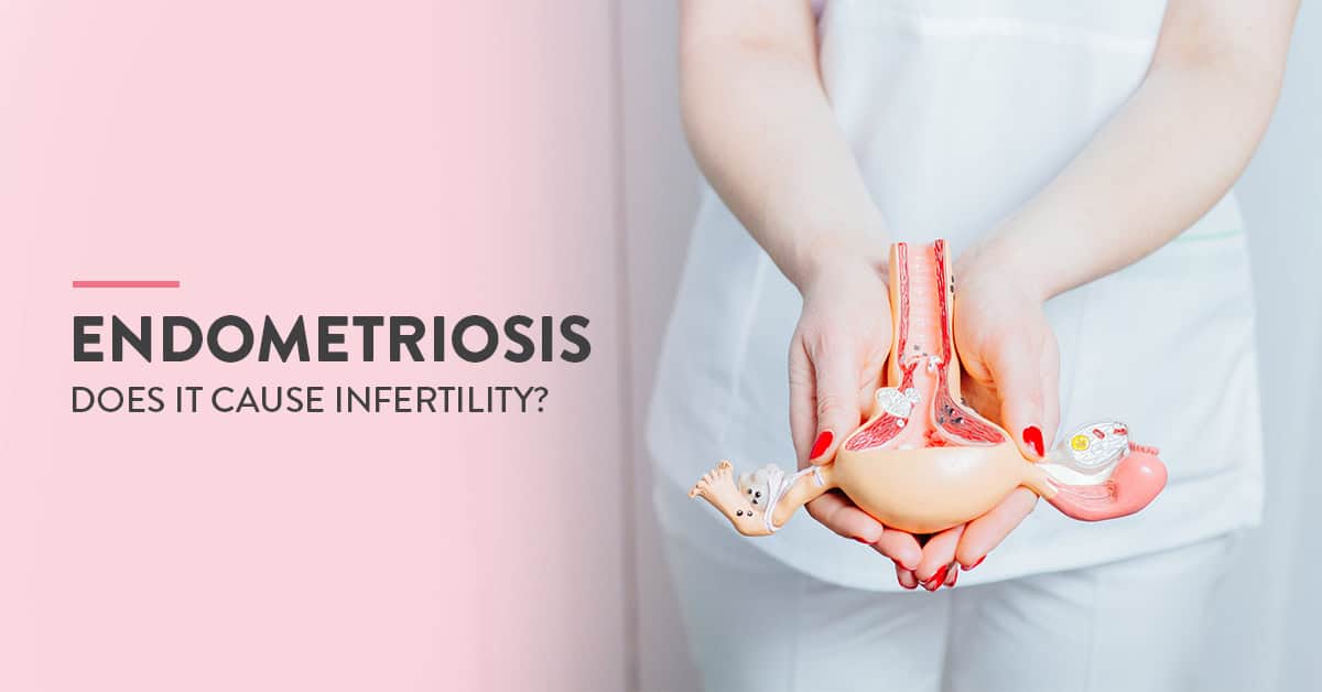
Your uterus is lined with a layer of tissue called the endometrium. During your menstrual cycle, this tissue builds up in preparation for pregnancy. If you don’t become pregnant, this layer is shed and is passed out during your period.
In the case of endometriosis, tissue similar to the endometrial tissue is formed outside the uterus. This generally occurs in other parts of your reproductive system or inside the abdominal cavity. Every month, this tissue also grows and sheds along with your menstrual cycle, causing swelling and scarring of the normal tissue in the affected regions.
While the exact causes of endometriosis are still unclear, some familiar causes of this condition are as follows:
While women with endometriosis can sometimes be infertile, but researchers do not know exactly why this is the consequence. A recent study declared that endometriosis could make natural conception tougher but it does not equal infertility. Endometriosis causes adhesions and scar tissue, which result in the internal organs to get stuck to each other. This may cause blockage of tubes or ovaries. With timely medical intervention and treatment, women may even be able to conceive naturally without requiring any assisted reproductive techniques.
Also, read: What to do when you suspect infertility
Some of the common symptoms of endometriosis include
Pain: Pain is the most significant and common symptom associated with endometriosis. Pain caused by endometriosis can be of different types such as
Bleeding or spotting: Light bleeding between your periods can indicate endometriosis
Infertility: Difficulties in becoming pregnant or infertility can be a result of endometriosis
Gastric problems: Diarrhoea, constipation, bloating, nausea etc during your periods can indicate endometriosis in some cases.
Painful periods are not the only unpleasant outcome of endometriosis. The abnormal growth of tissue can cause complications such as:
Endometriosis is generally treated by medication or surgery. Treatment protocols depends on the severity of the condition in that individual. In some cases, over the counter medications are enough to manage the symptoms. This is also combined with hormone therapy in case the patient is not aiming for a pregnancy.
In more severe manifestations of endometriosis, surgery might be required. If you are trying to become pregnant, the surgeon might perform a conservative surgery to remove the abnormal tissue. This surgery is usually done laparoscopically.
Unfortunately, there are no steps you can take to completely eliminate the risk of developing endometriosis. Following a healthy diet and lifestyle can go a long way in managing symptoms. Getting regular gynaecological check-ups can also go a long way in monitoring the health of your reproductive system.
If you are experiencing extreme menstrual pain, you should consider getting screened for endometriosis. Book your visit to meet Dr Astha Dayal and other top gynaecologists in Gurgaon.

In the year 2020, 297 cases of dengue were reported in Delhi itself (till Sept. 20’). Dengue is one of the diseases spread by the bite of an infected mosquito. These types of diseases are called mosquito-borne diseases. The five most common diseases in this category that exists in India include dengue, malaria, Chikungunya virus and Zika virus. These diseases are responsible for millions of deaths around the world. In this article, we will look at some of the common mosquito-borne diseases in the country, their symptoms, treatments and preventive steps.
Table of Contents
Dengue is a viral infection caused by the bite of an infected mosquito. Over the past few years, the rising incidence of dengue cases and deaths has become alarming. It is transmitted by female mosquitoes.
Dengue can affect people differently. In some cases, the infection is so mild that the affected person may not even realise that they were sick. It can also cause severe flu-like symptoms in other cases.
Severe manifestations of dengue can cause any number of complications such as severe bleeding, organ impairment and even death.
Symptoms are expected to last from 2-7 days with an incubation period of 4-10 days (days for symptoms to appear after exposure to the virus). WHO has classified dengue as “Dengue” and “severe dengue” to help doctors develop the required treatment plan.
The patient may develop severe dengue 3-7 days after the onset of initial symptoms. This stage can be fatal as well. Warning signs of severe dengue include
If these symptoms manifest, care needs to be taken to avoid further complications.
Malaria is a disease caused by “plasmodium parasites” which is spread by the bite of the female anopheles mosquito. Over the years, several initiatives have been taken by both international and domestic institutions to curb the spread of malaria around the world. According to the world malaria report (December 2019), approximately 228 million cases of malaria were reported around the world in the year 2018.
Climatic conditions and human immunity are two major factors which determine the spread of this disease. Symptoms of malaria include:
Some patients also experience malaria “attacks”. A malaria attack starts with shivering and chills, which is followed by a high fever and sweating. The patient then returns to normal temperature, before the cycle starts again.
Complications caused by malaria include:
Chikungunya is a virus that was first detected in 1952. The term “chikungunya” comes from a word in the Kimakonde language which translates to becoming contorted. It refers to the debilitating joint pain which causes the patient to have a stooped appearance. In the year 2020, 77 confirmed cases of chikungunya were reported in Delhi alone. Symptoms of chikungunya generally appear 3-7 days after the infected bite, they include:
Chikungunya is not usually fatal. However, the symptoms can be extremely severe and disabling.
Japanese encephalitis was diagnosed in India for the first time in 1955 (in Tamil Nadu). The most recent outbreak occurred in 2019, predominantly in regions of Bihar and Muzaffarpur. People of any age can contract this disease, however, it is more severe in children. Once infected, the individual is said to develop immunity from future infections.
Symptoms can manifest 5-5 days after contracting the infection. They include:
Zika virus is a mosquito-borne disease which is spread by the bite of an infected mosquito. One of the major risks of this disease is that it can be transmitted to the fetus in-utero, resulting in birth defects. The disease can also be transmitted through sexual intercourse.
While there is currently no evidence of an ongoing zika virus outbreak in India, it is pertinent to note that there is a history of previous zika virus transmission in the country. Hence, it is important to be prepared for any possible outbreak.
Symptoms of zika can last up to a week and fatalities associated with zika are rare. Most common symptoms of zika are:
The most important aspect of preventing these diseases is limiting the breeding of mosquitos. The National Centre for Disease Control has set the following guidelines to protect oneself from mosquito-borne diseases:
Prevent/control mosquito breeding by:
The advisory also states guidelines for personal protection against mosquito bites. It recommends the use of mosquito nets, mosquito repellents and full-sleeved clothing to prevent bites.
In most cases, outbreaks of mosquito-borne diseases are seasonal. Hence, prepare your home or locality before the expected outbreak by taking the aforementioned steps. Prevention of mosquito breeding is the best way to prevent any outbreak as there are no vaccinations for the same.
If you experience any of the symptoms associated with the above diseases or if you require more information regarding these diseases, you can contact Dr Tushar Tayal at the CK Birla Hosptial-Gurgaon.
Book an appointment with Internal Medicine specialist at the CK Birla Hospital, Gurgaon.

Throughout the ages, childbirth has been considered to be one of the most beautiful and life-changing experiences. Many expectant mothers, however, especially first-time mothers dread the infamous labour pains that come along with “the miracle of birth”.
With leaping advances in technology and medical care, women have a number of alternatives to choose from while deciding how to deliver their baby. These birthing techniques adopt different ways to manage labour pain and make childbirth easier for the mother as well as the baby. Here we will explore these birthing alternatives in greater detail, exploring the various pros and cons associated with each.
Benefits of vaginal delivery
Going into labour and opting for a vaginal delivery generally means spending several hours in the labour room, straining to bring your baby into this world. It is a physically gruelling process which involves a lot of hard work and pain.
However, many women opt for vaginal delivery in-spite of the labour pains because of its benefits in the long run. Vaginal delivery usually means significantly shorter hospital stays and recovery times. The mother also avoids life-long scarring that comes with surgical birthing techniques.
Women opting for vaginal deliveries also avoid the risks associated with major surgery such as bleeding, infection, post-op pain and complications due to anaesthesia. As the you would be more aware during vaginal birth (as compared to post surgery), you can start breastfeeding sooner.
Studies also indicate that vaginal birth is beneficial for the baby as it helps in squeezing out fluid from the baby’s lungs as he/she travels through the birth canal. The baby is also exposed to healthy bacteria in the birth canal which in turn boosts his/her immunity.
Risks of vaginal delivery
Even though vaginal delivery is often thought to be the “best” birthing technique, it does involve a number of risks that every expectant mother should be aware of. Today, medical authorities around the world are mandating that every birthing alternative should be discussed with the to-be parents in detail, enabling them to make informed decisions.
Natural vaginal delivery is associated with risks such as tearing, excessive bleeding (haemorrhaging), injury to pelvic floor muscles amongst others. In some cases, the doctor can make a small incision in the perineum (the area between the vagina and anus) to aid the delivery. This procedure is called an episiotomy. Vaginal delivery can also result in injuries to the baby, if the baby is too large or the labour too intensive and long.
Also, read: Normal delivery – tips & preparation
Benefits of caesarean section
Caesarean section is a surgical birthing technique in which the doctor delivers the baby through incisions in the abdomen and uterus. Opting for caesarean delivery often allows the parents to schedule the procedure. In some cases, it can be an emergency procedure or the only suitable birthing technique available. This is especially true for high risk pregnancies, pregnancies with complications such as gestational hypertension, preeclampsia etc.
Caesarean section is the safest option for mothers if there are any concerns about her health or her baby’s. This surgical birthing method reduces the risk of painful labour, vaginal injuries, heavy bleeding, loss of bladder control, pelvic organ prolapse etc. It also offers the doctor better control of the delivery.
Risks of caesarean section
Like all surgical procedures, caesarean section involves the risks of bleeding, infection, blood clots, surgical injury and complications from anaesthesia. Caesarean section is usually not recommended for first time mothers who want to have more children, as this procedure can cause complications in subsequent pregnancies. These complications include placenta previa, preterm birth etc.
The recovery time for patients who underwent caesarean section is also longer. They are usually kept under observation in the hospital for a few days, post which they can continue recovering at home.
Read: Caesarean section delivery – What to expect
Benefits of assisted vaginal delivery
When vaginal delivery is aided by forceps or a vacuum device to pull the baby out, it is called an assisted vaginal delivery. In some cases, such as when the mother becomes too tired to push, forceps or vacuum is used to help her deliver the baby. In case of a forceps assisted delivery, the forceps are inserted into the vagina to gently hold the baby’s head which is then pulled out of the birth canal while the mother is pushing. A vacuum assisted delivery is similar with the exception of a suction cup being used instead of forceps.
Assisted vaginal delivery is usually done if there are any concerns about the baby’s heart rate, if the mother is too tired, the baby is not moving forward in the birth canal and if the mother has any medical condition which limits her ability to push safely.
The main benefit of assisted vaginal delivery is that it helps avoid the need for a caesarean section and the risks involved in the same.
Risks of assisted vaginal delivery
Risks associated with assisted vaginal delivery are very similar to those of normal vaginal delivery. These include injury to the vaginal tissues, perineum and anus. It can also cause injury to the pelvic floor muscles. For the baby, there is an extremely small risk of injury due to improper use of forceps and suction.
Benefits of water birthing
This birthing technique has been around for centuries however, the past few years has seen an exponential rise in the popularity of water birthing. Simply put, it is the process of giving birth inside temperature-controlled water. Some women choose to give birth to their baby while immersed in water, while some women may choose to step out of the birthing pool before giving birth to the baby.
There are numerous benefits of water birthing. Reduction of labour pains is one of the most well-known benefits of water birthing. Many women also say that they felt calmer and more in control while they were immersed in water. They also had a greater sense of privacy. The warm water helps in making the perineum more elastic and relaxed, reducing the risk of injury or tear during birth.
Water birthing is also thought to be less stressful for the baby as he/she enters an environment similar to the womb (amniotic sac).
Risks of water birthing
Like every medical procedure, water birthing has a few risks as well. In case of emergencies such as a baby in distress or increased labour pains, it can become difficult to get the mother out of the birthing pool. Another disadvantage of water birthing is that fetal monitoring is not possible. Water birthing also limits the usage of pain management techniques such as epidurals. Due to such reasons, water birthing is recommended only for healthy non-complicated pregnancies.
There is also a risk of infection if the baby passes stool during delivery. However, these are extremely rare cases.
Today, there are a number of birthing options available to women. Many factors such as personal opinion, maternal and fetal health, pregnancy complications etc have to be considered while deciding which birthing technique to opt for. Remember to have this discussion with your maternity doctor early on in the pregnancy so you can start preparing mentally and physically for childbirth.
Also, read: Water Birth: The Most Natural Form of Normal Birth
Ques 1: Can I have normal delivery after caesarean?
Ans: Yes, these deliveries are termed as VBACs (Vaginal delivery after caesarean section). You can consult an obstetrician who specialises in VBACs to find your suitability for this procedure.
Ques 2: Can my baby be injured during normal vaginal delivery?
Ans: There are a few risks for the baby during normal vaginal delivery. This process is quite painful and long for many women. They can get tired mid labour, causing the baby to get stuck in the birthing canal, the baby can also get hypoxia if he/she gets stuck in their umbilical cord etc. While choosing a maternity hospital, make sure the team is experienced in handling pregnancy emergencies. In such cases, the doctor usually aids the delivery with forceps or vacuum suction cups.
Ques 3: Can caesarean section make it tougher for me to get pregnant again?
Ans: Caesarean sections are known to make future pregnancies difficult, hence it is not recommended for first-time mothers or for women desiring to have more children.
Read: Normal Vaginal Delivery – What Should ‘Mothers-to-Be’ Expect