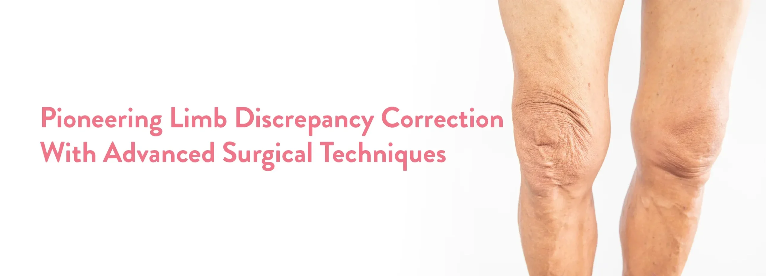
A 26-year-old patient, Abdullah, from Saudi Arabia, presented at CK Birla Hospital, Gurugram, grappling with a significant limb length discrepancy that had affected his mobility and quality of life for years. Despite undergoing Total Hip Replacement (THR) surgery two years prior, Abdullah continued to experience considerable pain and relied on crutches.
On consulting with Dr Debashish Chanda, it was determined that the underlying cause of Abdullah’s continued discomfort was a limb length discrepancy not properly addressed in his previous surgery. The imbalance measured a significant 1.5 cm, surpassing the medically acceptable maximum of 1 cm.
Dr Chanda explains, “Limb length discrepancy can cause severe physical complications, including joint pain, abnormal gait, and potential spinal issues, which necessitated a precision-driven corrective procedure for Abdullah.” The surgery aimed to equalize Abdullah’s leg lengths to restore balance and function. The team employed advanced surgical techniques to adjust the limb lengths meticulously, ensuring symmetry and alignment.
Abdullah’s recovery was closely monitored by the medical team. His post-operative progress was significant-he was able to walk without crutches for the first time in years following the removal of his stitches, marking a major success in his long journey toward full mobility.
This case exemplifies our commitment to clinical excellence and innovative surgical solutions. Abdullah’s successful outcome highlights the importance of addressing limb discrepancies thoroughly to enhance patients’ overall quality of life. The hospital continues to be a beacon of hope for patients requiring complex orthopedic interventions.

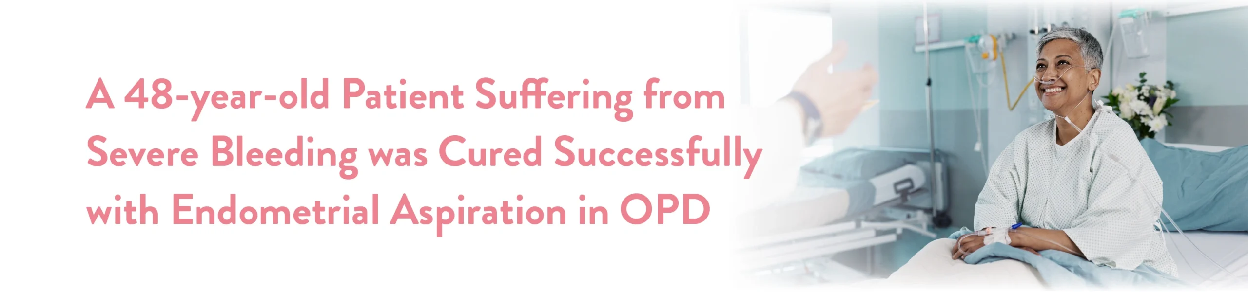
Six months ago, Dr Manjusha Goel in the Gynae Department of the CK Birla Hospital was visited by a 48-year-old female patient named Neeru who had been suffering from severe bleeding for a year.
After pelvic examination of patient Dr Manjusha Goel advised her ultrasound to check uterus lining & uterine pathology. Further history indicated that the patient was on multiple medications.
High blood pressure medicine for 5 years
Diabetes medicine for 2 years
Hypothyroid medicine for 10 years
Ultrasound showed that her uterus size was large with a small fibroid. Her endometrial lining was around 16mm thick. The patient had been bleeding continuously for 15 days and her Haemoglobin had dropped to 8gm percent.
As the patient was approaching her menopause, Dr Manjusha recommended an endometrial aspiration in OPD. With proper medical consultation, Mirena was inserted when no atypia was seen in the biopsy report. Based on her past medical records, an option of conserving the uterus and surgical procedures was discussed with her. She was given oral iron and other guidance to ensure that her other medical conditions remained in check.
Thanks to a quick and thorough OPD diagnosis from our medical expert, the patient conserved her uterus successfully. Now, she is happy and doing well.
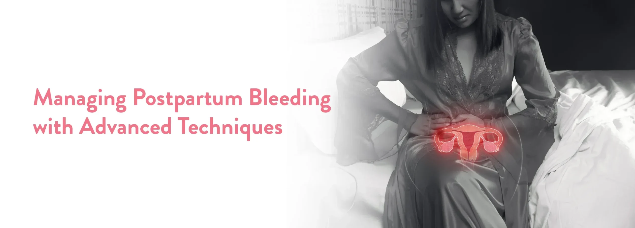
A 39 years-old-female presented at the CK Birla Hospital. The patient had normal vaginal delivery 15 days back at a reputed hospital in south Delhi. She had a history of caesarean section in her first pregnancy and had multiple episodes of heavy bleeding (Postpartum Haemorrhage) since her delivery & now was severely anaemic despite receiving blood transfusion.
The patient was prescribed detailed investigations including blood tests and ultrasound. After evaluation it was found that the bleeding was caused by retained tissue of placenta. This can be a serious issue if not treated promptly.
Dr Tripti Raheja immediately decided to perform a minimally invasive procedure using both laparoscopy and hysteroscopy, which allowed her to look both inside the uterus and inside the abdomen to assess the situation. During the procedure, She discovered that the right half of the scar from her previous caesarean section was ruptured and placental tissue was adherent to the inner wall of the uterus in the surrounding area.
Using the guidance from laparoscopy, She performed a suction and evacuation to remove the remaining tissue safely. Afterward, the ruptured caesarean scar was also stitched laparoscopically. The patient recovered well after the procedure.
This case highlights the importance of timely intervention and the use of advanced Minimally Invasive Techniques (MIS) like Laparoscopy & Hysteroscopy for better outcomes in postpartum complications without the need of an extensive laparotomy.
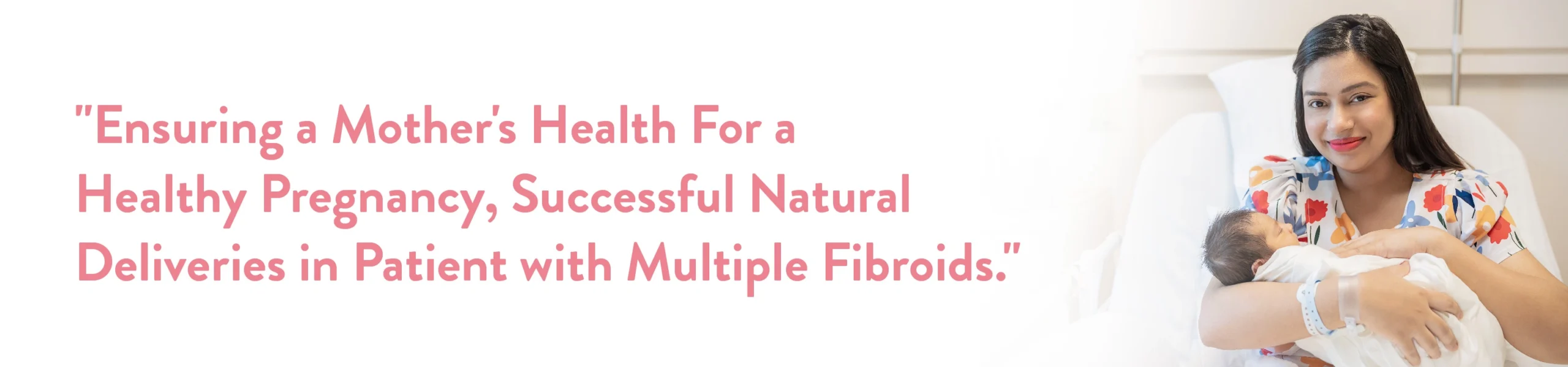
A pregnant woman with multiple large uterine fibroids, presented at the CK Birla Hospital, Gurgaon. These fibroids, measuring 6×6 cm, 6×7 cm, and 7×8 cm, posed a considerable challenge to her pregnancy. Typically, such cases are complicated and carry a high risk to both the mother and the unborn child.
The patient’s journey began with concerning symptoms of delayed periods and vaginal spotting on & off. Her initial presentation revealed a woman who was severely anaemic, pale, and thin, with a BMI of only 20. Detailed investigations, including ultrasound, were conducted, which unveiled a 6-week pregnancy with multiple uterine fibroids of significant sizes. Anaemia further complicated her antenatal condition, as she struggled with abdominal cramps due to these growing fibroids. Despite limited space for the foetus to grow, her body managed to gain 2.4 kg of weight, battling for adequate blood supply. The pregnancy progressed to 39 weeks, and she delivered vaginally through natural birth, which proved to be a lifesaving decision, sparing her from blood loss and ensuring a faster recovery compared to a caesarean section.
Dr Aruna Kalra, Director of Obstetrics & Gynaecology at CK Birla Hospital, Gurgaon, played a pivotal role in the patient’s journey. To mitigate the risks associated with fibroid-complicated pregnancies, the medical team initiated a comprehensive management plan. Anaemia was addressed through iron therapy, and preterm labour was prevented with uterine relaxant and anti-inflammatory medications. Despite the challenges posed by the fibroids, the patient’s determination, coupled with careful medical intervention, allowed for a successful vaginal delivery.
3 months post this delivery, the patient underwent a laparoscopic myomectomy to remove the uterine fibroids. Remarkably, uterine reconstruction was performed perfectly without the need to open the uterine cavity. This surgical procedure ensured that the patient could consider a second pregnancy naturally, and she did so after six months.
During her second pregnancy, rigorous monitoring and follow-up were implemented due to the scar from the myomectomy. As expected, the second baby displayed healthier growth in terms of weight. Continuous surveillance and care were provided for the myomectomy scar. Ultimately, the patient.
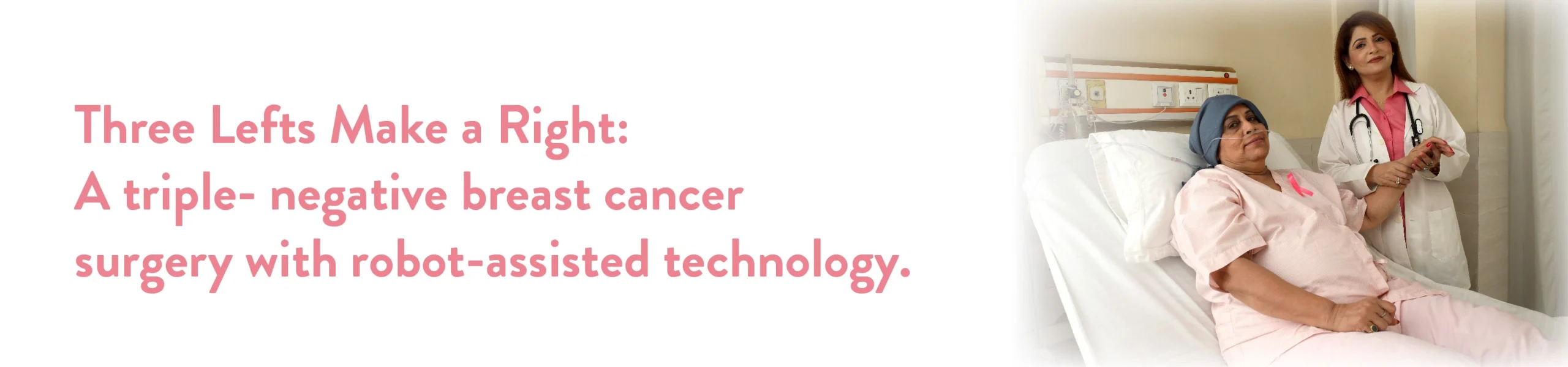
A few months ago, we diagnosed a 37-year-old woman with an aggressive form of breast cancer, medically categorized as Triple Negative Cancer. The cancer was located in the upper outer part of her breast and had spread to one lymph node in her armpit
Upon careful examination and subsequent investigative tests, it was found that the cancer was in its early stages. She began Neo Adjuvant Systemic treatment, which included chemotherapy, immunotherapy, high doses of IV vitamin C, and natural supplements. For Triple Negative breast cancer, systemic therapy is given before surgery to evaluate the treatment response
Patients who respond well usually have a better prognosis. Performing surgery first would prevent this assessment. Fortunately, she responded very well to the treatment, with scans showing no significant cancer activity in her breast and armpit.
Dr Mandeep S. Malhotra and his team at the CK Birla Hospital, Gurgaon, performed a robot-assisted functional breast preservation surgery in the following steps to eliminate the cancer tissue completely:
We supported the patient with subsequent dressing and precautionary measures to consider post-surgery, helping her resume her quality of life with a speedy recovery. At this point in her life, the patient feels incredibly relieved. She started with Triple Negative Breast Cancer, but now she is cancer-free and classified.
Her breast looks similar to how it did before. She is looking forward to enjoying a happy family life with her child and partner. With the dexterity and precision provided by robotic systems, it is easy to operate on delicate areas with complete ease

A 28-year-old new mom was diagnosed with an advanced stage of breast cancer. She was treated with systemic therapy consisting of chemotherapy with Anti Her 2 Neu targeted drugs, supported by high-dose IV Vitamin C and natural supplements. She had a very good response to systematic therapy and was keen to be cured of her breast cancer with minimal mutilation.
Post the Neo Adjuvant treatment, a PET CT scan was performed. Upon review, the primary tumour site as well as the regional lymph nodes showed no active disease. The surgery was subsequently planned.
Dr Mandeep S Malhotra and his team from the CK Birla Hospital, Gurgaon, meticulously planned the surgery in the following steps to eliminate the cancer tissue and reconstruct the breast back to its original size and shape:
Step 1: Sentinel Lymph Node evaluation of axillary disease
Step 2: Robot-assisted skin-sparing resection of the primary tumour site
Step 3: Robot-assisted Latissimus tissue harvesting for reconstruction
Step 4: Latissimus tissue inserted in Sentinel Lymph Node (SLN) evaluation helps determine if cancer has spread to the lymph nodes in the axilla (armpit).
The sentinel lymph node is the first lymph node to which cancer cells are likely to spread from the primary tumor. A skin-sparing mastectomy is a surgical procedure commonly used in breast cancer treatment. The breast tissue is removed while preserving as much of the overlying skin as possible. Using robotic assistance, we were able to perform the surgery with minimal scarring, higher precision, and no skin necrosis.
Robotic assistance allowed us to harvest Latissimus dorsi tissue through an axillary incision only, thus avoiding any scar on the back and saving a lot of time in repositioning. The latissimus dorsi is a large muscle in the back, and its tissue, along with its blood vessels, can be relocated to the chest to reconstruct a natural-looking breast Using this step-by-step process, the team performed the surgery successfully, maintaining complete cosmetic aesthetics
We assisted the patient with subsequent dressing and precautionary measures to be considered post-surgery, helping her resume her motherhood sooner without compromising her appearance.
Robotic assistance was instrumental in performing this highly delicate surgery. It preserved the original breast skin without any ischemia and loss of sensation. It avoided the unsightly incision on the back. The latissimus tissue was harvested from the axilla and brought to fill the breast defect. The patient came in with breast cancer and walked out cancer-free with the same functional breast in just 24 hours.
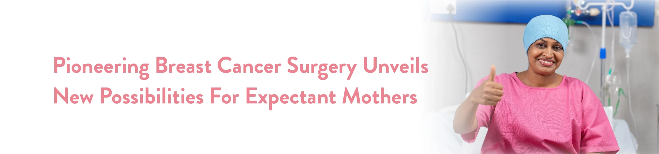
A pregnant lady sought guidance at the CK Birla Hospital Gurgaon, upon discovering a growing lump in her breast during the early stages of pregnancy. Dr Aruna Kalra initially examined her and subsequently referred her to Dr Rohan Khandelwal, our distinguished onco-surgeon specializing in breast cancer treatment.
Dr Khandelwal’s evaluation revealed a sizable 7 x 8-centimeter lump in the patient’s right breast. Biopsy results indicated a Phyllodes tumour, a type of growth differing from typical breast cancer, yet potentially malignant.
Despite being in her first trimester, surgical intervention was cautiously, considering anaesthesia risks and limited medication options during pregnancy.
Continuous monitoring and routine follow-ups occurred throughout the pregnancy across all trimesters. Remarkably, the patient experienced an uneventful delivery and successfully breastfed from the treated breast post-surgery.
Six months ago, Dr Manjusha Goel in the Gynae Department of the CK Birla Hospital was visited by a 48-year-old female patient named Neetu who had been suffering from severe bleeding for a year.
After pelvic examination of patient Dr Manjusha Goel advised her ultrasound to check uterus lining & uterine pathology. Further history indicated that the patient was on multiple medication.
As the patient was approaching her menopause, Dr Manjusha recommended an endometrial aspiration in OPD. With proper medical consultation, Mirena was inserted when no atypia was seen in the biopsy report. Based on her past medical records, an option of conserving the uterus and surgical procedures was discussed with her. She was given oral iron and other guidance to ensure that her other medical conditions remained in check.
Thanks to a quick and thorough OPD diagnosis from our medical expert, the patient conserved her uterus successfully. Now, she is happy and doing well
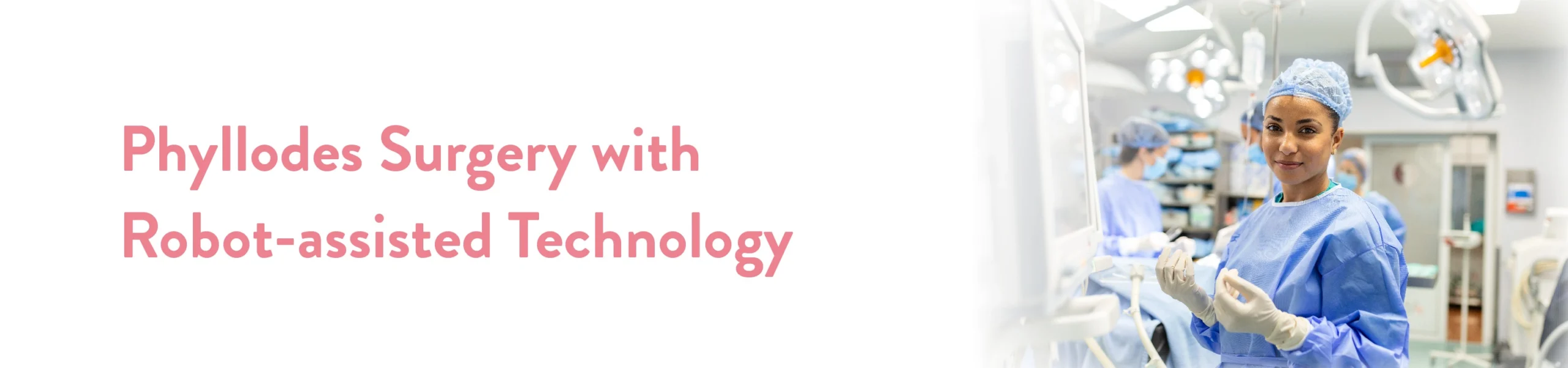
A 36-year-old woman with a 6 cm lump in her breast presented at the CK Birla Hospital Gurgoan. A biopsy revealed that it was a phyllodes tumour. Unlike breast cancer that starts in the breast ducts, phyllodes tumours originate from the connective tissue in the breast. This type of tumour is classified as a sarcoma, differing from the more common breast carcinoma.
Upon careful examination and subsequent investigative tests, it was confirmed to be a sarcoma. The treatment for a phyllodes tumour involves surgical excision with clear margins, meaning the tumour is removed along with some healthy tissue around it.
The patient, being young, was very concerned about losing her breast and how it would look afterward. This psychological concern is common among patients who fear the disfigurement of their breast. During the examination, it was found that the tumour was in the upper part of the breast and extended to the nipple, but the overlying breast skin and nipple were not involved in the disease.
Dr Mandeep S. Malhotra and his team at the CK Birla Hospital, Gurgaon, performed a robot-assisted functional breast preservation surgery in the following steps to completely eliminate the tumour:
A circumareolar incision (a small cut around the areola) was made Robotic arms were used to reach the upper part of the breast and mobilize the tissue, including the tumour.
The tumour was then removed with ample margin space, ensuring enough healthy tissue around it. Latissimus tissue from the back was harvested using the precision of robotic arms. The tissue from the back was moved with the robot, without making any cuts in the back, to fill the space where the tumour was removed.
We supported the patient with subsequent dressing and precautionary measures post-surgery, helping her resume her quality of life with a speedy recovery. Based on the response from her follow-up visits, the patient is very satisfied and happy to have retained her aesthetic feminine form With robotic surgery, we can perform fine precision-based surgery on delicate malignant tumours that are dangerous unless completely removed. Upon recovery, the patient’s breast is similar to its original state with the same functionality and sensation.
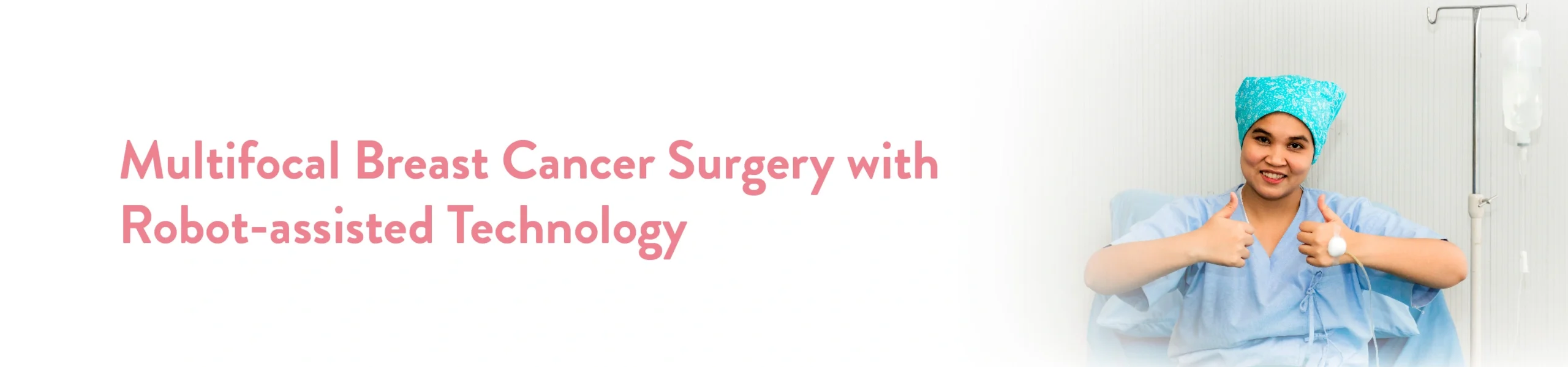
A female patient in her late 60s came to the CK Birla Hospital with a primary diagnosis of breast cancer. Her initial test reports revealed two cancers in her left breast, one at the 1 o’clock position and another at the 3 o’clock position. This required further investigation to determine the stage of the cancer, as breast cancer in older patients can be particularly deceptive.
Upon careful examination and subsequent investigative tests, it was found that the cancer was fortunately in its early stages. Importantly, the skin was not involved, and further evaluation showed no apparent signs of axillary spread in the armpits, which is the first place breast cancer typically manifests. Based on these findings, the team planned the patient’s surgery.
Dr Mandeep S. Malhotra and his team at the CK Birla Hospital, Gurgaon, performed robot-assisted functional breast preservation surgery in the following steps to eliminate the cancer tissue completely:
An incision was made in the lateral mammary crease, extending into the axilla Using robotic assistance, the breast tissue from the 12 o’clock to 4 o’clock positions was excised, enabling a nipple-sparing approach A sentinel lymph node biopsy was performed through the same incision Robotic assistance enabled the team to harvest latissimus tissue from the back and fill the breast defect.
This single 5-6 cm lateral mammary crease incision allowed for the precise and complete removal of multifocal breast cancer. It also facilitated the harvest of latissimus tissue from the back, reconstructing the bres size, shape, and functionality.
We supported the patient with subsequent dressing and precautionary measures to consider post-surgery, helping her resume her quality of life with a speedy recovery. The patient came to us with a life-threatening condition and walked out cancer-free in just 24 hours. Delicate, life-saving surgeries rely heavily on precision, and in this case, the dexterity of the robotic arms and small incisions helped achieve successful clinical outcomes.
The patient had a history of 8-10 admissions in 4-5 months at various hospitals in various regions for the aforesaid
complaints. She has been receiving treatment for complicated pneumonia, non-resolving pneumonia, organising
pneumonia, long-haul COVID, complicated pneumonia & septicemia. The treatment protocols included broad-spectrum IV and nebulised antibiotics, nebulisation, steroids, antifibrotics, oxygen therapy, BIPAP, anticoagulants, antitussives, and symptomatic medication.
However, she remained unrelieved of her symptoms, and her clinical condition continuously worsened. She was referred to us for further management in a critical state.
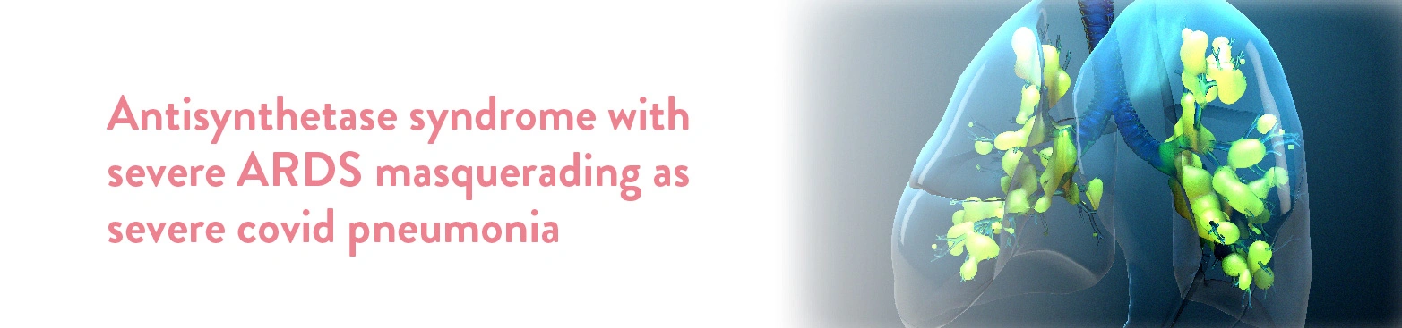
A 39-year-old female patient from Madhya Pradesh was admitted to the Department of Pulmonology at the CK Birla Hospital. The patient complained of severe breathlessness – Grade 5 MRC even on high flow oxygen support at 15 lit/min. She was also affected with severe Type 1 respiratory failure and severe paroxysmal dry cough, headache, severe generalised body ache and high grade fever for the last four to five months. Patient autoimmune profile was strongly positive for anti jo antibody which is specific for patients suffering from inflammatory myopathies which is seen very rarely and commonly misdiagnosed.
Anti synthetase syndrome is a rare autoimmune disorder that can affect multiple body parts. In this condition, certain antibodies target specific proteins in the body that lead to the abnormal functioning of the immune system. As a result, this condition causes diverse symptoms, including inflammation of the muscles and interstitial lung disease.
ARDS stands for acute respiratory distress syndrome. In this condition, excess fluid fills up in the air sacs (alveoli) of the lungs causing less oxygen intake. Common symptoms of ARDS include severe shortness of breath, low blood pressure and abnormally rapid breathing.
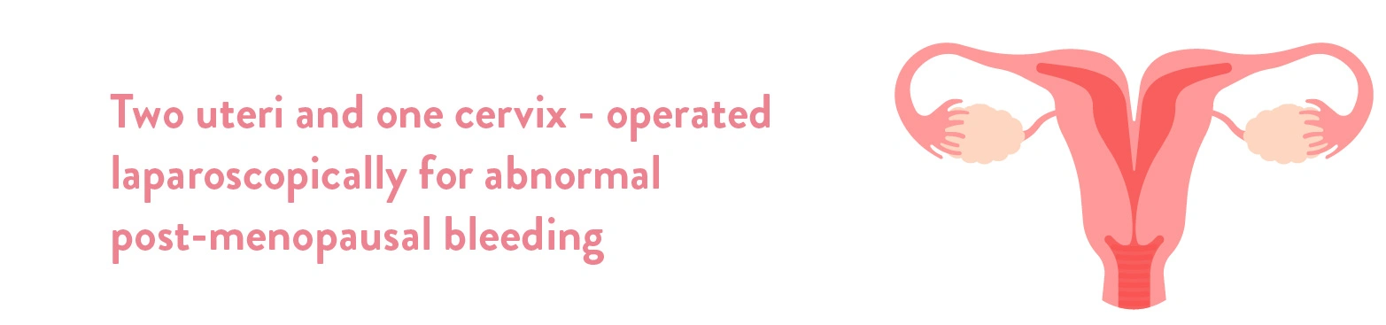
A 56-year-old female patient presented at the Department of Gynaecology at the CK Birla Hospital®, Gurugram. She presented with chief complaints of post-menopausal bleeding. After extensive examination and investigations, it was
discovered that the patient had a double uterus.
Uterus didelphys or double uterus is a rare anomaly. It accounts for 8% of all female reproductive tract congenital anomalies. It occurs in 0.3% of the total population.
Repeated miscarriages, mid-trimester abortions, cervical incompetence, preterm labour or stillbirth are a few of the poor obstetric outcomes in females with a double uterus. In addition, these abnormalities are often associated with abnormal positions of kidneys such as pelvic kidney or single kidney.
The patient reported having birthing and pregnancy complications in the past. She lost her first child after preterm labour. The pregnancy must have occurred in the smaller, less developed uterus, which could not provide space and nutrition to the developing foetus. And due to a weak uterus (womb), intrauterine death of the baby occurred, which is a very common complication with uterine abnormalities.
Her second and third pregnancies were in the more developed left uterus, which carried till 9 months. However, vaginal delivery was not possible. Hence, both the deliveries were caesarian sections through a vertical incision on the abdomen.
A 47-year-old international patient came to Dr Mayank Madan for a consultation with the following problems:
Based on the patient’s age and condition, further investigative procedures were carried out to get a clear picture of the problem.
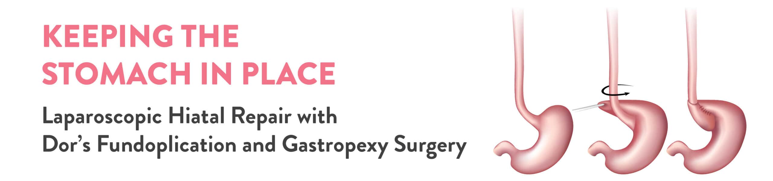
An Upper GI Endoscopy was performed which revealed Hiatal Hernia with a dilated stomach and mesentero-axial volvulus. Simply put, there was an abnormal rotation of more than 180 degrees, around a transverse axis connecting the middle of the greater and lesser curvatures of the stomach. This was due to the protrusion of the stomach, through the oesophagal opening in the diaphragm.
Further, a Barium meal study was performed which revealed mild luminal attenuation with mucosal irregularity in the middle of the stomach. There was no existence of any significant obstruction most likely as a result of previous GIST surgery. Based on these findings the patient was diagnosed with Hiatal Hernia with gastric volvulus.
Upon careful assessment of the findings, Dr Mayank Madan and his team decided on performing the Laparoscopic Hiatal Repair with Dor’s Fundoplication and Gastropexy done under general anaesthesia.
During surgery, it was found that there were dense omental and bowel adhesions due to the existence of a previous midline scar. To avoid causing any bowel injury, an adhesiolysis was performed to meticulously avoid any complications due to injury.
Further, the whole of the greater curvature of the stomach was stuck to the left lobe of the liver, causing the volvulus. To repair this, a meticulous dissection of adhesiolysis was performed to separate the organs, which led to the derotation of the volvulus formed. There was approximately 3 cm of hiatal defect noted. This was repaired by pulling back the derotated stomach into the abdominal cavity during gastropexy.
During the gastropexy, an anterior wrap of 180 degrees was made and a bougie (36F) was manoeuvred into the stomach cavity. It was suspected that the middle of the stomach might be narrowed due to the hernia, but the bougie could be successfully cleared past the suspected area. Finally, three transfascial sutures were made to secure the stomach to the abdominal wall or the diaphragm. This will further prevent the stomach from moving up into the chest cavity.
The long and eventful surgery was executed successfully without any further injury or operative complications thanks to the laparoscopic approach. Post-surgery, the patient was managed with IV fluids, IV antibiotics, IV antacids, analgesics and other supportive measures to ensure the digestive system is back on track.
Diet was a vital part of the post-operative care, which was mostly liquid from the next day of the surgery. The patient responded well to the surgery and no further complications surfaced. Within 48 hours of the surgery, the patient was discharged as he was hemodynamically stable and showed good signs of recovery.
During the first follow-up after one week, his port site staples were removed and he was sustaining his liquid diet with no complaints of regurgitation. We are happy to have helped another patient regain their health and attain proper relief from their prolonged discomfort.
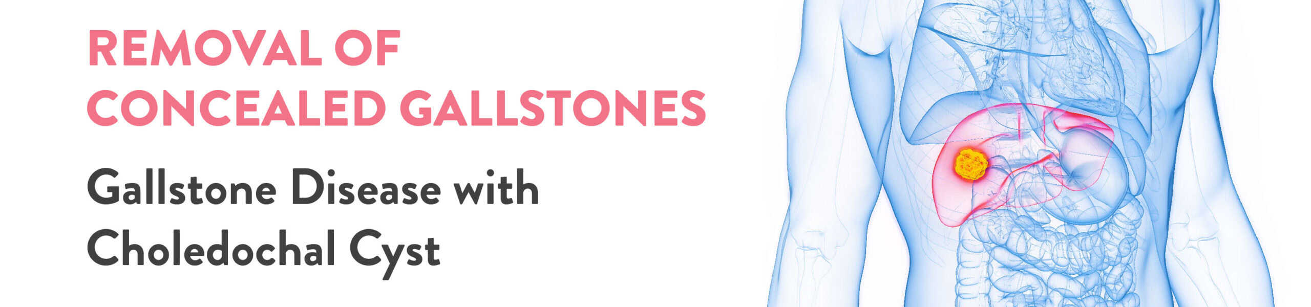
Recently we found ourselves in a unique case of Gallstone disease. An elderly lady came to seek consultation with Dr Mayank Madan & Dr Saurabh Garg, at the CK Birla Hospital, Gurugram, with a dilated common bile duct (CBD) and pain. The most common causes for these are CBD stones, chronic pancreatitis, and periampullary diverticulum.
Based on her age and condition, CBD stone had the highest probability, and it could prove to be fatal. To confirm this further investigative procedures were carried out.\
Women are at a higher risk of getting gallstones due to hormone imbalance and obesity. A Magnetic resonance cholangiopancreatography (MRCP) was carried out to obtain accurate imaging of the main area of concern. MRCP results showed no calculus in CBD. Biliary calculi suggest the presence of stones in the main biliary pathway which transmits the bile. This did not rule out gallstones but was indicative of choledochal cysts. To relieve the pain this needed to be surgically removed.
Intraoperatively it was seen that the gallbladder was distended with omentum parietal adhesions. A distended gallbladder implies that it was swollen or enlarged and does not function properly. Adhesions signify the presence of inflamed scar tissues that need to be repaired.
In this case, the CBD was dilated in a fusiform way throughout the whole extrahepatic course. Finally, it was confirmed that the patient had a Choledochal cyst (type 1). To repair this, the cystic duct was flushed along with the dilated cyst. And this contained a large stone.
Post menopause the levels of oestrogen and progesterone fall leading to the perfect cause for developing gallstones. This patient unfortunately ticked all the boxes. Given the presence of a large stone, it was decided that the gallbladder would be removed.
The stone was milked back into the Gallbladder and the organ was extracted after suturing the lateral CBD wall with sutures. The surgery was a resounding success.
Post-operative care in this case is vital and needs ample support from a good diet to help the body adjust to the changes post-removal of the gallbladder. We are happy that we could give the patient another chance to lead a happy, healthy and painless life.
Mr Botor from Uzbekistan came to us with an advanced case of Rheumatoid Arthritis. At the young age of just 33 years, he had already undergone total hip replacement surgery. The surgery was performed five years ago but the patient showed no improvement in his condition.
When he met Dr Ashwani Maichand at the CK Birla Hospital(R), Delhi, his knees were severely deformed, and the pain was unbearable. Due to this, he was walking with the help of crutches. He was on a high dose of steroids to manage the pain.
Based on the patient’s current condition and medical history, further investigations were carried out for a more precise diagnosis. The results revealed that the patient was suffering from osteoporosis which led to further deformity. With all these aspects kept into consideration, a thorough evaluation was carried out. Dr Maichand also discussed the findings with the patient’s wife who is a gynaecologist and understood the technicalities of the case.
Dr Ashwani Maichand concluded that Mr Botor will be greatly benefitted from Robotic M.I.S. knee replacement.
Robotics is the latest technology that has revolutionised high-precision surgical procedures worldwide. They not only help surgeons execute surgeries with precision, but also provide real-time AI-based backup to deal with and manage any kind of problems that might crop up during the surgery.
The primary reason Dr Ashwani Maichand opted for this procedure was to ensure quick recovery for a persisting problem that was only deteriorating with time. With robotic surgery patients can be assured of:
Given the amount of pain and discomfort the patient had endured over time, this was the best fit for him. Needless to say, the advanced skill set of Dr Ashwani Maichand helped ensure that the surgery was successful.
Post Operative Care
Post-surgery it was observed that the previous deformities were fully corrected. The patient was walking without support within 48 hours of his surgery. By day 3 he was also able to climb stairs.
Our commitment to delivering the highest standards of clinical excellence reflects in the combination of latest robotic systems with specialised surgical techniques which ensure a good quality of life for the patient in the long run.
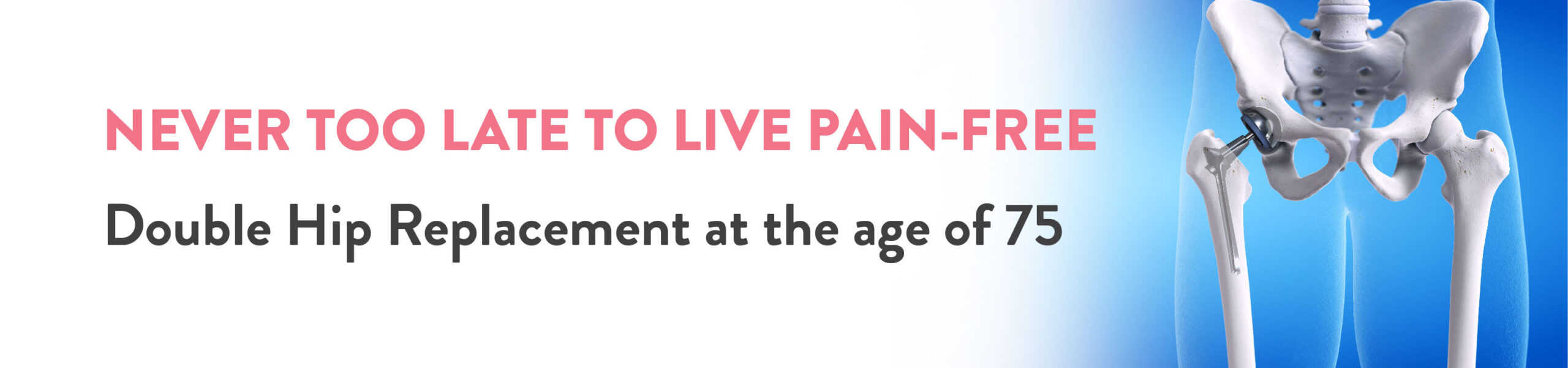
A 75-year-old male patient came to the Orthopaedic Department at the CK Birla Hospital, Punjabi Bagh with an ongoing ailment of Ankylosing Spondylitis. He showed up with all the complications of Ankylosing Spondylitis which had enhanced due to age.
The significant physical symptoms that are visible in patients with this condition are:
The optimum solution for this, based on physical examination, was surgery. But given the age of the patient, before deciding on surgery a thorough screening process was required to make sure the patient would be able to sustain the treatment.
The patient got a full screening done to help the surgical team understand the potential risks involved with respect to the patient’s current comorbidities and age. The patient got imaging and blood tests to arrive at a comprehensive view of the patient’s existing overall health conditions. Upon careful evaluation, the team decided to proceed with Total Hip Replacement (THR) Surgery.
Since the surgical team was planning a Total Hip Replacement (THR) Surgery, for both hips, the potential surgical complications need to be addressed. In this case, it was essential to weigh the pros and cons of the surgical techniques: open technique or MIS technique. But there are several complications to using the Open Technique for Total Hip Replacement Surgery.
But this was not the ideal approach, given the patient’s age. To provide prompt treatment and manage the condition better, Dr Ashwani Maichand and his team opted for the Minimally Invasive Surgical method for Total Hip Replacement Surgery. The surgery was successfully performed and the patient did not have to experience unnecessary complications.
Using the MIS technique, the surgical outcomes for the patient were well noticed. Compared to the more laborious technique, this gave the patient no complications that would lead to blood transfusion. Therefore no ICU stay was required either. Post-surgery the patient was able to get back on his feet and walk within the next 24 hours. With effective rehab exercises, the patient was able to climb stairs in the next 48 hours. With the right technique, we were able to preserve the functionality of the patient’s legs which was imperative to maintain a good quality of life.
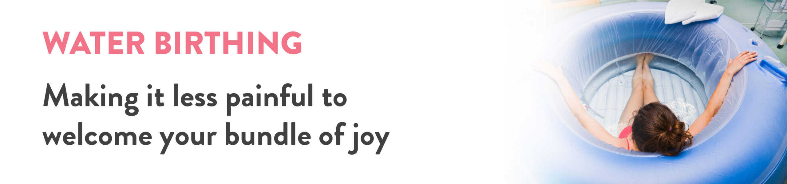
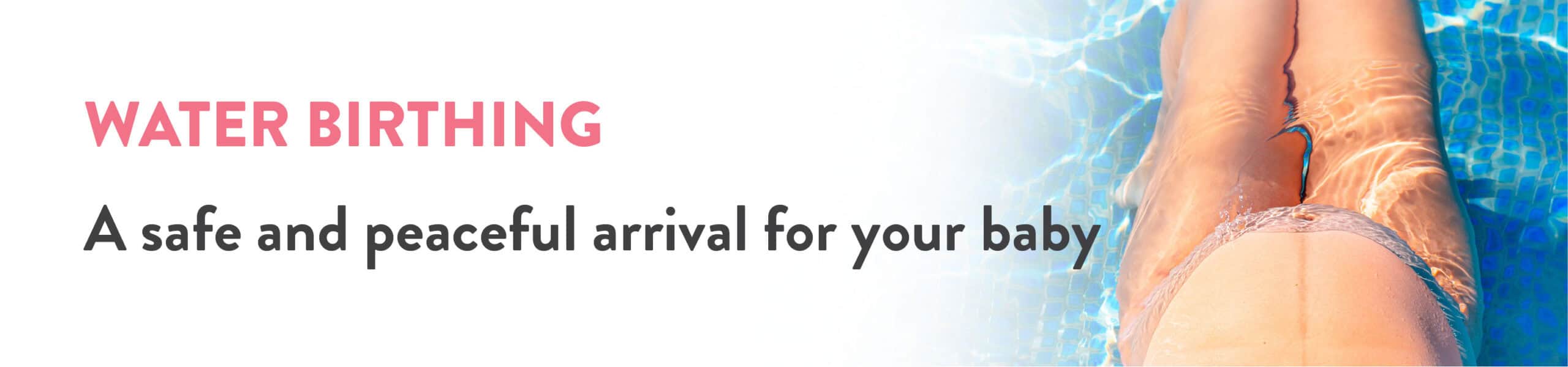
The CK Birla Hospital has been the pioneer in establishing the water birthing technique of delivering babies in Delhi NCR. But why do they prefer such a unique birthing technique? Read on to know more…
The technique is safe for babies and they are as healthy as others born using traditional birthing techniques. But the following needs to be addressed while planning for a water birth.
Medical guidelines is paramount under all circumstances. Make sure the procedure is carried out in the presence of the expert doctor who has experience & training in water birthing and trained nurses. Care must be taken so that the baby does not swallow water and is pulled out at the right time.
A firm believer in the lamaze techniques, Shruti discovered water birthing while she was attending a group yoga session while pregnant with her first child. She was keen on opting for water birthing, and was searching for a reliable healthcare partner and a doctor who would be fully trained in all the nuances of water birthing. “What got me intrigued were the potential benefits of water birthing in managing labour pain”, she said as she held her healthy newborn.
This is when a fellow expecting mother told her about Dr Deepika Aggarwal at the CK Birla Hospital, Gurugram.
Shruti was in her 8th month of the third trimester, with no complications in her pregnancy. This made her a suitable candidate to opt for water birthing. “When I confirmed her health conditions, I discussed in detail regarding water birthing process & her birth plan. I then suggested that she look into my videos about water birthing before incorporating the necessary changes in her customised birthing plan. Patient’s comfort level is a primary indicator for selecting this birthing procedure”, said Dr Deepika Aggarwal.
Further, she interacted with a water birth specialist nurse who was assigned to her and built the initial rapport necessary to facilitate the entire process. Now all she has to do is visit the hospital when she starts labour.
After a successful water birth under Dr Deepika Aggarwal at the CK Birla Hospital, she was delighted with the outcome. Speaking about the procedure, she said, “my Indian friends in London and California have opted for water birthing and I am grateful that I found the same in India. I believe more and more people in India should know about it and the practice definitely needs more awareness.”
On the day she came around 2 AM but was found to be in latent phase at 2 cm dilatation. She was advised to go home and re attend hospital with hood contractions (3 to 4 in 10 mins). She then returned at 5.30 AM nearly fully dilated. Under Dr Deepika’s direct supervision, she went into the pool and successfully delivered a healthy baby girl.
Speaking about Dr Deepika, she added that, “She has been an absolute blessing for me and my baby. I changed my doctor when we were supposed to book the hospital for delivery in my 8th month with a host of apprehensions.But I couldn’t have made a better choice. From the first day she made me comfortable and prioritised my choices and needs. Her team is also very sweet and co-operative. Would definitely recommend her to every expecting mother seeking a reliable doctor to guide her through her pregnancy journey.”
New mom, Neha Sood discovered water birthing during the 2nd trimester of her first pregnancy. She was undergoing a hypnobirthing course where she first got to learn about the pain alleviation mechanism and its benefits in managing labour pain. Her first baby was born via water birthing in Mumbai.
Two years later she moved to Gurgaon and was expecting again. “Given how beautiful my first water birthing experience was, I started searching for healthcare providers in NCR who specialise in water birthing. During my research I found that hypnobirthing was more easily accessible in Mumbai, Hyderabad and parts of Kerala. However, in NCR, the only credible name I could find was Dr Deepika Aggarwal at the CK Birla Hospital, Gurugram.”
Recalling their first interaction with Dr. Deepika, she said, “The first meeting itself gave us the confidence to proceed with her, especially because the CK Birla Hospital has always been known for their keen focus on women’s health, backed by an elite doctor panel and preference for natural birthing methods. This just seemed like the perfect fit for us.”
This technique is gentle for the babies. Since the transition is in a fluid mode itself.
Additionally the skin to skin contact with the mother is quickly being established which helps initiate breastfeeding easily.
The technique is safe for babies and they are as healthy as others born using traditional birthing techniques. But the following needs to be addressed while planning for a water birth.
Medical guidelines are paramount under all circumstances. Make sure the procedure is carried out in the presence of the expert doctor who has experience & training in water birthing and trained nurses. Care must be taken so that the baby does not swallow water and is pulled out at the right time.
After a successful water birth under Dr Deepika Aggarwal at the CK Birla Hospital, she was very happy with the outcome. Speaking about the procedure, she said, “my Indian friends in London and California have opted for water birthing and I am grateful that I found the same in India. I believe more and more people in India should know about it and the practice definitely needs more awareness.”
Speaking about Dr Deepika, she added that, “I loved the fact that she has always been very assuring throughout our pregnancy and birthing journey. As a knowledgeable expert she always gave patient and detailed explanations backed with medical research on hundreds of our questions with a smile. We felt safe and comfortable going for a Water birth with her and she was always accommodating to my pursuits of a perfectly natural delivery. She made some acute recommendations on my birthing position to ease out the baby and reduce tears, which was another big win for me. I believe she’s a great combination of an astute doctor and a warm friendly figure whose diagnosis you can completely trust. I wish her more success in her mission to make water birthing more mainstream and help women enjoy the joys of natural birthing.”
A 71-year-old male patient came to Dr Debashish Chanda at the Orthopaedic Department of the CK Birla Hospital, Gurugram complaining about pain in his Right leg. The patient had undergone total knee replacement in both his legs: the left leg surgery was done in 2011 and the right leg surgery was done in 2013. The Right knee was causing more pain making it difficult for the patient to walk without the help of painkillers.
When the patient came to us, he had already undergone knee replacement, but despite his problems in alignment, he was discouraged to not undergo a revisional surgery saying that it makes the joint more fragile and weak. Also, there’s a myth that 2nd surgery is risky in redo cases. But that is not the case at all.
Given the age and previous history of Total Knee Replacement, the patient got a full screening done to help the surgical team isolate the problem. A varus knee alignment test was also performed to check for alignment errors. Upon physical examination, the alignment error was confirmed as the patient was experiencing limping and shorting on the right leg. With proper evaluation and due respect to the patient’s age, the team decided to proceed with Joint Transplant Surgery or Revised Total Knee Replacement Surgery.
Varus Knee can happen when the natural alignment of the bones shifts or moves by as low as a millimetre which shows up in the patient’s stride when he limps or uses a stick to walk. So, the whole approach to revised knee replacement was simple: rectify the alignment defects, and transplant the knee joint completely.
From the standpoint of an aged patient, the benefits of this joint transplant surgery are:
Joint replacement over time has become more safer and effective. The advancements in this procedure are known to lead to a better level of postoperative outcomes. The operation was successful and the team finally performed alignment correction and redid the joint transplant.
Post-surgery, the patient did not need any ICU stay. He was able to stand & walk within 24 hours of surgery & was safely discharged as normal primary (1st time) knee replacement within 48 hours.With regular physiotherapy and rehab exercises, his reviews revealed that his problem had completely healed within 3 weeks of the surgery. Now he leads a happy and content life doing everything he always missed, he can now even sit with crossed legs after this surgery.
A 45-year-old male patient came to the Orthopaedic Department at CK Birla Hospital, Gurugram with persisting pain in his legs. The patient had a fracture in his leg 10 years ago, and the same area was now causing pain again. He could not sit for too long in one stride, as his leg would get locked and jammed, making it difficult for him to walk. His previous orthopaedic had suggested total knee replacement, but the patient waited another year before finally approaching Dr Debashish Chanda.
The patient underwent a full screening, including an alignment scanogram and a Cartilage Sequence MRI. The report revealed a very mild varus alignment issue and severe cartilage damage.
In such cases, healthcare providers often ignore cartilage damage and suggest knee replacement at a later, more mature age. However, it is always desirable to preserve one’s natural knee and tissue, even if this is a more laborious procedure requiring a skillful hand. The outcome of this procedure has a more lasting impact if executed successfully. Upon careful evaluation, the team decided to proceed with Cartilage Transplant Surgery along with other reconstructive procedures depending on the outcome of the first procedure.
Varus Knee is a condition where the alignment of the bones in the leg and knee are affected. Even a millimetre shift can result in a lot of pain. During the cartilage transplant procedure, the patient’s own cartilage cells were taken from the same knee, grown in a lab for six months, and then placed back in the final surgery with an alignment change. This implanted cartilage adhered to the bone and formed connective tissue, filling the defect completely within a few weeks.
The operation was successful, and the team finally performed alignment correction and cartilage transplant through grafting.
Post-surgery, the patient did not require an ICU stay. The review revealed that his cartilage defect had grown back in three months. The patient was able to get back on his feet and walk within the next 24 hours. With effective rehab exercises, he was able to move his leg around comfortably, even with light weights. With the right technique, the team restored the functionality of the patient’s legs and completely healed his condition in under 6 months.
The surgery was performed to ensure that the patient could retain their own natural body and get rid of the pain that prevented them from leading a normal life. We are glad our efforts have given the patient a new lease on life. He is back to his active life and is really happy with the procedure.
A 52-year-old male patient from Iraq visited the Department of Oncology at the CK Birla Hospital, Gurgaon. The patient presented with a history of jaundice. He had also undergone ERCP and a stenting procedure.
ERCP, Endoscopic retrograde cholangiopancreatography, is a procedure to diagnose and treat conditions affecting the gallbladder, liver, pancreas and bile duct. This procedure combines an X-ray and endoscope (a long, thin, lighted tube) to visualise the insides of these organs.
Stenting is a procedure in which a hollow tube made up of a fine, thin tube is used to keep an obstructed area of the GI tract open.
The patient was indicated to undergo a PET scan to produce a precise diagnosis along with other routine investigations. The findings of the investigations revealed that the patient was diagnosed with localised pancreatic cancer. The cancer was located at the head of the pancreas.
The oncological team at the CK Birla Hospital led by Dr Vinay Gaikwad counselled the patient about his recommended treatment protocol. The patient was indicated to have a laparoscopic Whipple’s pancreaticoduodenectomy operation.
Whipple’s procedure, also known as pancreaticoduodenectomy, is a complex procedure to surgically remove the head of the pancreas. The head of the pancreas is its widest part which is located on the right side of the abdomen.
Whipple’s pancreaticoduodenectomy is an effective and safe procedure for the treatment of pancreatic cancer. After removing the head of the pancreas, the surgeon performed an anastomosis with which they connected the remaining organs for normal digestion.
The patient underwent the minimally invasive surgery successfully and was discharged on the 6th postoperative day on a normal diet. The overall surgical approach enhanced the patient’s recovery and minimised pain.